University of Maryland School of Medicine logo
Emergency Medicine
All Departments
UMSOM Assistant
UMEM Educational Pearls
  • Home
  • Departments
  • Emergency Medicine
  • CME
  • UMEM Educational Pearls
  • CME
  • Courses and Conferences
  • Free Open Access Medical Education
  • UMEM Educational Pearls

Search

101-120 of 245 results with category "Visual Diagnosis"

Previous  |  1 |  2 |  3 |  4 |  5 |  6 |  7 |  8 |  9 |  10 |  11 |  ... |  13 |  Next

Title: What's the Diagnosis? Case by UMEM alumni Dr. Ari Kestler

Category: Visual Diagnosis

Posted: 6/30/2014 by Haney Mallemat, MD

Question

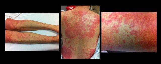
49 year-old female on trimethoprim/sulfamethoxazole presents with a rash & lesions on her oral mucus membranes. What's the diagnosis?
Show Answer

Answer: Steven-Johnson Syndrome (SJS)

  • SJS: involvement of <10% body-surface area (BSA) PLUS mucus membrane involvement (e.g., ocular, oral, genital)
  • Toxic Epidermal Necrolysis (TEN): involvement of >30% BSA PLUS mucus membrane involvement
  • Occurs secondary to immune-complex hypersensitivity reaction from viral, bacterial, or chemical exposure (Sulfonamides > PCN > Cephalosporins)
  • Management:
    • Remove possible trigger(s)
    • Manage like a severe burn (supportive care with hemodynamic support, electrolyte management, analgesia)
    • Strongly consider admission to an ICU
 

Show References

Follow me on Twitter (@criticalcarenow) or Google+ (+criticalcarenow)


Title: What's the Diagnosis?

Category: Visual Diagnosis

Posted: 6/23/2014 by Haney Mallemat, MD (Updated: 6/23/2014)

Question

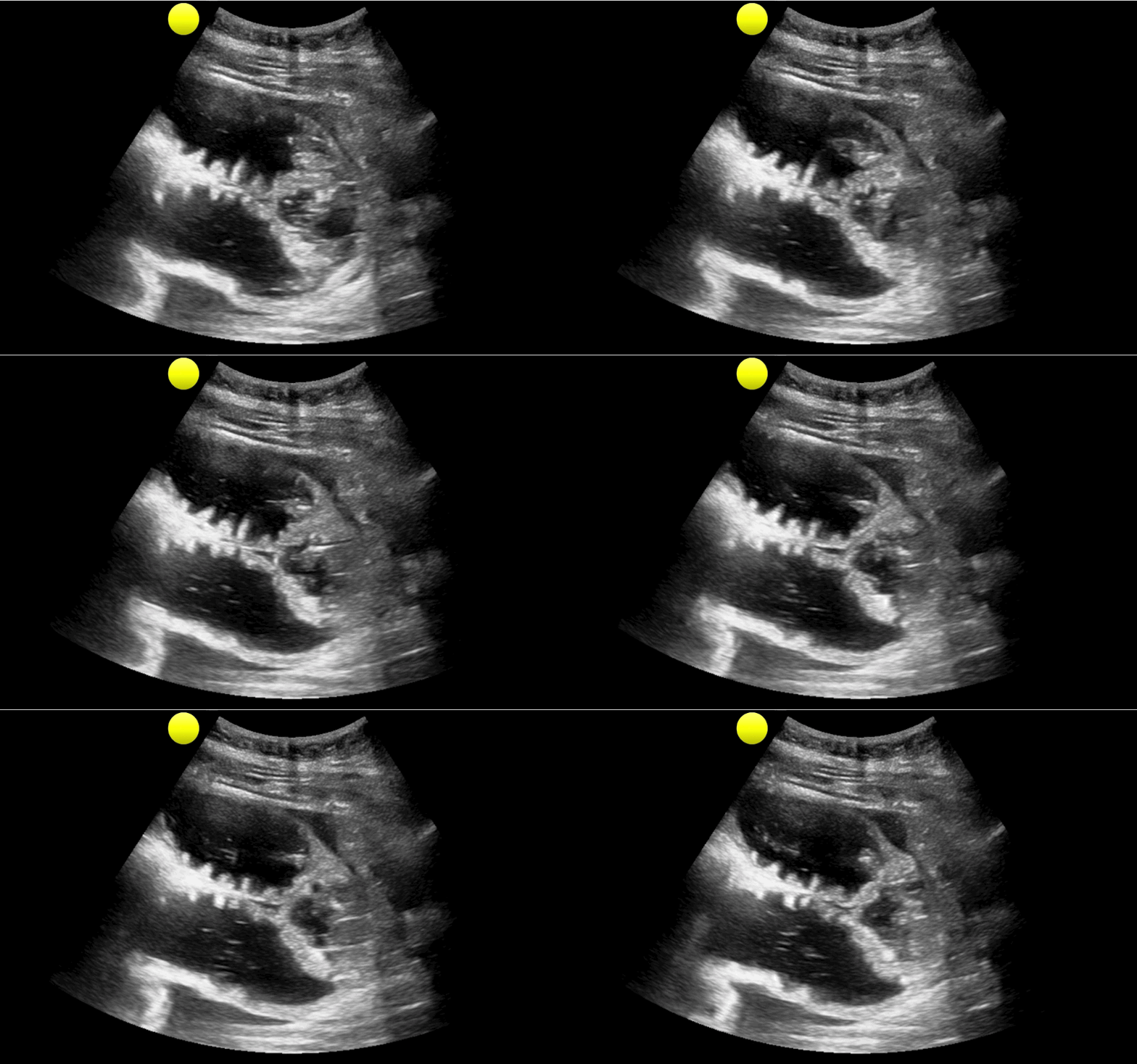
35 year-old female presents with nausea and vomiting 1 week post-op for an abdominal surgery. Abdominal ultrasound is below; what's the diagnosis? 
Show Answer

Small bowel obstruction (SBO)

Ultrasound for Small bowel obstruction (SBO)

  • Wait….ultrasound can be used to detect an SBO? Yes, it can!
  • Historically, abdominal XRay has a 77% and 57% (sensitivity and specificity, respectively) for SBO; ultrasound has 88% and 96% sensitivity and specificity, respectively.
  • Want to learn more about ultrasound for SBO? Click here for a podcast describing the technique by Mike and Matt from the Ultrasound Podcast (@ultrasoundpodcast)
  • Bottom line: Consider using ultrasound as your initial screening test for SBO, rather than an abdominal XR

Show References

Follow me on Twitter (@criticalcarenow) or Google+ (+criticalcarenow)


Title: What's the Diagnosis? Case by Dr. Swati Singh, Amanda Quiller, P.A.

Category: Visual Diagnosis

Posted: 6/16/2014 by Haney Mallemat, MD (Updated: 6/16/2014)

Question

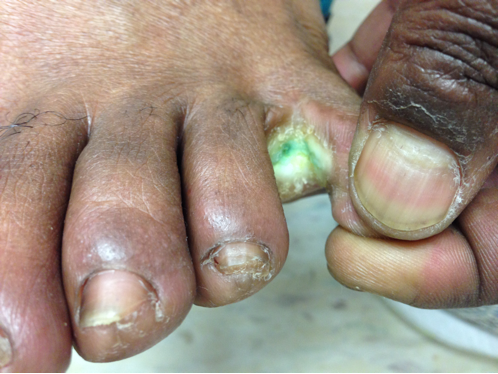
41year-old male without past medical history presents with the image below. What's the diagnosis and what's the most likely causative organism?
Show Answer

Gram-Negative web-space infection; most common etiologic organism is Pseudomonas

Gram Negative Web-space Infections

Gram-negative infections may occur in toe web-spaces of healthy patients, especially in areas with hyperhidrosis (excessive perspiration) and macerated skin.

Common organisms include Pseudomonas; others include Corynebacterium minutissimum (causes erythrasma, chronic superficial infection of the intertriginous areas of the skin), and other gram-negative bacteria

Woods-light examination may be clinically helpful if Pseudomonas is suspected (fluoresce green) and erythrasma will fluoresce coral red. KOH direct examination for fungal elements may show the presence of dermatophytes or Candida. 

Treatment:

  • Moisture reduction of web-spaces by keeping feet dry (e.g., open-toed shoes, drying powders, etc.)
  • Uncomplicated, superficial infections respond well to topical agents active against the causative organisms (e.g., gentamycin ointment)
  • Severe or complicated infections may require oral / parenteral antibiotics (i.e. third-generation cephalosporin or quinolone or aminoglycoside) plus antifungals; tissue removal/surgical debridement may be required.

Show References

Follow me on Twitter (@criticalcarenow) or Google+ (+criticalcarenow)

Wolff, K., & Johnson, R. J. (2009). Fitzpatrick's Color Atlas and Synopsis of Clinical Dermatology. (6th ed., pp. 662-664;698). McGraw-Hill.

http://www.aafp.org/afp/2005/0901/p833.html

http://emedicine.medscape.com/article/1055306-overview


Title: What's the Diagnosis? Case by Dr. Maite Huis in 't Veld

Category: Visual Diagnosis

Posted: 6/10/2014 by Haney Mallemat, MD (Updated: 6/9/2014)

Question

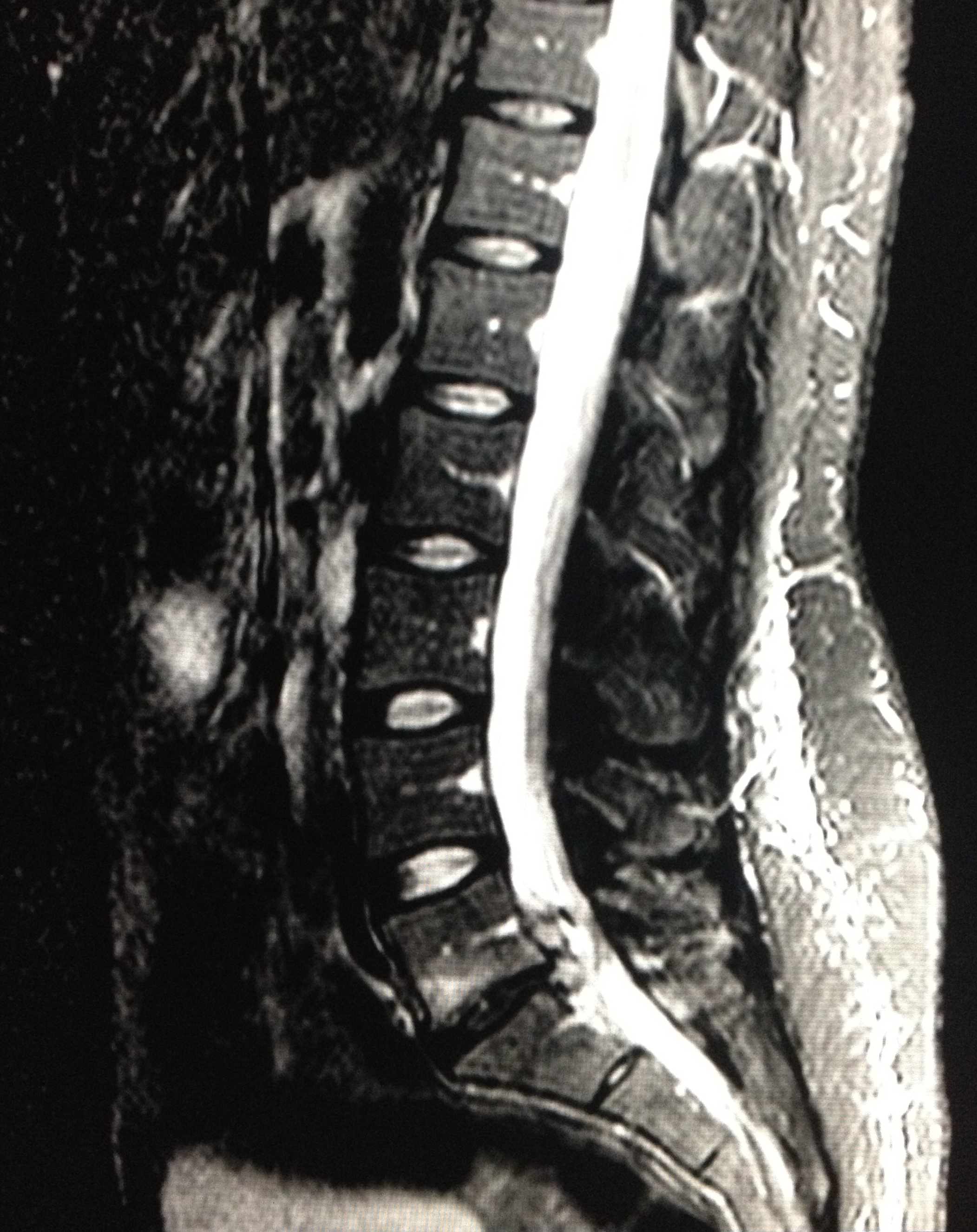
28 year-old female with history of chronic back pain presents with right-foot numbness and inability to move her foot at the ankle joint. What’s the diagnosis and what neurologic finding would you expect to find?
 

Show Answer
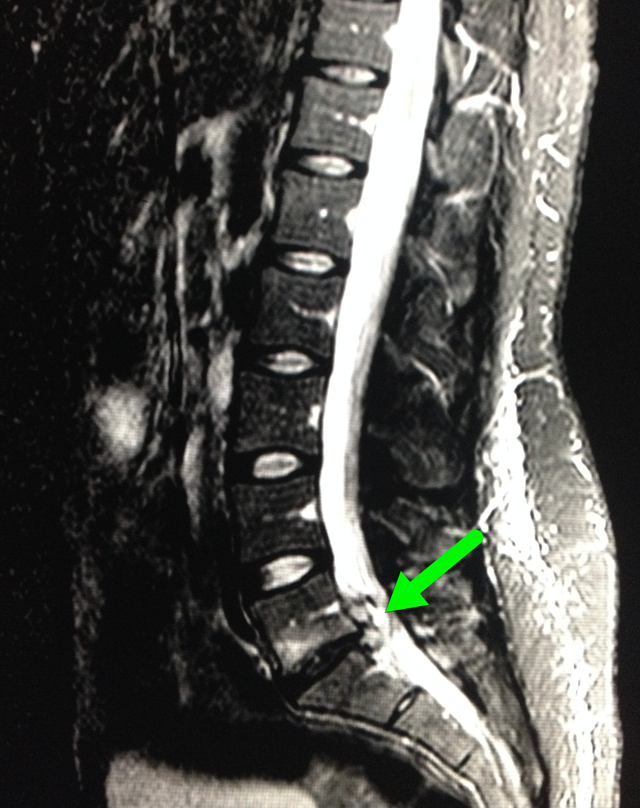
Large disc bulge at L5-S1

  • Patient in the case was found to have reduced:
    • Dorsiflexion (2/5)
    • Plantar flexion (4/5)
    • Reduced sensation in L5-S1 dermatome
  • Neurosurgery emergently took the patient to operating room for an L5-S1 discectomy.
  • Patient discharged the following day with improved motor and sensory findings

Show References

Follow me on Twitter (@criticalcarenow) or Google+ (+criticalcarenow)


Title: What's the Diagnosis? Image by Dr. Brandi Knight

Category: Visual Diagnosis

Posted: 6/2/2014 by Haney Mallemat, MD

Question

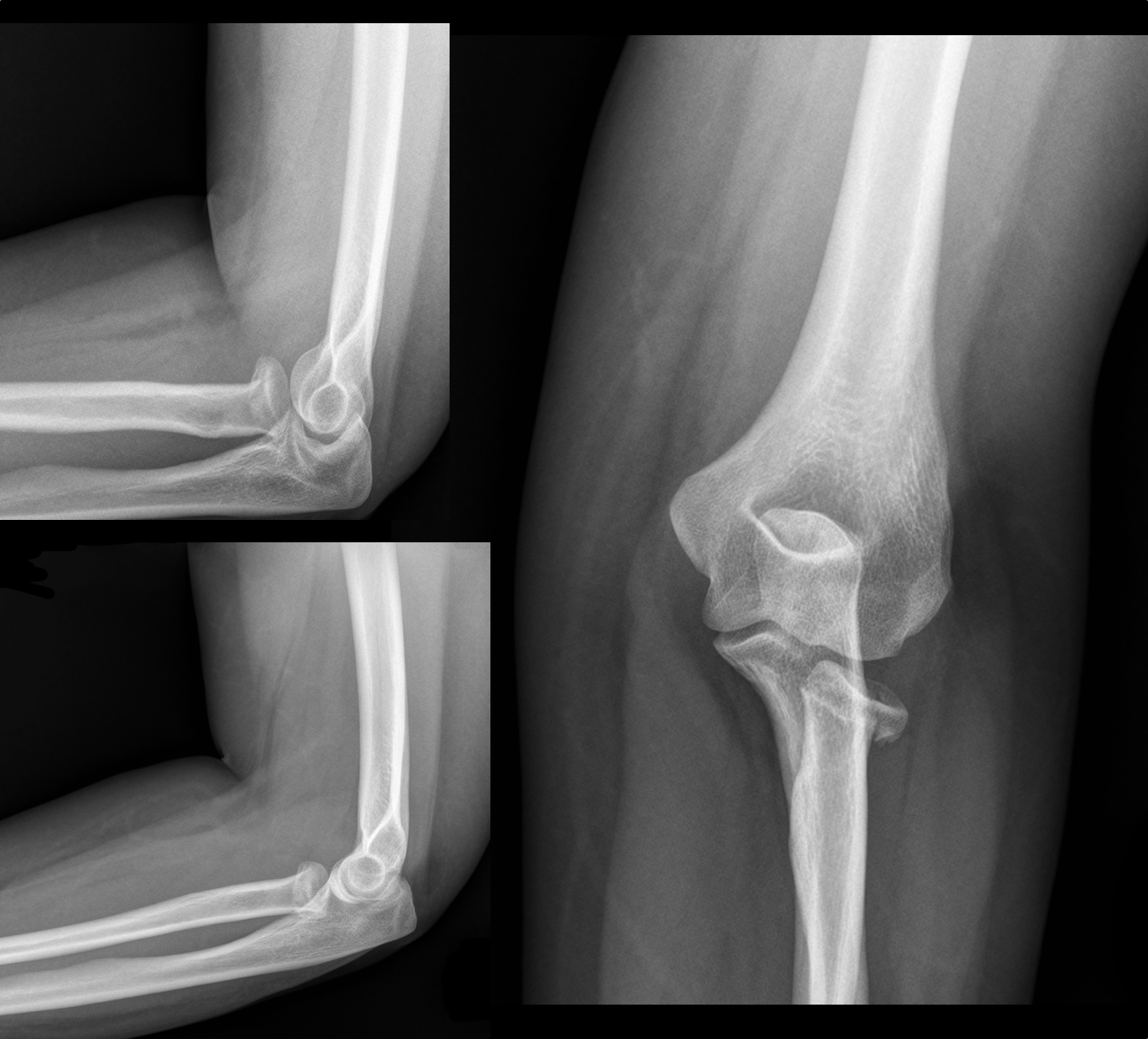
30 year-old female with elbow pain following a fall. What's the diagnosis? (bonus points if you name the fracture with an associated radio-ulnar joint dislocation)
Show Answer
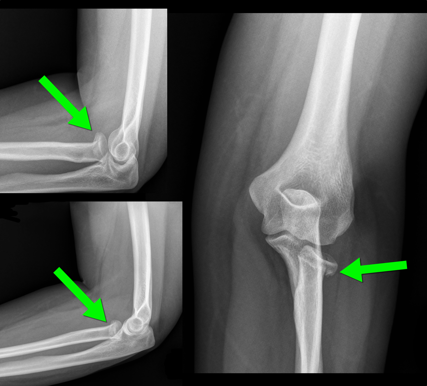
Radial Head Fracture           


  • Typically secondary to fall on an outstretched hand; head of radius collides axially with capitulum of humerus
  • Most common type of adult elbow fracture; radial neck fractures more common in children; 
  • Must be on the lookout for the Essex-Lopresti fracture (bonus answer)
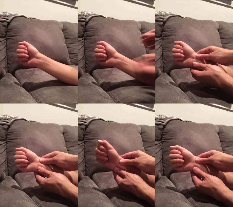
    • Radial head fracture + dislocation of the radio-ulnar joint (typically disruption of interosseous membrane
    • Repair early (typically ORIF) otherwise there may be loss of forearm extension, pronation, and supination
    • Look for distal radio-ulnar tenderness / instability by grabbing distal radius and ulnar and moving in opposite directions (see video below)

d


Show References

http://www.wheelessonline.com/ortho/radial_head_frx

Follow me on Twitter (@criticalcarenow) or Google+ (+criticalcarenow)


Title: What's the Diagnosis? Images by Dr. Michael Abraham

Category: Visual Diagnosis

Posted: 5/26/2014 by Haney Mallemat, MD

Question

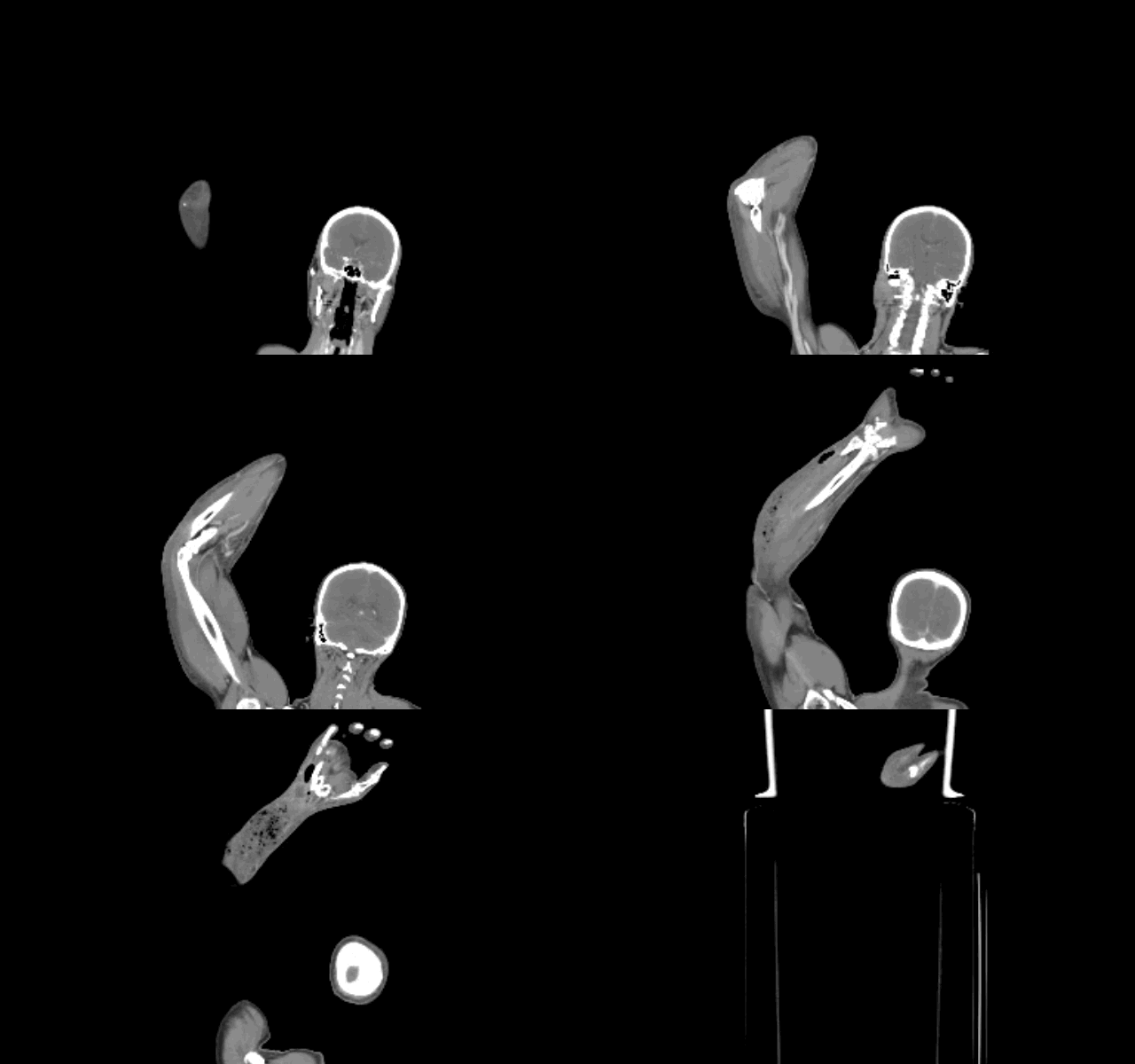
Diabetic patient with active intravenous drug use presents with hypotension, fever, and tenderness of right arm. What's the diagnosis and what antibiotic(s) would you start?
Show Answer

Right arm with necrotizing fasciitis

  • Necrotizing fasciitis (NF) may occur secondary to either mono-microbial (e.g., Strep, Staph, Clostridial species, etc.) or poly-microbial (above in addition to anaerobes and/or gram negative) bacteria; etiology may be unclear from history alone.
  • Urgent surgical debridement is the key to management but because the infection can spread rapidly, antibiotics are essential while awaiting surgery.
  • Empiric antibiotics should provide broad-spectrum coverage as well as cover MRSA if the patient is at increased risk (e.g., recent hospital admission).
  • Clindamycin is often been added for its theoretical anti-toxin effects from certain streptococcal species. Although there is little in vivo data to support this practice, it is still a reasonable approach considering the high-morbidity and mortality from NF
  • Other adjunctive therapies to consider are IVIG and hyperbaric oxygen

Show References

http://lifeinthefastlane.com/education/ccc/necrotising-fasciitis/

Follow me on Twitter (@criticalcarenow) or Google+ (+criticalcarenow)


Title: What's the Diagnosis? Case by Dr. Ashley Strobel

Category: Visual Diagnosis

Posted: 5/19/2014 by Haney Mallemat, MD

Question

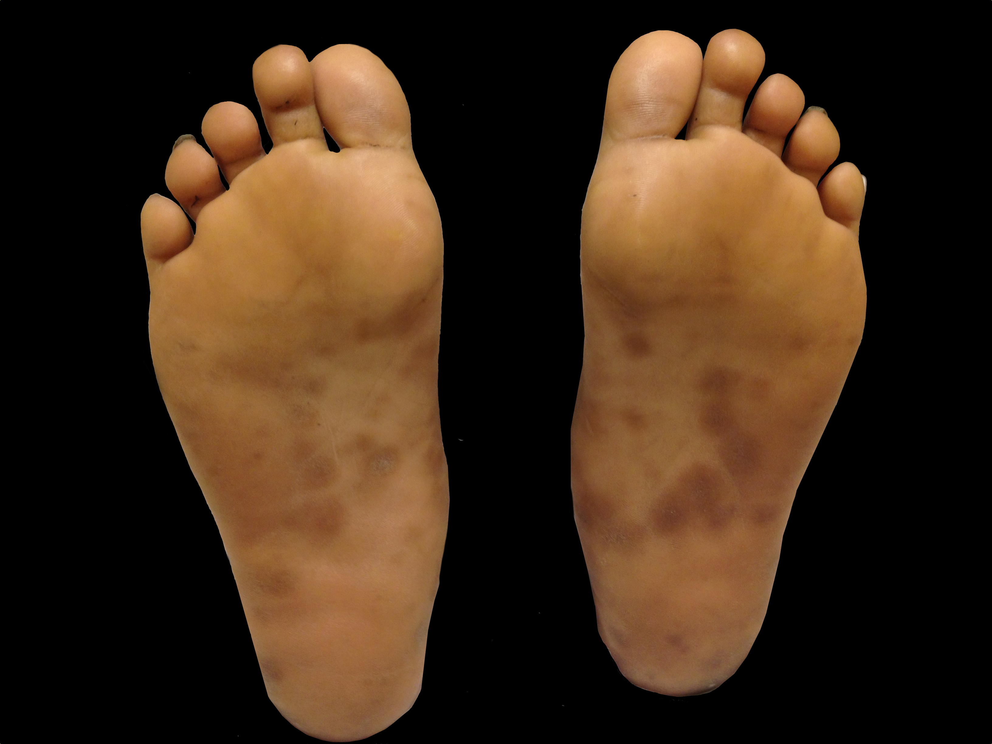
A patient presents with the rash shown below and is treated with penicillin. Fever, headache, and myalgia develop four hours later. What’s the diagnosis?
Show Answer

Answer: Jarisch-Herxheimer reaction

  • Reaction observed 2-8 hours following antibiotic treatment of syphilis as a result of endotoxin released from dying from organisms; it is not an allergy to penicillin, nor a dose related phenomenon.
  • Symptoms may include:
    • Fever / chills / rigors
    • Headache
    • Flushing / Vasodilation
    • Worsening rash
    • Hypotension
  • Can occur when following treatment of any stage of syphilis although most commonly occurs following treatment of primary syphilis; also with other illnesses caused by spirochetes (Lyme, Leptospirosis, etc.)
  • Reaction is typically self-limiting and treatment is supportive care

Show References

Follow me on Twitter (@criticalcarenow) or Google+ (+criticalcarenow)


Title: What's the Diagnosis?

Category: Visual Diagnosis

Posted: 5/5/2014 by Haney Mallemat, MD

Question

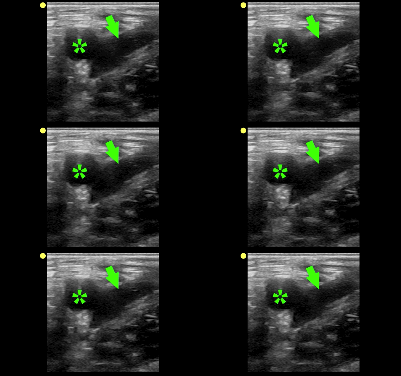
The clip below demonstrates normal right femoral anatomy. The structure with the asterisk is the right common femoral vein and the arrow is pointing to a branch of the right femoral vein. What is the name of the branch and what is its importance during lower extremity ultrasound?
Show Answer

Answer: Greater Saphenous Vein; it is one of the two regions that should be compressed when evaluating for a lower extremity DVT in the Emergency Department (the other is at the trifurcation of the popliteal vein).

Here is a podcast from the Ultrasound Podcast describing the entire bedisde DVT exam http://www.ultrasoundpodcast.com/2011/08/dvt/

Show References

Follow me on Twitter (@criticalcarenow) or Google+ (+criticalcarenow)a


Title: What's the Diagnosis? Case by Dr. Christina Tupe

Category: Visual Diagnosis

Posted: 4/28/2014 by Haney Mallemat, MD

Question

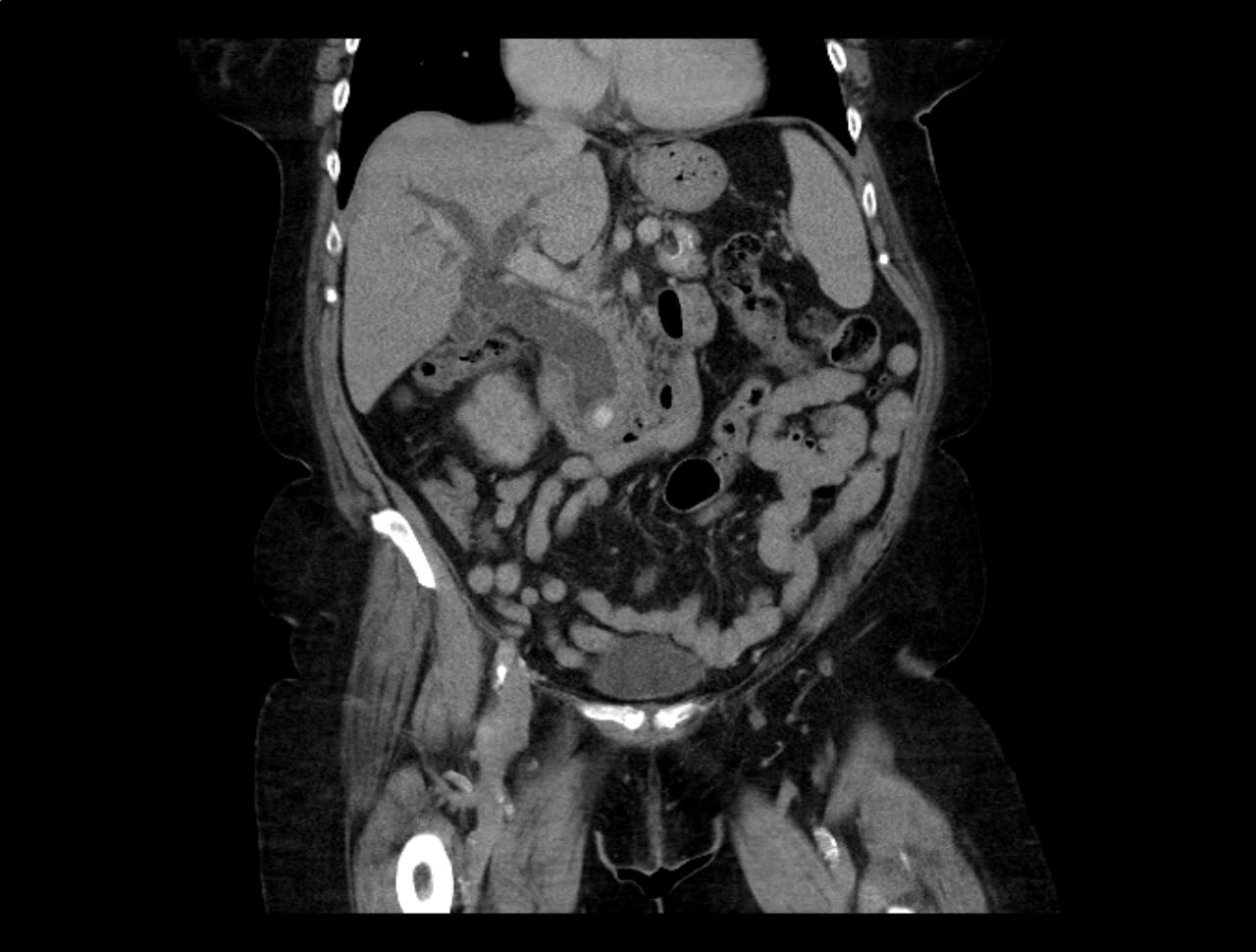
66 year-old female presents with one week of epigastric and right flank pain. Urinalysis was normal. What’s the diagnosis?
Show Answer
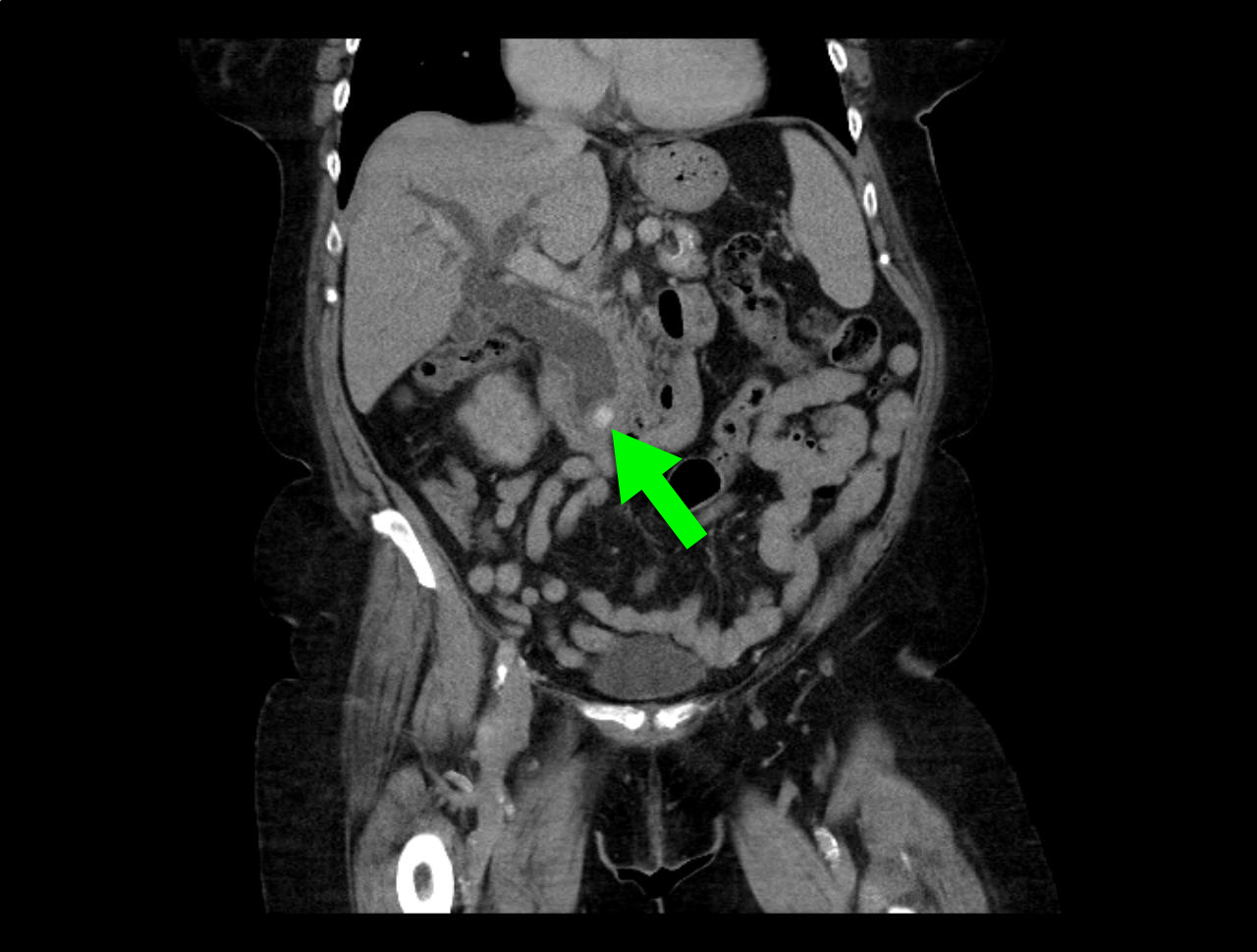
Answer: Choledocholithiasis with pancreatitis secondary to 2.2 cm common bile duct stone

Show References

Follow me on Twitter (@criticalcarenow) or Google+ (+criticalcarenow)


Title: What's theDiagnosis? Images by Dr. Ari Kestler

Category: Visual Diagnosis

Posted: 4/14/2014 by Haney Mallemat, MD

Question

25 year-old female (G1P1) presents with 3 weeks of vaginal bleeding. Her serum beta-HCG is 65,000. Her bedside ultrasound is below; what's the diagnosis? 
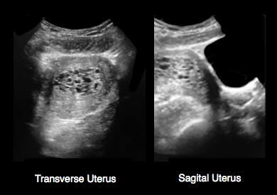
Show Answer
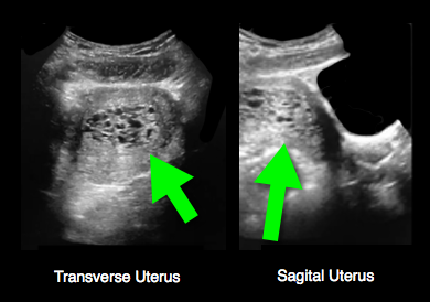
Answer: Hydatidiform mole (molar pregnancy)

Show References

Follow me on Twitter (@criticalcarenow) or Google+ (+criticalcarenow)


Title: What's the Diagnosis? Case by Dr. Ali Farzad

Category: Visual Diagnosis

Posted: 4/7/2014 by Haney Mallemat, MD

Question

23 year-old female presents complaining of progressive right lower quadrant pain after doing "vigorous" pushups. CT abdomen/pelvis below. What’s the diagnosis? (Hint: it’s not appendicitis)
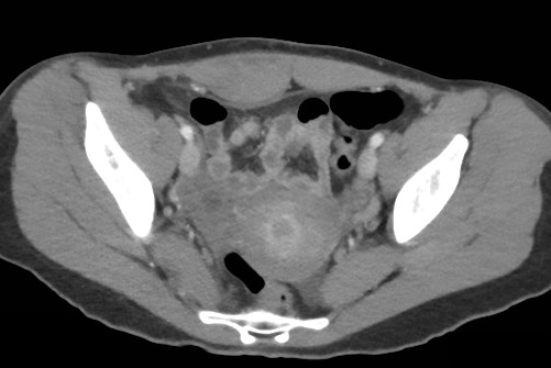
 

Show Answer
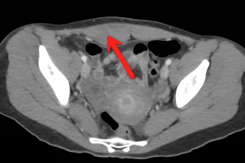
Answer: Rectus sheath hematoma

Rectus Sheath Hematoma (RSH)

Rectus muscle tear causing damage to the superior or inferior epigastric arteries with subsequent bleeding into the rectus sheath; uncommon cause of abdominal pain but mimics almost any abdominal condition.

May occur spontaneously, but suspect with the following risk factors:

  • Coagulopathy (#1 cause); acquired (e.g., warfarin) or inherited disorder
  • Rectus muscle trauma
  • Vigorous or sudden contraction of rectus muscle
  • Increased intra-abdominal pressure from vigorous coughingPregnancy (gestation, labor, or post-partum)

Typically a self-limiting condition, but hypovolemic shock may result from significant hematoma expansion.

  • Hemodynamically stable (non-expanding hematoma): conservative treatment (rest, analgesia, and ice)
  • Hemodynamically unstable (expanding hematoma): treat with fluid resuscitation, reversal of coagulopathy, and transfusion of blood products.

Show References

Follow me on Twitter (@criticalcarenow) or Google+ (+criticalcarenow)


Title: What's the Diagnosis? Case by Dr. Ali Farzad

Category: Visual Diagnosis

Posted: 4/7/2014 by Haney Mallemat, MD (Updated: 7/22/2026)

Question

23 year-old female presents complaining of progressive right lower quadrant pain after doing "vigorous" pushups. CT abdomen/pelvis below. What’s the diagnosis? (Hint: it’s not appendicitis)


Show Answer

Answer: Rectus sheath hematoma

Rectus Sheath Hematoma (RSH)

Rectus muscle tear causing damage to the superior or inferior epigastric arteries with subsequent bleeding into the rectus sheath; uncommon cause of abdominal pain but mimics almost any abdominal condition.

May occur spontaneously, but suspect with the following risk factors:

  • Coagulopathy (#1 cause); acquired (e.g., warfarin) or inherited disorder
  • Rectus muscle trauma
  • Vigorous or sudden contraction of rectus muscle
  • Increased intra-abdominal pressure from vigorous coughingPregnancy (gestation, labor, or post-partum)

Typically a self-limiting condition, but hypovolemic shock may result from significant hematoma expansion.

  • Hemodynamically stable (non-expanding hematoma): conservative treatment (rest, analgesia, and ice)
  • Hemodynamically unstable (expanding hematoma): treat with fluid resuscitation, reversal of coagulopathy, and transfusion of blood products.


Title: What's the Diagnosis? Image by Dr. Ashley Strobel

Category: Visual Diagnosis

Posted: 3/31/2014 by Haney Mallemat, MD (Updated: 4/1/2014)

Question

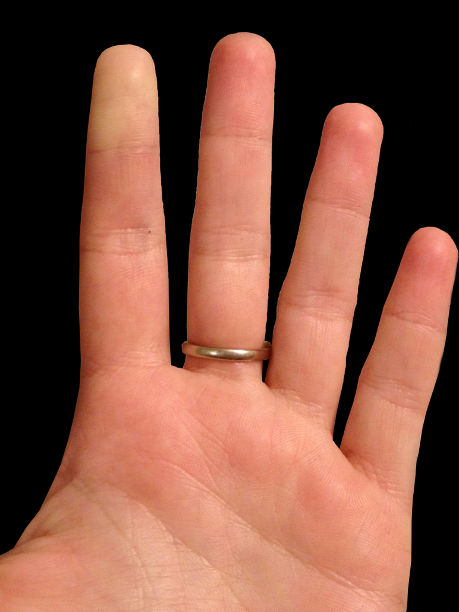
25 year-old female presents with the following. It seems to have occurred spontaneously and spontaneously resolves during her ED evaluation.
 

Show Answer
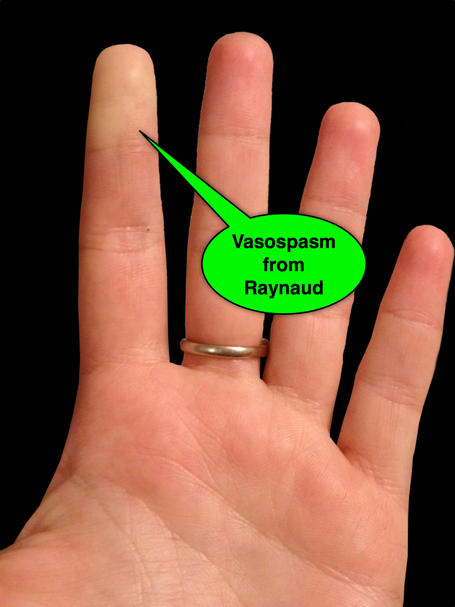
Answer: Raynaud phenomenon

  • Raynaud phenomenon is recurrent vasospasm of fingers or toes
  • Causes
    • Primary (e.g., external factors; such as cold temperature or stress)
    • Secondary to underlying illness, typically autoimmune disease (e.g., progressive systemic sclerosis or SLE)
  • Treatment
    • Avoid known triggers
    • Low-dose calcium channel blockers (e.g., nifedipine)
    • Topical nitroglycerine applied to areas of vasoconstriction
    • Identify and treat secondary disorders

Show References

Follow me on Twitter (@criticalcarenow) or Google+ (+criticalcarenow)


Title: What's the Diagnosis? Case by Dr. Ali Farzad

Category: Visual Diagnosis

Posted: 3/17/2014 by Haney Mallemat, MD

Question

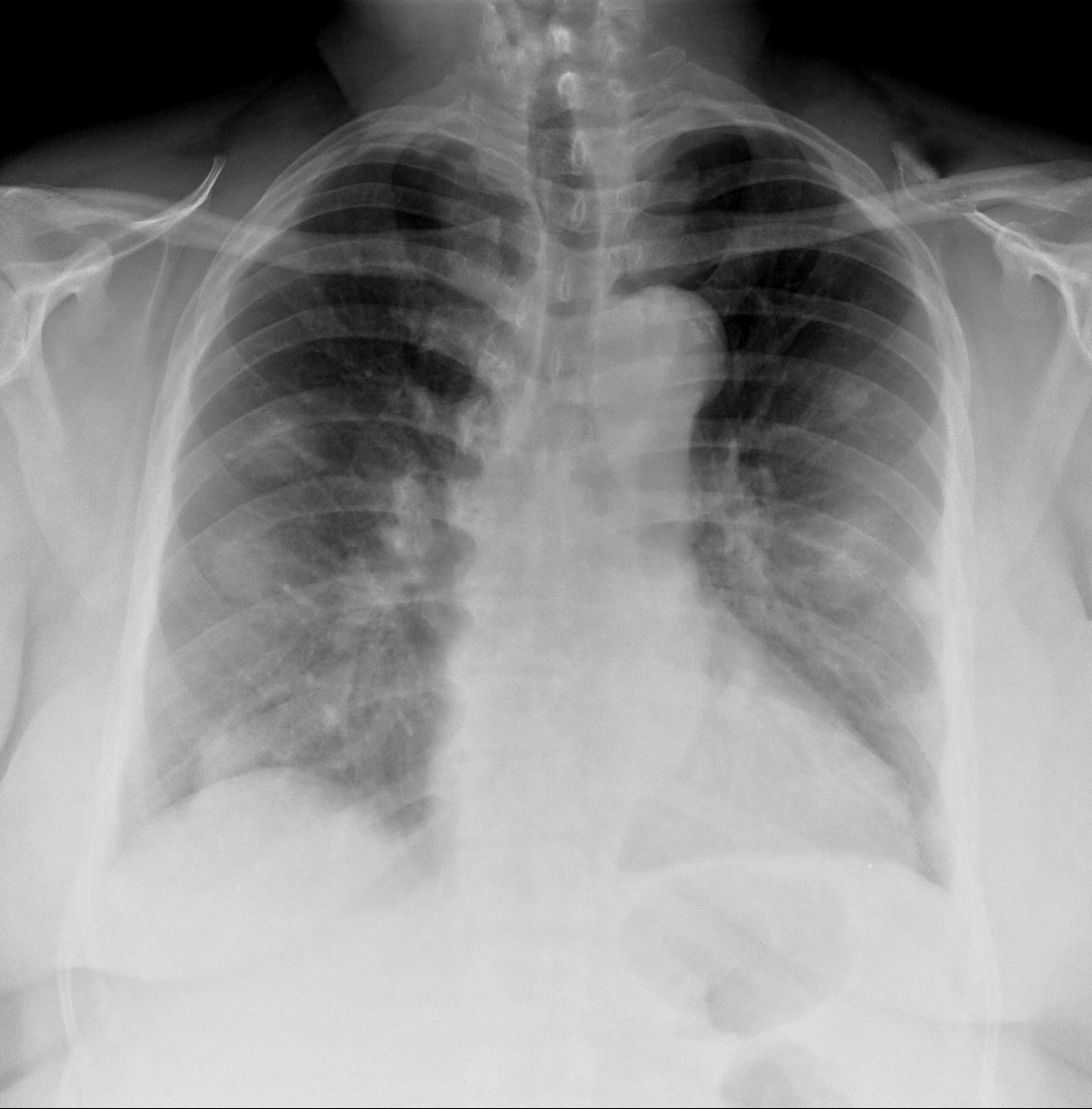
62 year-old male presents with weakness, chills, cough, and malaise. Recently, he had four teeth extracted but felt fine immediately after the extraction. Past medical history includes diabetes and hypertension; CXR is below. What’s the diagnosis?
 

Show Answer
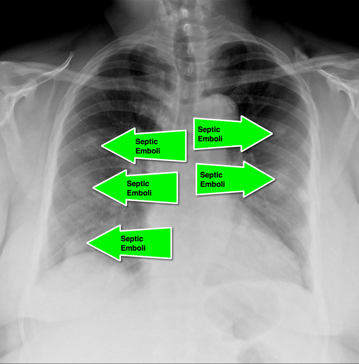
Answer: Septic pulmonary emboli (a.k.a. pulmonary puffballs); he was eventually diagnosed with bacterial endocarditis.

  • This history with the presence of "puffballs" on CXR, should clue the practitioner to the diagnosis of septic pulmonary emboli secondary to endocarditis
  • CT of the chest will confirm the diagnosis and possibly demonstrate cavitary lesions.
  • Blood cultures should be promptly obtained and the patient should be started on broad-spectrum antibiotics.

Show References

Follow me on Twitter (@criticalcarenow) or Google+ (+criticalcarenow)


Title: What's the Diagnosis? Image by Dr. Kami Hu

Category: Visual Diagnosis

Posted: 3/14/2014 by Haney Mallemat, MD

Question

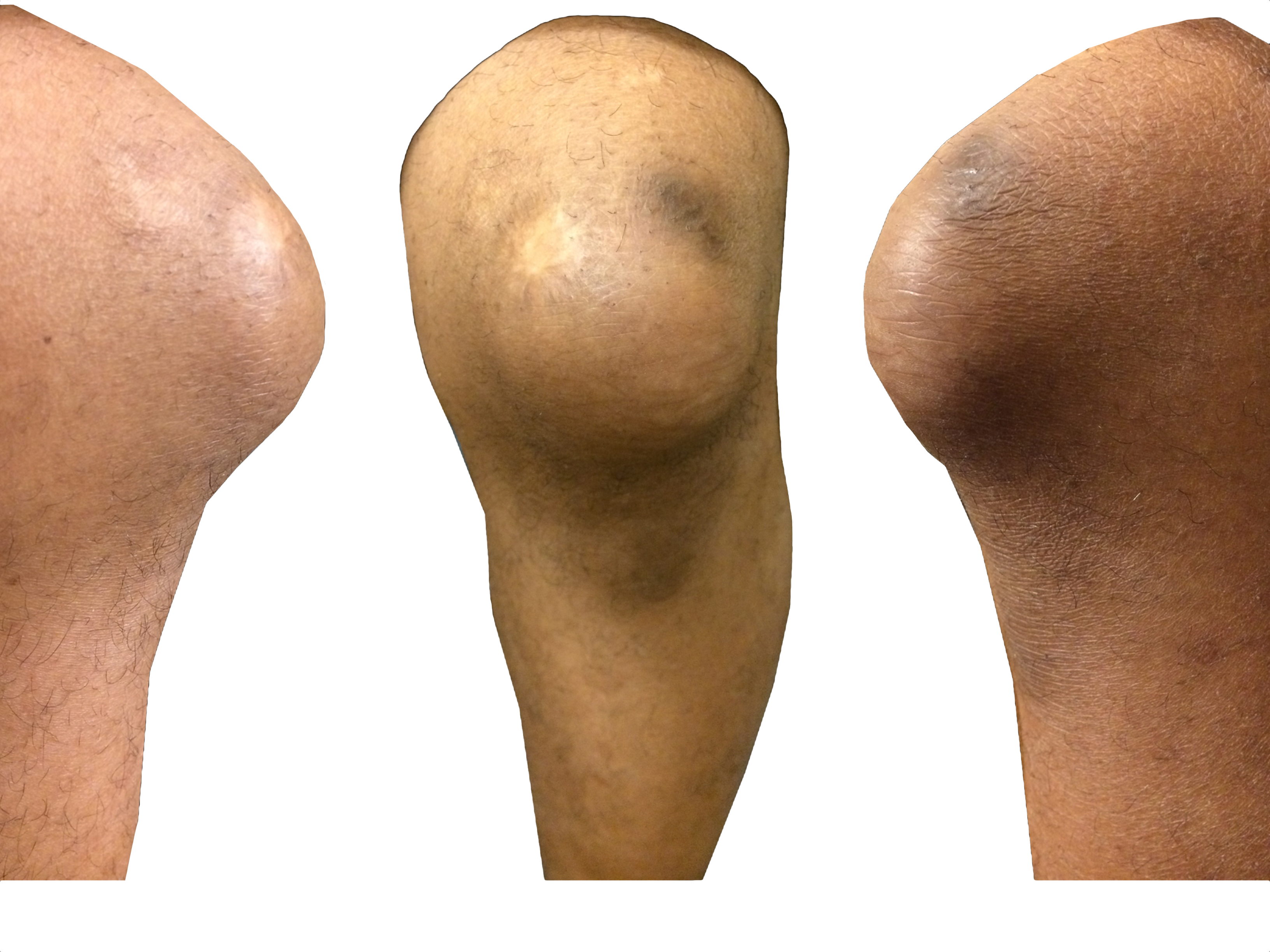
35 year-old carpet-layer presents with swelling of the superior portion of his knee that has progressively gotten worse over one week. He has no fever and has full range of motion (although pain is worse with movement). The knee is not tender to touch and the area is not erythematous or warm. What's the diagnosis?
 

Show Answer

Answer: Pre-patellar bursitis

  • The pre-patellar bursa is a superficial and synovial-lined space that reduces friction at the knee, separating the patella from the skin and the patellar tendon.
  • Inflammation to the bursa occurs secondary to direct knee trauma (either acute or chronic) and is typically benign.
  • It is associated with occupations that have a high incidence of trauma to the knee or repeated kneeling; hence the condition’s many names (carpet-layer's knee, coal-miner's knee, housemaid's knee, nun's knee)
  • Caution must be taken not to miss patients with septic bursitis (e.g., bacterial), especially in immunocompromised patients. If septic bursitis is suspected, fluid should be aspirated from the bursa and sent for analysis
  • Treatment for non-septic bursitis includes rest, (i.e., reduce trauma), NSAIDs, and physical therapy. If it remains persistent, incision and drainage by a qualified clinicians may be performed.

Show References

Follow me on Twitter (@criticalcarenow) or Google+ (+criticalcarenow)


Title: Visual Diagnosis

Category: Visual Diagnosis

Keywords: international, global, hypoxia, clubbing (PubMed Search)

Posted: 3/12/2014 by Andrea Tenner, MD

Question

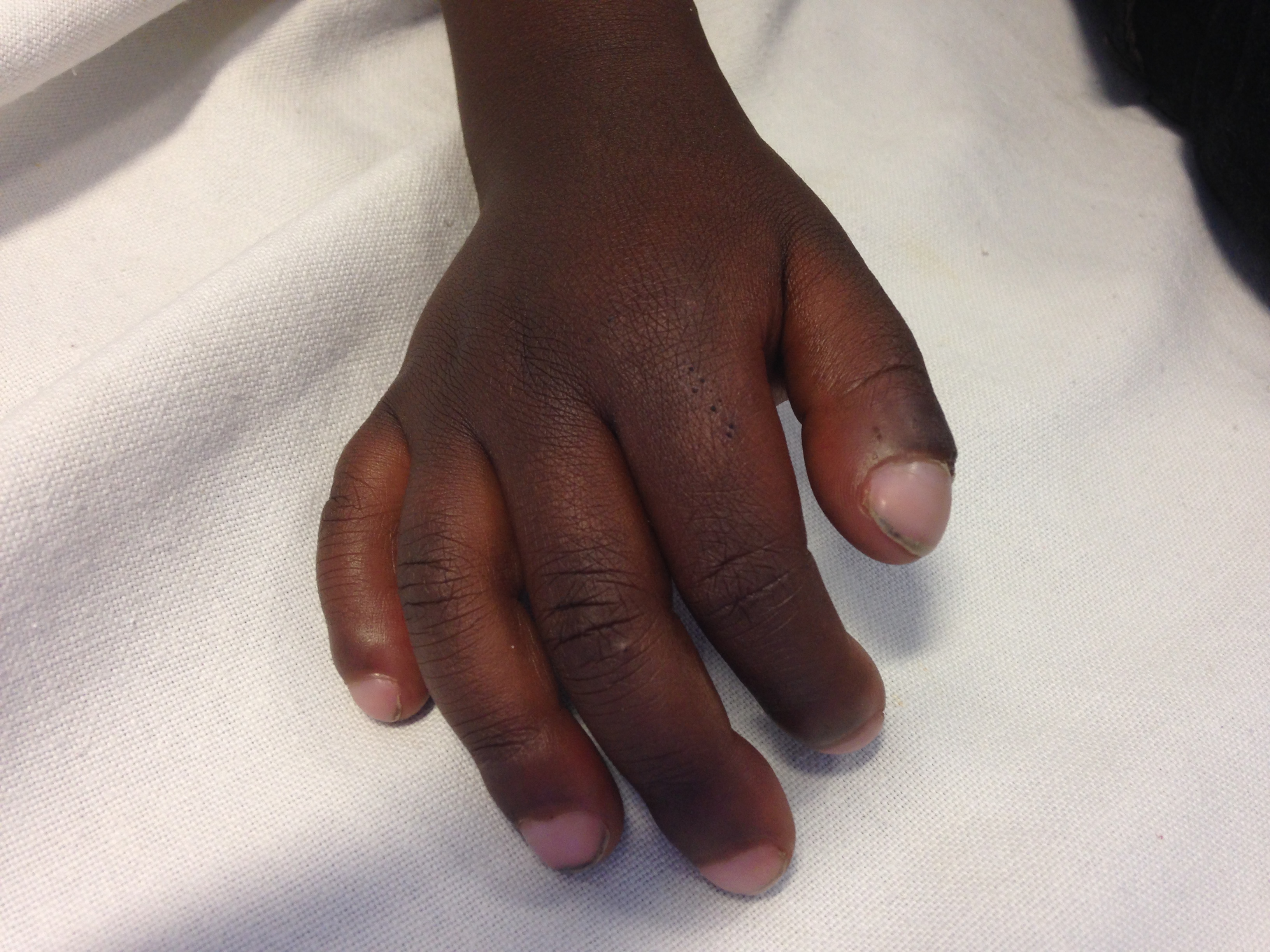
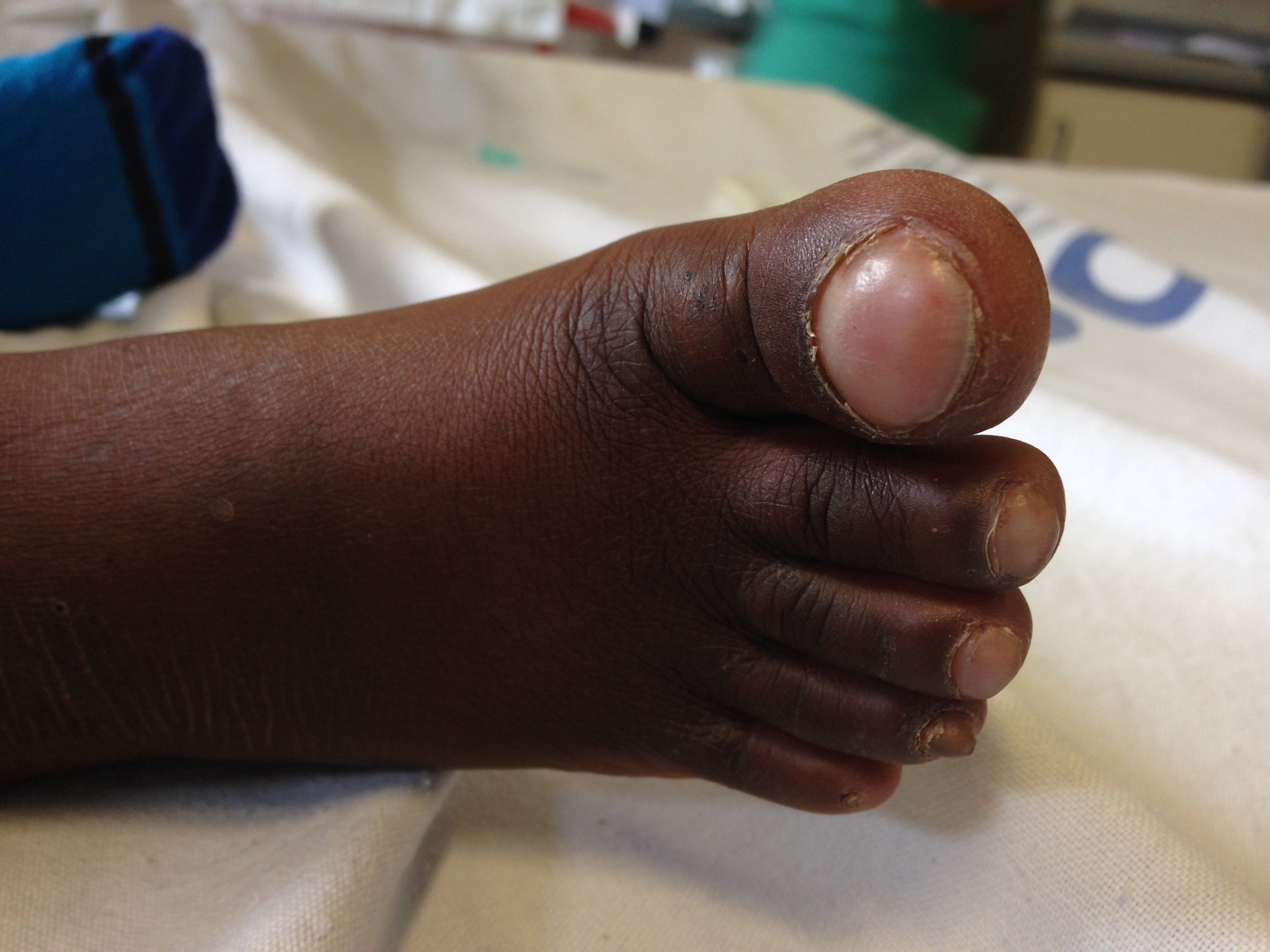
What is this physical finding?

Show Answer

Answer: Clubbing

Differential Diagnosis of Clubbing in Children:

  • Cystic Fibrosis
  • TB
  • Sarcoid
  • Cyanotic congenital heart disease
  • Thyrotoxicosis
  • HIV
  • Malnutrition
  • Empyema
  • Hepatobiliary Disease

Clubbing was first documented by Hippocrates in a patient with empyema. It has been associated with cardiopulmonary disease, but also found in other disease processes such as hepatobiliary, infectious, and endocrine diseases. It can also occur without an underlying pathology. The pathophysiology behind these physical findings remain unknown. The workup and treatment should be aimed at managing the underlying cause.

University of Maryland Section for Global Emergency Health

Author: Van Pham, MD


Show References

Schwartz, Robert. Clubbing of the Nails. 3.12.14. http://emedicine.medscape.com/article/1105946-overview#showall


Title: What's the Diagnosis? Image by Dr. Thuy Pham

Category: Visual Diagnosis

Posted: 3/3/2014 by Haney Mallemat, MD

Question

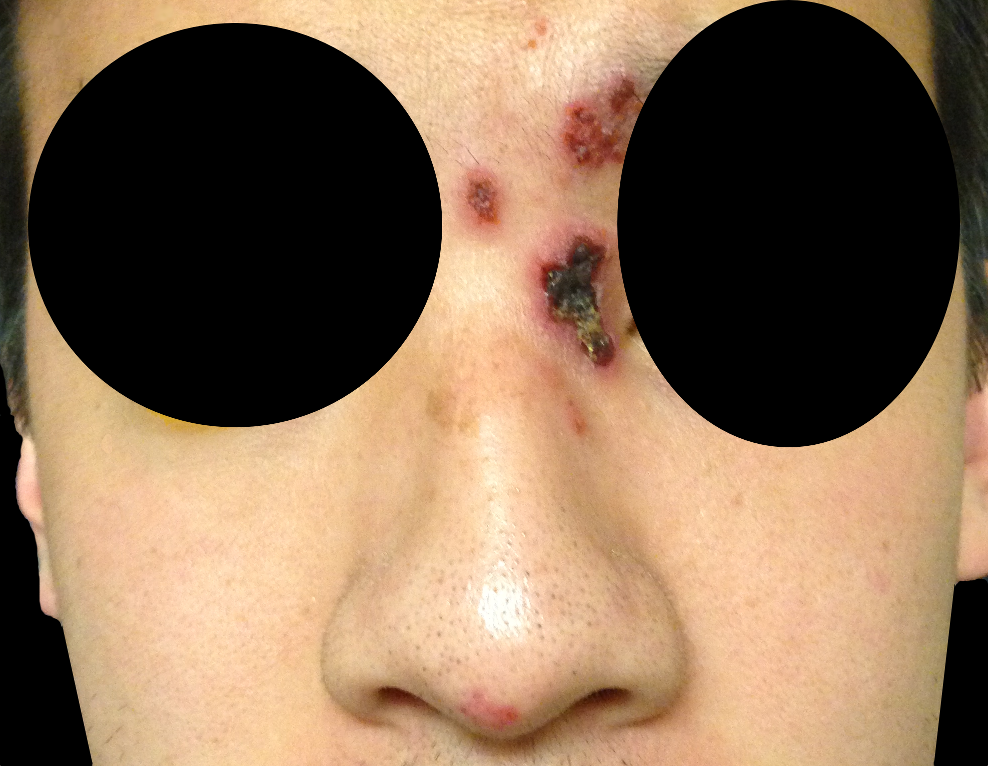
32 year-old male presents with the following. What's the diagnosis?
 

Show Answer
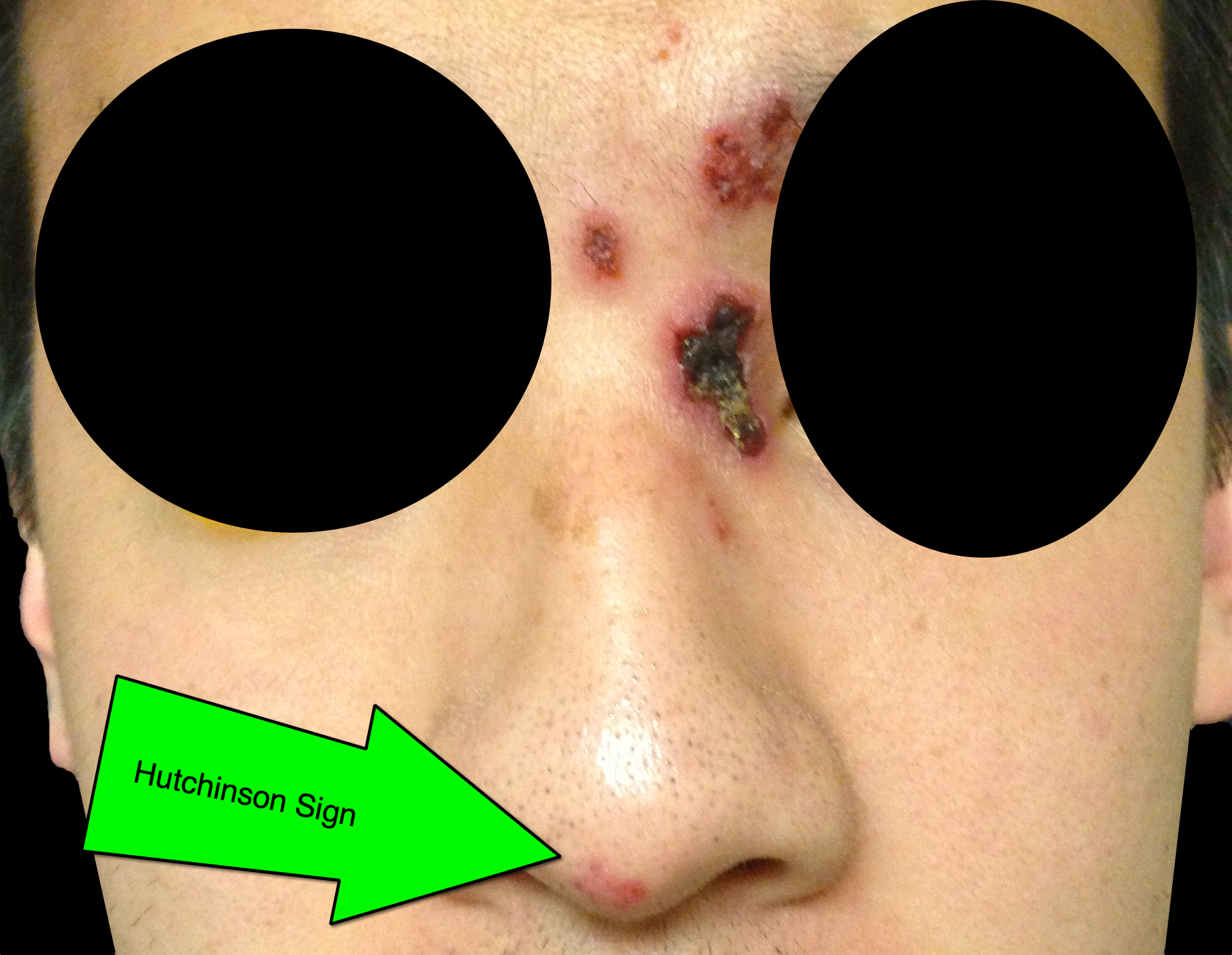
Answer: Hutchinson's Sign

Herpes zoster ophthalmicus (HZO)

  • HZO occurs when varicella-zoster, becomes reactivated in the ophthalmic division (V1) of the trigeminal nerve; any of the V1 branches may be affected (supraorbital, lacrimal, and nasociliary branches); nasocilliary innervates the globe.
  • Most patients develop only a periorbital rash, but a minority of patients have corneal involvement; the risk is increased in immunocompromised patients.
  • Hutchinson’s sign is when the rash occurs at the tip of the nose; patients with the sign have twice the incidence of ocular involvement, but 33% with HZO do not develop the sign.
  • Corneal involvement can result in significant visual complications and vision loss. Slit lamp exam with fluorescein is required when corneal involvement is suspected; a dendritic pattern may be seen.
  • Patients with eye involvement should be referred for urgent ophthalmologic evaluation following initiation of antiviral medications.

Show References

Follow me on Twitter (@criticalcarenow) or Google+ (+criticalcarenow)


Title: What's the Diagnosis? Images by UMEM Alumni Dr. Ari Kestler

Category: Visual Diagnosis

Posted: 2/24/2014 by Haney Mallemat, MD

Question

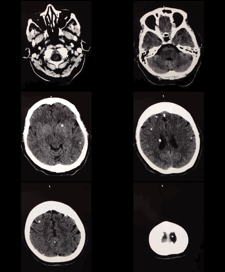
50 year-old with facial weakness and dysarthria. What's the diagnosis?
Show Answer
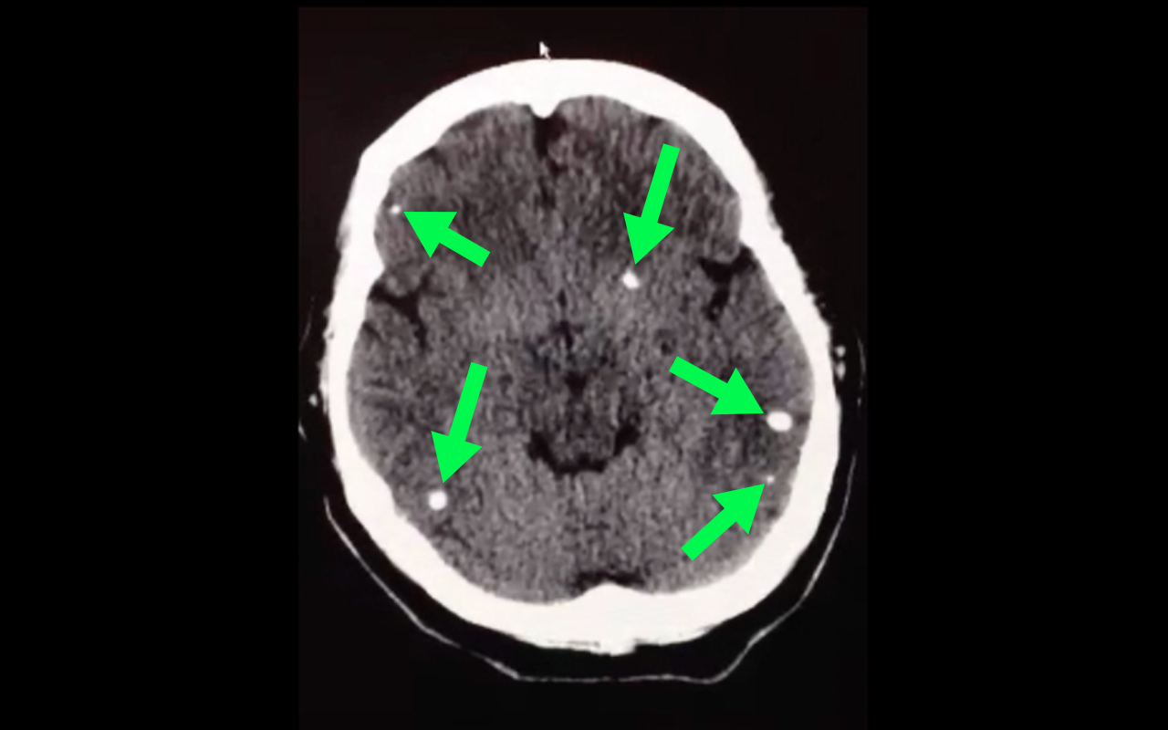
Stroke secondary to neurocysticercosis

Neurocysticercosis

Neurocysticercosis occurs secondary to ingestion of eggs from the Taenia solium tapeworm; ingestion typically occurs from ingestion of contaminated food.

Most patients are asymptomatic but symptoms of neurocysticercosis may include

  • Seizures (most common presentation)
  • Headache
  • Stroke
  • Neuropsychiatric dysfunction

Diagnosis is made via imaging such as CT scan; findings may vary depending on what stage of life the parasite is in (e.g., larvae); lumbar puncture should also be performed if the diagnosis is suspected.

Management should be symptomatic (e.g., seizures receive anticonvulsants) as well as eradication of the active parasites using steroids followed by albendazole (anti-parasitic drug)

Show References

Follow me on Twitter (@criticalcarenow) or Google+ (+criticalcarenow)


Title: What's the Diagnosis? Image by Dr.Heather Mezzadra

Category: Visual Diagnosis

Posted: 2/17/2014 by Haney Mallemat, MD

Question

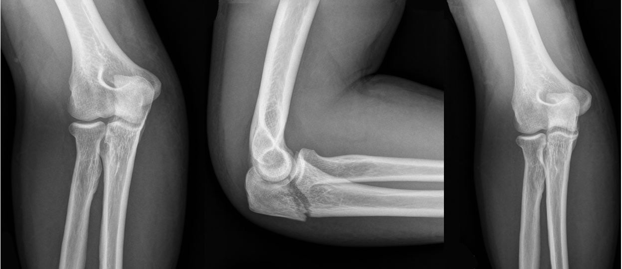
44 year-old construction worker fell off a ladder and presents with elbow pain. What's the diagnosis and what is the most commonly associated nerve injury? 
 

Show Answer
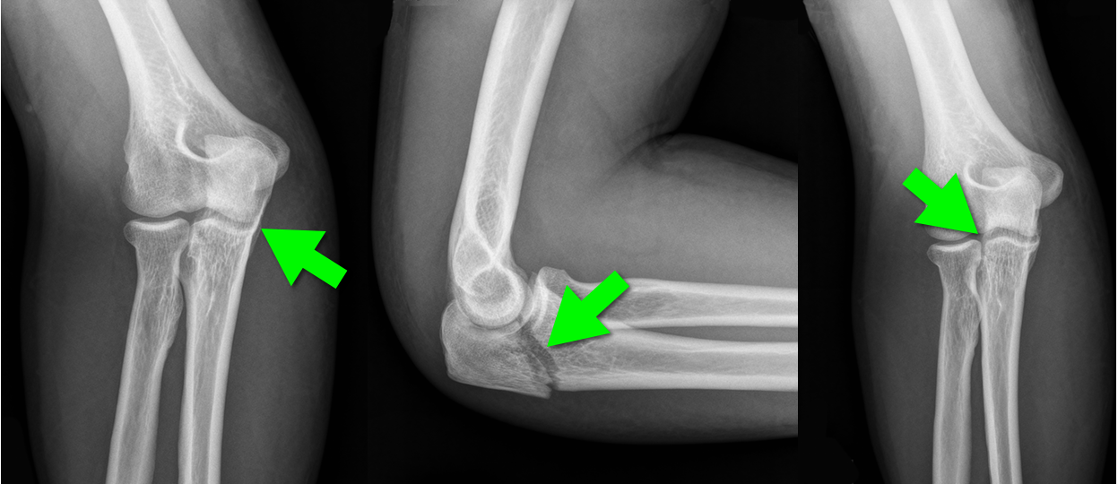
Olecranon fracture; the ulnar nerve is most commonly injured

Pearls for Olecranon Fracture

  • Typically occurs secondary to direct blow or fall on an outstretched hand
  • Patients present with a complaint of pain along the olecranon with an inability to extend the elbow
  • Ulnar nerve injury is common.
  • Non-displaced fractures are typically managed conservatively with placement in a long-arm posterior splint; elbow must be placed in flexion of 70° and forearm is neutral
  • Other types of olecranon injuries require surgical repair 

Show References

Follow me on Twitter (@criticalcarenow) or Google+ (+criticalcarenow)


Title: What's the Diagnosis? Case by Dr. Ryan Spangler (@Emterp2)

Category: Visual Diagnosis

Posted: 2/10/2014 by Haney Mallemat, MD (Updated: 2/10/2014)

Question

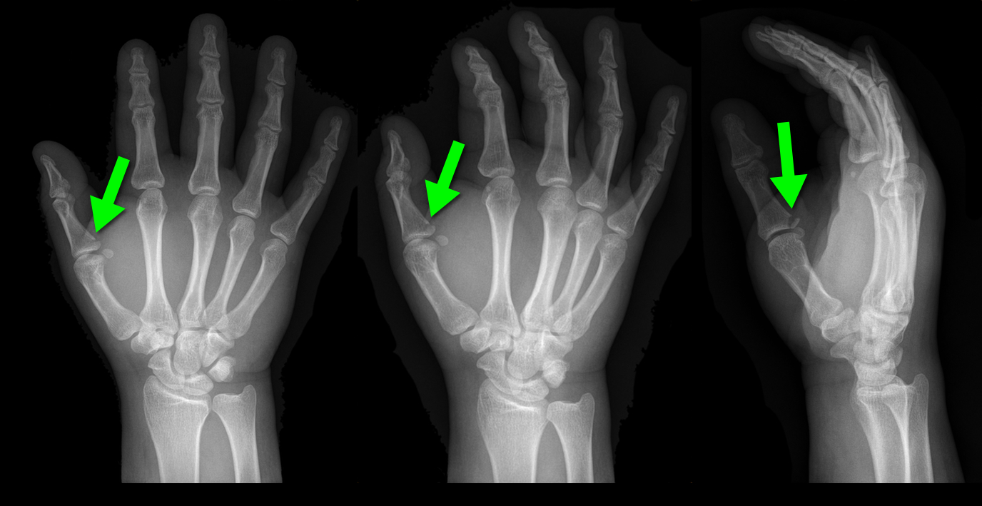
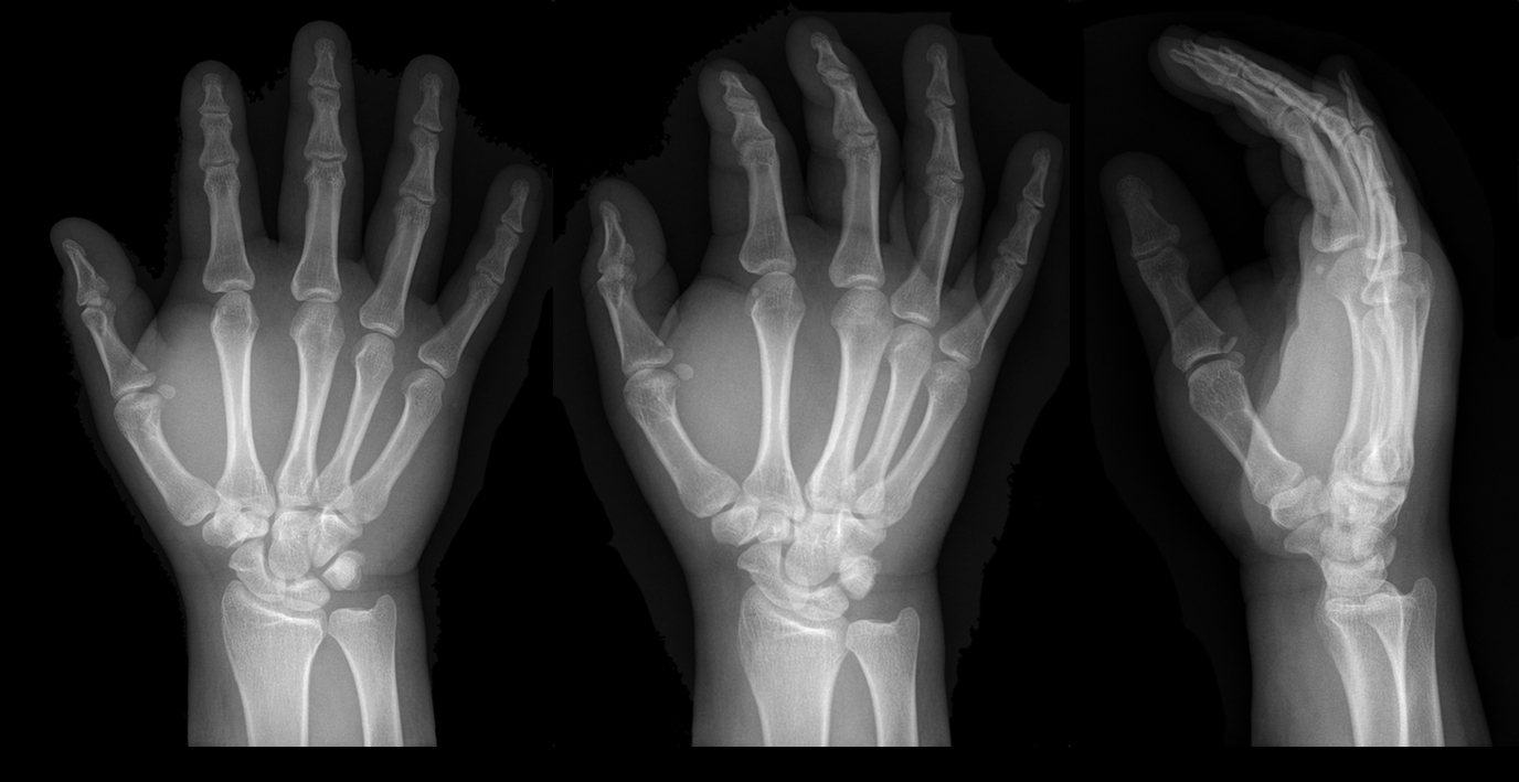
25 year-old male presents after falling off his bicycle. He complains of pain in his right-hand (he is right-hand dominant). What's the diagnosis? 
Show Answer

Answer: Gamekeeper's thumb or Skier's thumb

Injury to the ulnar collateral ligament (UCL) secondary

  • repetitive injury to hand
  • acute hyper-abduction injury to the thumb (i.e., during a skiing injury or fall on outstretched hand); most common cause if injury. 

Called “Gamekeeper Thumb” because injury was originally described in gamekeepers who job was to break the necks of captured game (or animals). The act of breaking necks led to a repetitive stress and injury secondary to the valgus forces on the thumb.

UCL can become torn or avulsed from the insertion on the proximal phalanx; the UCL is an important stabilizer of the thumb; improper management can lead to future disability.  

Treatment is with a short-arm thumb-spica cast and follow up with a hand surgeon to determine whether management will be conservative or operative.

Chuter GS, Muwanga CL, Irwin LR (June 2009). "Ulnar collateral ligament injuries of the thumb: 10 years of surgical experience". Injury 40 (6): 652–6 Wheeless' Textbook of Orthopaedics Online: Surgical treatment of acute gamekeeper's thumb

Show References

Follow me on Twitter (@criticalcarenow) or Google+ (+criticalcarenow)


Previous  |  1 |  2 |  3 |  4 |  5 |  6 |  7 |  8 |  9 |  10 |  11 |  ... |  13 |  Next
University of Maryland School of Medicine logo
  • Departments
  • Programs
  • Research Centers
  • Institutes
  • Graduate Education
  • CIBR Cores
  • Offices of the Dean
    • University of Maryland, Baltimore
    • University of Maryland Medical Center
    • Medical Alumni Association of the University of Maryland
    • Health Sciences and Human Services Library
University of Maryland School of Medicine
655 W. Baltimore Street
Baltimore, MD 21201
Contact Us

© University of Maryland School of Medicine

    • Webmaster
    • Web Accessibility
    • Site Index
    • UMB Hotline
    • Employment
    • Map