University of Maryland School of Medicine logo
Emergency Medicine
All Departments
UMSOM Assistant
UMEM Educational Pearls
  • Home
  • Departments
  • Emergency Medicine
  • CME
  • UMEM Educational Pearls
  • CME
  • Courses and Conferences
  • Free Open Access Medical Education
  • UMEM Educational Pearls

Search

61-80 of 245 results with category "Visual Diagnosis"

Previous  |  1 |  2 |  3 |  4 |  5 |  6 |  7 |  8 |  9 |  ... |  13 |  Next

Title: What's the Diagnosis?

Category: Visual Diagnosis

Posted: 4/27/2015 by Haney Mallemat, MD

Question

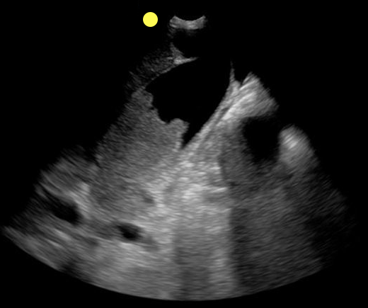
70 year-old female presents from a nursing home with fever and abdominal pain. A right upper quadrant ultrasound is shown, what's the diagnosis?
Show Answer
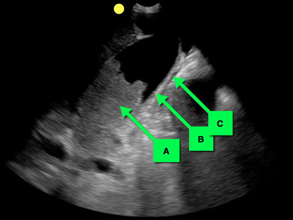
Acalculous cholecystitis

Acalculous Cholecystitis

  • Acalculous cholecystitis is due to gallbladder (GB) wall ischemia and GB stasis (i.e., from reduced GB contraction secondary to reduced PO intake).
  • The result is gallbladder "sludge" (A), thickened GB walls (B), and pericholecystic fluid (C).
  • Risk factors for acalculous cholecystitis include
    • critical illness (especially sepsis)
    • acute cholecystitis
    • chronic immobility
    • total parenteral nutrition
    • diabetes and immunosuppression
  • Treatment includes antibiotics covering enteric / biliary pathogens and cholecystectomy (if a surgical candidate) or percutaneous cholecystostomy if unsuitable for the OR.
  • Complications include perforation, GB gangrene, and extra-biliary abscess.

Show References

Follow me on Twitter (@criticalcarenow) or Google+ (+criticalcarenow)


Title: What's the Diagnosis?

Category: Visual Diagnosis

Posted: 4/20/2015 by Haney Mallemat, MD

Question

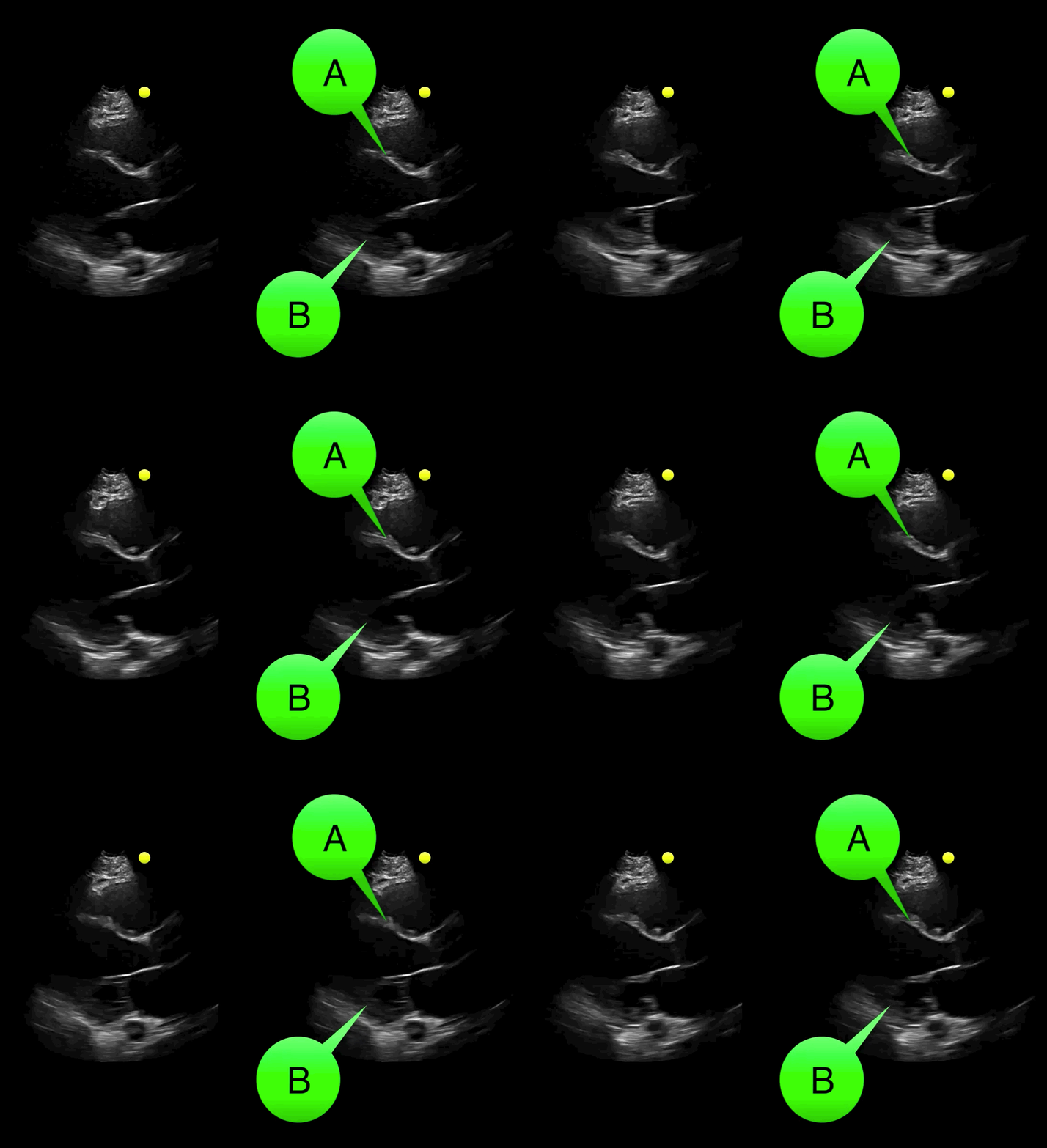
You see the following on a parasternal long-axis view; what's the diagnosis and what coronary distribution is involved?
Show Answer

Severe hypokinesis with anterioseptal wall motion abnormality.

  • A: Anteroseptal wall (perfused by left anterior descending)
  • B: Inferolateral wall (perfused by left circulmflex artery and variable contributions from posterior descending artery)
  • Click on the links to read more about cardiac wall segments and their coronary distribution
    • Parasternal view and Apical four chamber view

Show References

Follow me on Twitter (@criticalcarenow) or Google+ (+criticalcarenow)


Title: What's the Diagnosis? Case by Dr. Leen Alblaihed

Category: Visual Diagnosis

Posted: 4/13/2015 by Haney Mallemat, MD

Question

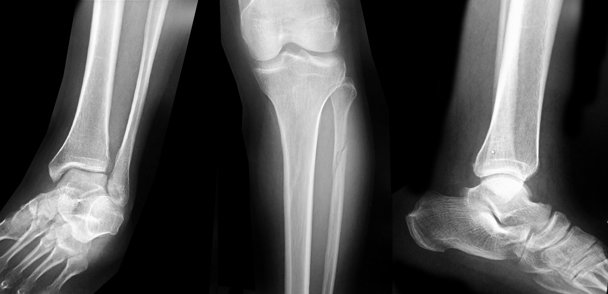
Patient presents with leg and ankle pain after a fall 3 weeks earlier. Initial ankle Xrays were negative. Patient presents today with persistent leg and ankle pain. What's the diagnosis and what other imaging would you perform and why?
 

Show Answer
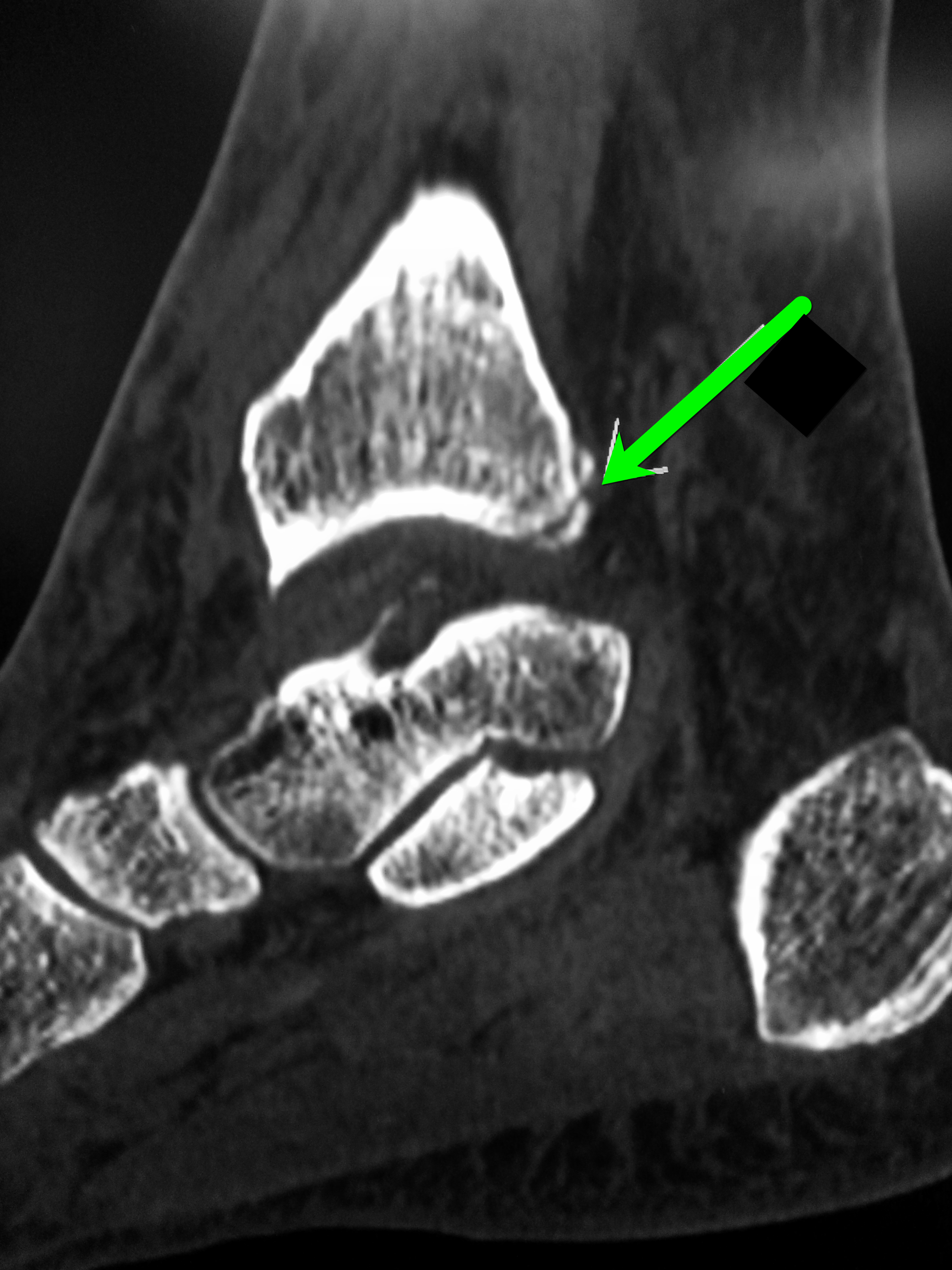
Proximal fibula fracture; have a high-clinical suspicion for a Maisonneuve fracture and consider CT scan of the ankle

Maisonneuve fracture

  • Fracture of proximal third of the fibula with associated injury of the tibiofibular syndesmosis and interosseous membrane; there is typically an associated fracture of the medial malleolus or ruptured deltoid ligament.
  • This patient had a normal ankle X-ray, but the proximal fibular fracture and the persistent ankle pain lead to an ankle CT scan demonstrating a non-displaced fracture at the medial aspect of the posterior malleolus, extending into the tibiotalar joint (see below). 
  • The take home point is to always look for an associated ankle injury when a proximal fibular fracture is discovered and Xray above and below the fracture. If Xrays are negative but there is still high clinical suspicion consider advanced imaging such as CT scan.

Show References

Follow me on Twitter (@criticalcarenow) or Google+ (+criticalcarenow)


Title: What's the Diagnosis?

Category: Visual Diagnosis

Posted: 4/6/2015 by Haney Mallemat, MD

Question

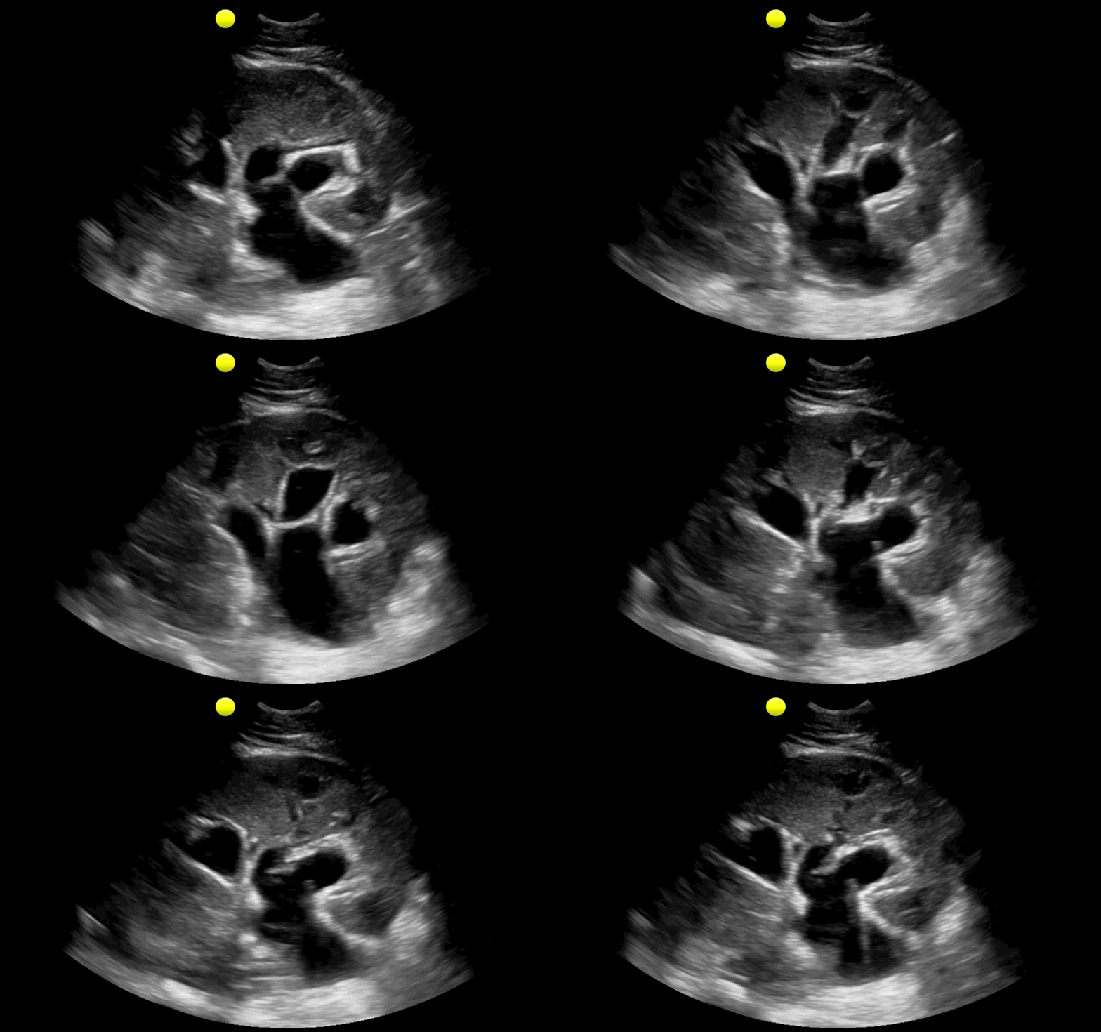
25 year-old male with the acute onset of right flank pain. Ultrasound of the right flank is shown. What's the diagnosis?
Show Answer

  • This is an ultrasound of the kidney demonstrating moderate to severe hydronephrosis seondary to an obstructing renal stone (not shown)
  • The areas that are anechoic (or black) are the blocked ureter, renal pelvis, and calyces

Show References

Follow me on Twitter (@criticalcarenow) or Google+ (+criticalcarenow)


Title: What's the Diagnosis? Image by Dr. Tu Carol Nguyen & Dr. R. Gentry Wilkerson

Category: Visual Diagnosis

Posted: 3/30/2015 by Haney Mallemat, MD

Question

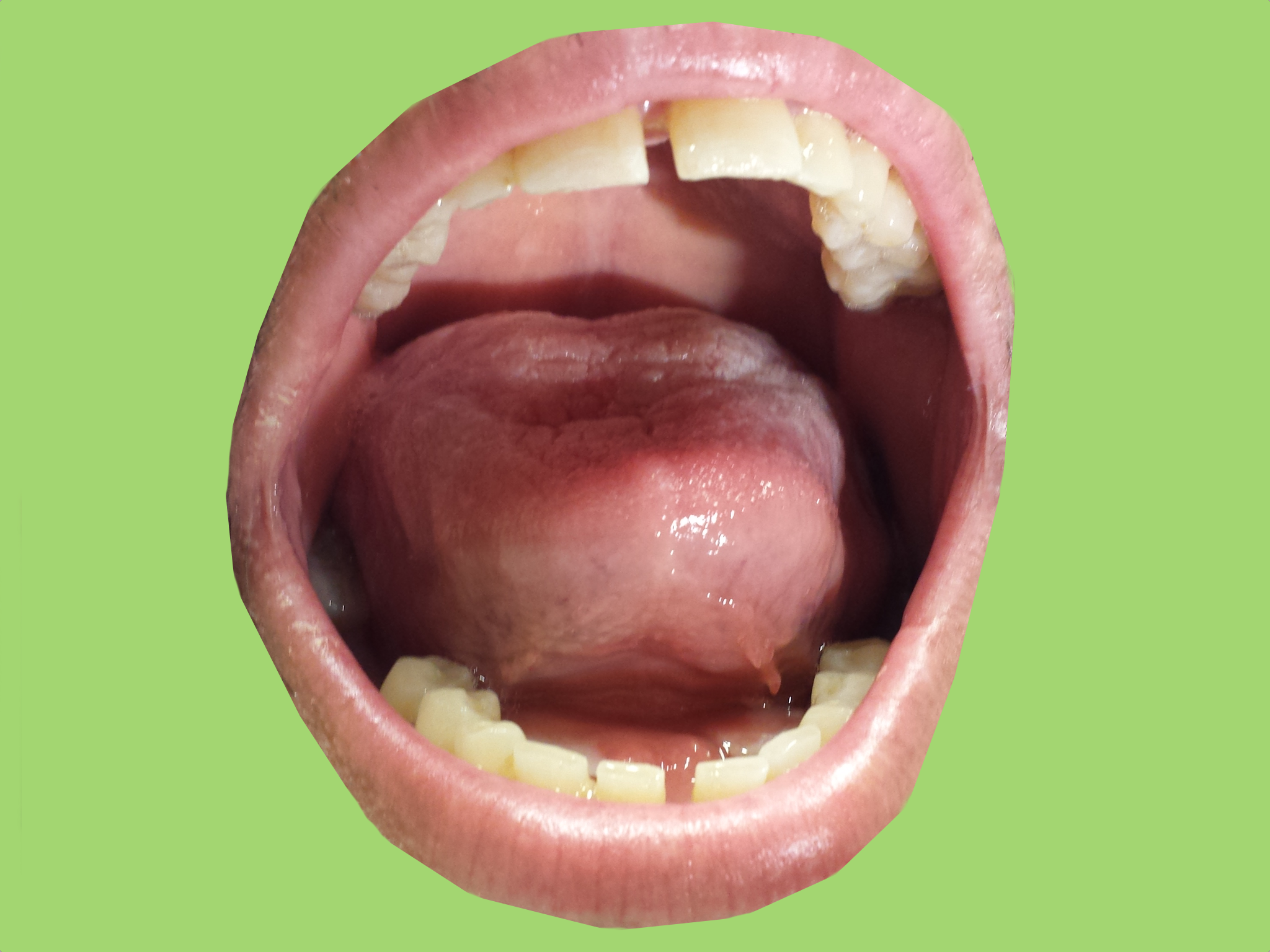
35 year-old male presents with increasing difficulty swallowing and tenderness in the floor of him mouth. What's the diagnosis?
Show Answer

Ludwig's angina

Show References

Follow me on Twitter (@criticalcarenow) or Google+ (+criticalcarenow)


Title: What's the Diagnosis? Case by Dr. David Bostick and Dr. Gentry Wilkerson

Category: Visual Diagnosis

Posted: 3/23/2015 by Haney Mallemat, MD

Question

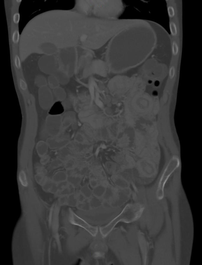
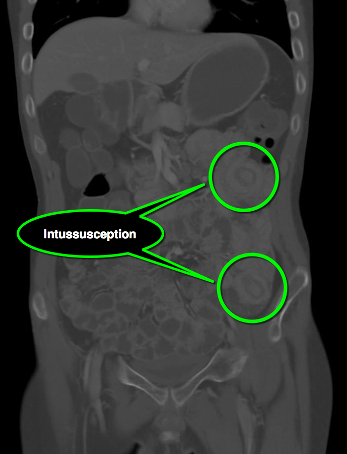
25 year-old male with autoimmune enteropathy presents with intractable vomiting and diarrhea for 7 days. What's the diagnosis?
Show Answer
Show References
Follow me on Twitter (@criticalcarenow) or Google+ (+criticalcarenow)


Title: Take the #whatsthedx challenge! Image by Dr. Joseph Martinez

Category: Visual Diagnosis

Posted: 3/16/2015 by Haney Mallemat, MD

Question

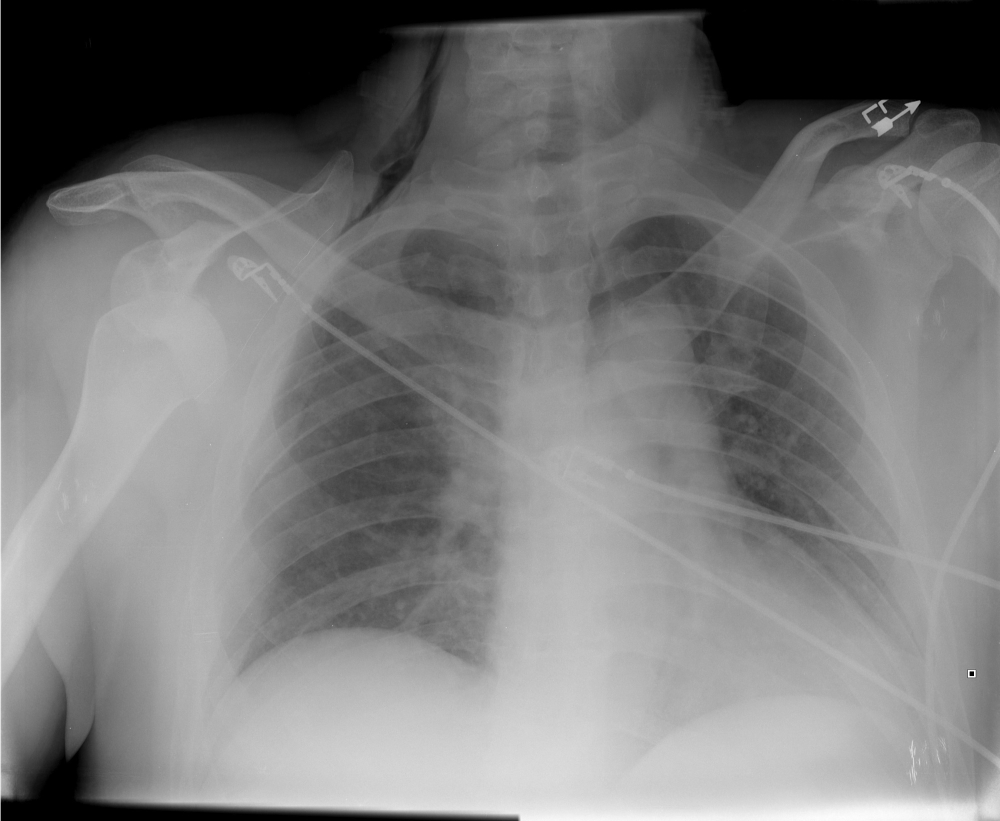
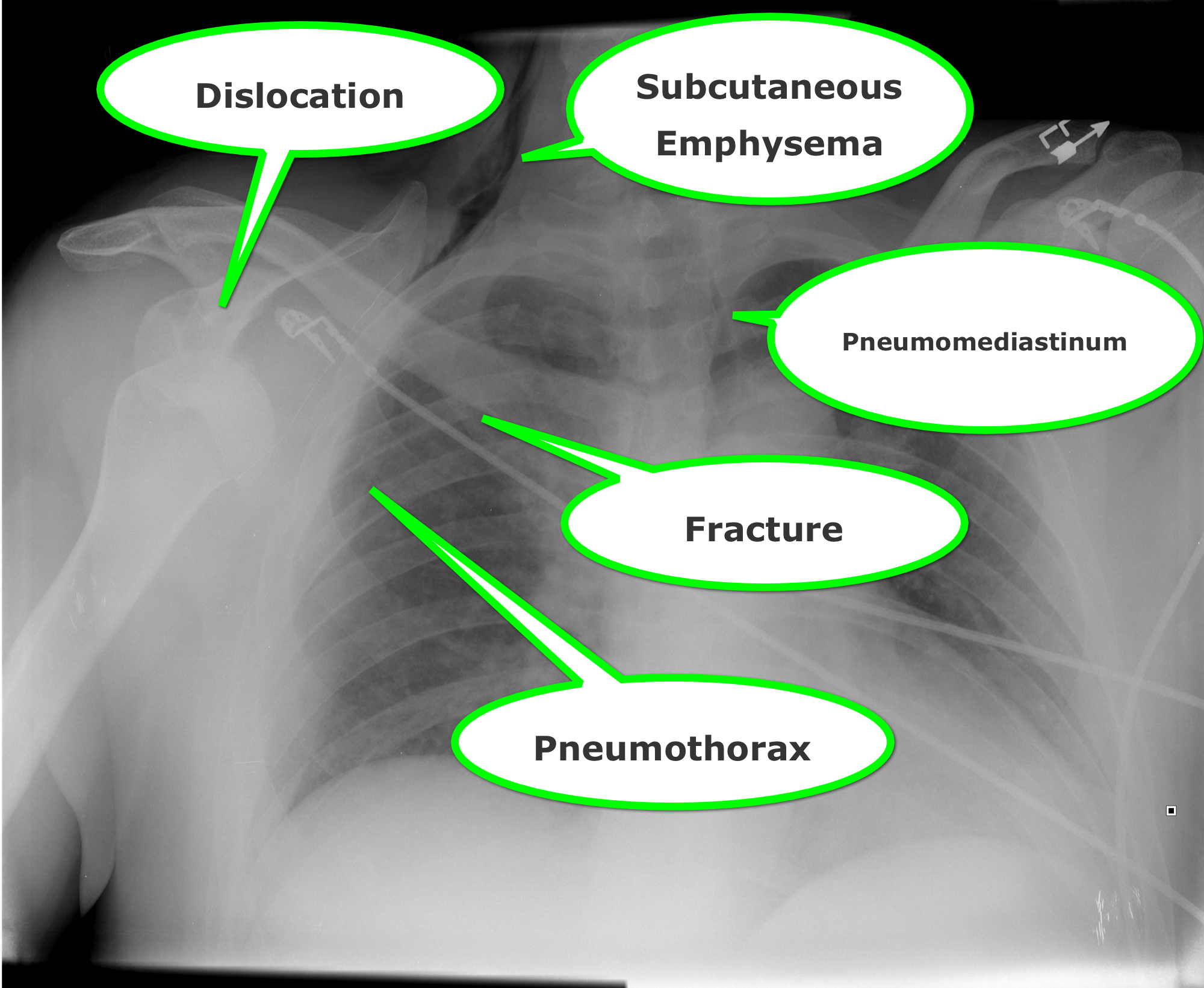
How many abnormalities can you find below?

Show Answer

Show References

Follow me on Twitter (@criticalcarenow) or Google+ (+criticalcarenow)


Title: What's the Diagnosis? Image by Dr. Connor Lundy

Category: Visual Diagnosis

Posted: 3/9/2015 by Haney Mallemat, MD

Question

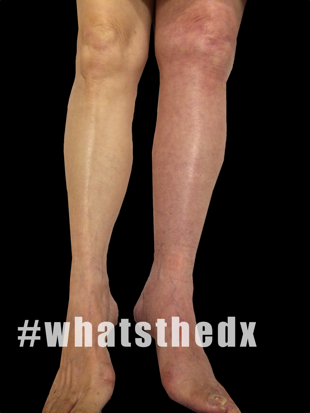
35 year-old female presents with acute leg pain and swelling. What's the diagnosis?
Show Answer

Phlegmasia cerulea dolens; there was extensive clot found in the left external iliac, common femoral, superficial femoral, and popliteal veins.

  • Phlegmasia alba dolens (literally: swollen, white, and painful) is a thrombosis of the major deep veins of the lower extremity. No collateral veins are involved so venous drainage of the leg is reduced but not completely compromised.
  • Phlegmasia cerulea dolens (literally: swollen, blue, and painful) thrombosis of major deep veins with extension to collateral veins. The result is venous congestions, edema, and can progress to gangrene when arterial flow is compromised.
  • Anticoagulation with heparin is recommended, although catheter directed thrombolytics or surgical thrombectomy may be recommended when circulation is threatened.

Show References

Follow me on Twitter (@criticalcarenow) or Google+ (+criticalcarenow)


Title: What's the Diagnosis? Image by Dr. Ahmed Alrasheedi

Category: Visual Diagnosis

Posted: 3/2/2015 by Haney Mallemat, MD

Question

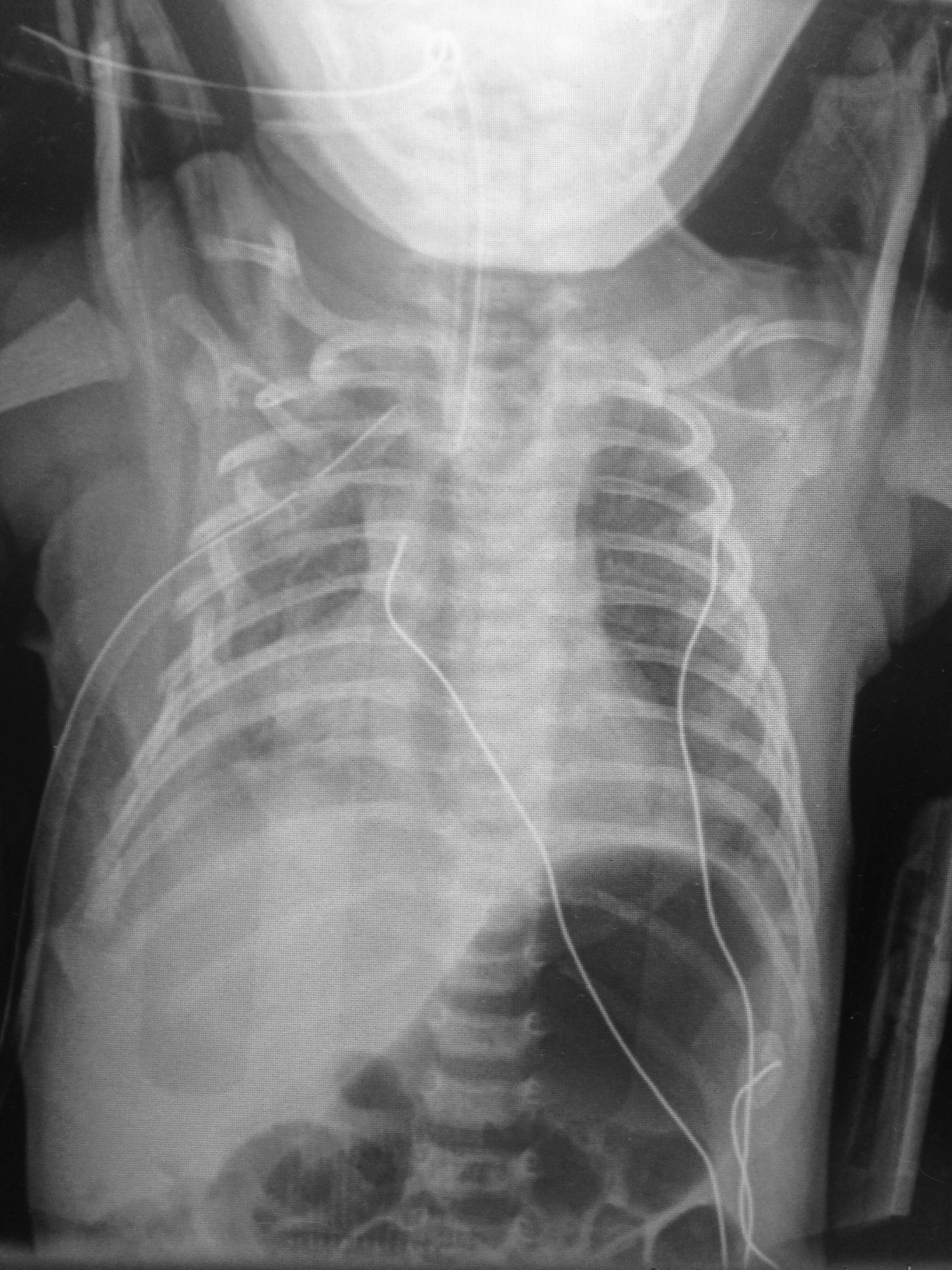
6 day-old child is brought in by parents with 1 day of reduced oral intake and 4 hours of rapid breathing. The child has no fever and no significant birth history. The child is tachycardic, hypotensive, and hypoxic. What’s the diagnosis? 
Show Answer

  • Multiple rib fractures, bilateral clavicle fracture, and hemothorax.
  • This child was ultimately diagnosed with osteogenesis imperfect, but child abuse should be quite high on your differential.

Show References

Follow me on Twitter (@criticalcarenow) or Google+ (+criticalcarenow)


Title: What's the Diagnosis? Image by Dr. Anand Swaminathan

Category: Visual Diagnosis

Posted: 2/23/2015 by Haney Mallemat, MD

Question

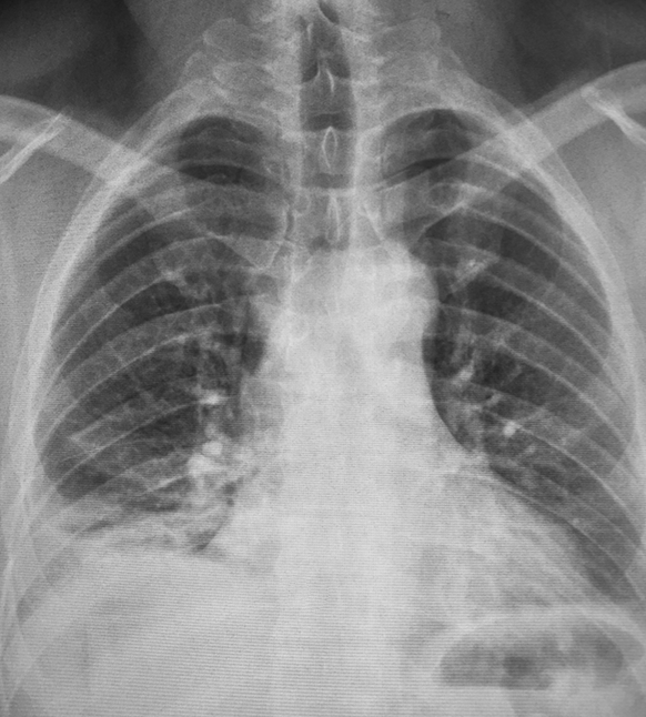
45 year-old male complains of pleuritic chest pain following a "long" flight. What's the diagnosis and what's this sign called?
Show Answer
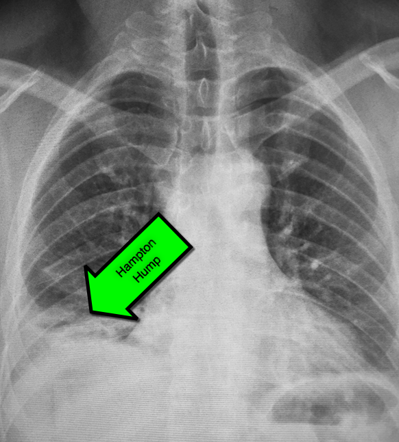
First described by Dr. Aubrey Hampton in 1940, hampton hump is a peripheral (pleural) based opacification found on chest X-ray; it occurs secondary to infarction of the lung

Although commonly associated pulmonary embolism it may occur secondary to other causes of lung infarction

Here are some other infrequent, but not rare signs, of pulmonary embolism on chest X-ray:

  • Westermark sign: area of vascular cutoff distal to a pulmonary embolism
  • Fleishner sign: an enlarged pulmonary artery
  • Pleural effusion

Show References

http://radiopaedia.org

Follow me on Twitter (@criticalcarenow) or Google+ (+criticalcarenow)


Title: What's the Diagnosis? Case by Dr. Michael Allison (@mgallison15)

Category: Visual Diagnosis

Posted: 2/16/2015 by Haney Mallemat, MD

Question

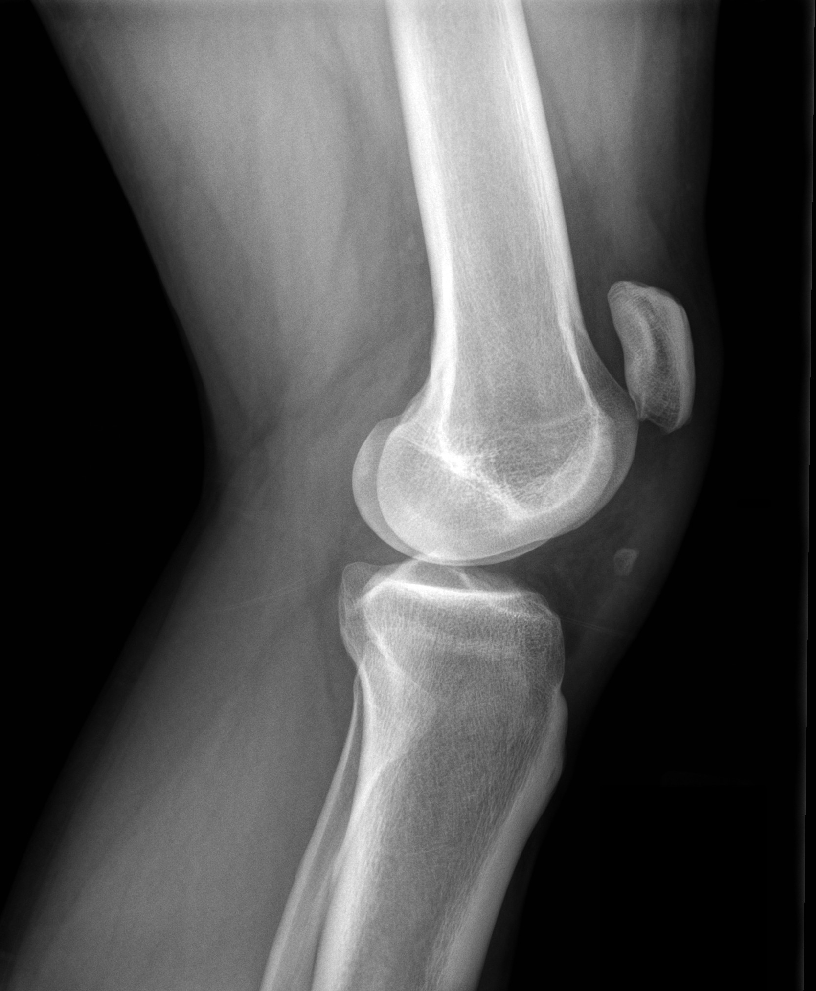
28 year-old male felt his left knee "pop" after landing from a jump. He has limited ability to extend his knee. Xray shown. What's the diagnosis?

Show Answer

Patella Alta secondary to patellar tendon rupture
Patella Alta
  • Mechanism is knee flexion combined with forceful contraction of the quadriceps
  • Swelling, patellar tenderness, and limited extension of knee
  • Radiographs are helpful in diagnosis when using the Insall-Salvatti ratio (see references)

Reference

  • Insall J, Salvati E. Patella position in the normal knee joint. Radiology 1971;101:101
  • Sibley T, Algren D, Ellison S. Bilateral patellar tendon ruptures without predisposing systemic disease or steroid use: a case report and review of the literature. American Journal of Emergency Medicine 2012; 30: 261

Show References

Follow me on Twitter (@criticalcarenow) or Google+ (+criticalcarenow)


Title: What's the Diagnosis? Case by Dr. Jackie Wnek and Dr. Jonathan Hoover

Category: Visual Diagnosis

Posted: 2/2/2015 by Haney Mallemat, MD

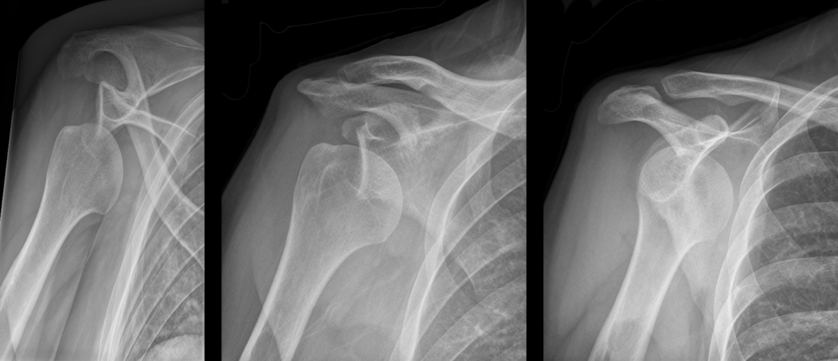
Question

Patient presents with right shoulder pain following minor trauma. What's the diagnosis....and what's the Cunningham technique?
Show Answer

Anterior shoulder dislocation

Cunningham Technique

  • A technique for shoulder reduction that typically does not require sedation.
  • Relies on proper positioning of the patient and gentle massage of the biceps muscle while gentle traction is applied to the antecubital fossa
  • Click here for a step by step tutorial on the technique and click here for videos on the technique

Show References

Follow me on Twitter (@criticalcarenow) or Google+ (+criticalcarenow)


Title: What's the Diagnosis? Image by Dr. Zachary Dezman

Category: Visual Diagnosis

Posted: 1/26/2015 by Haney Mallemat, MD

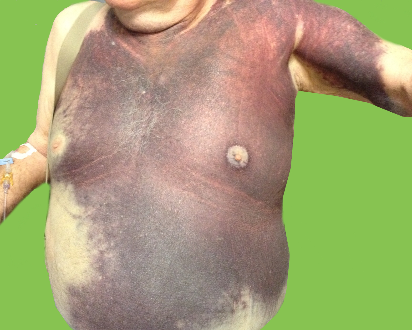
Question

Elderly male presents with the skin findings below. He is also on a medication for atrial fibrillation. What's the diagnosis?
Show Answer

Severe ecchymosis from coumadin overdose

Show References

Follow me on Twitter (@criticalcarenow) or Google+ (+criticalcarenow)


Title: What's the Diagnosis?

Category: Visual Diagnosis

Posted: 1/19/2015 by Haney Mallemat, MD

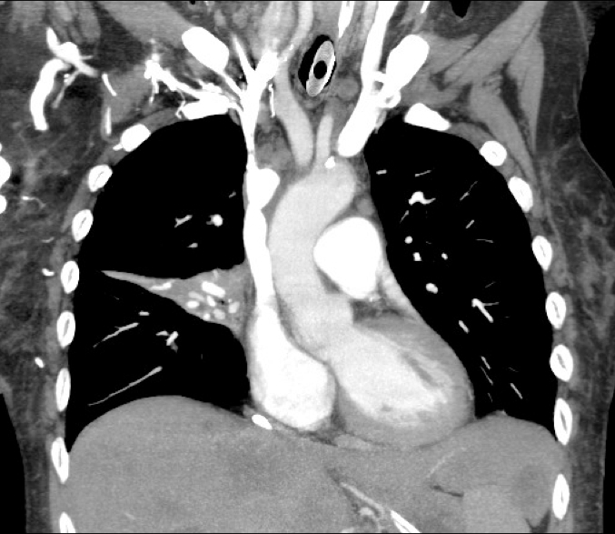
Question

60 year-old male presents with 6 months of weight loss,epistaxis, and increased headache when bending over. What's the diagnosis?
Show Answer
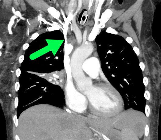
SVC Syndrome

  • SVC syndrome is caused by compression or obstruction of the superior vena cava blocking anterograde flow.
  • Most cases are secondary to extrinsic compression by malignancy. Other causes are secondary to internal obstruction (e.g., thrombosis).
  • Symptoms present sub-acutely, worsen with bending over, and are secondary to increased venous pressure in the head and neck (e.g., epistaxis, headache, tinnitus, conjunctival injection, neck swelling, etc.).
  • Treatment focuses on reversing the underlying cause (e.g., radiation or chemotherapy if due a sensitive tumor) and symptomatic treatment:
    • Steroids, diuretics, and hyperosmolar agents to reduce edema
    • Head of bed elevation to reduce brain edema
    • Anti-emetics to prevent vomiting-associated increases in intracranial pressure

Rice TW, Rodriguez RM, Light RW. The superior vena cava syndrome: clinical characteristics and evolving etiology. Medicine (Baltimore). Jan 2006;85(1):37-42.

Nunnelee JD. Superior vena cava syndrome. J Vasc Nurs. Mar 2007;25(1):2-5

Show References

Follow me on Twitter (@criticalcarenow) or Google+ (+criticalcarenow)


Title: What's the Diagnosis? Case by Dr. Ahmed Alrasheedi @aalrasheedi

Category: Visual Diagnosis

Posted: 1/12/2015 by Haney Mallemat, MD

Question

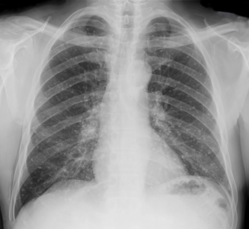
60 year-old male presents with rhinorrhea, body aches, and dry cough for two days. He recently moved here from Kentucky. What’s the diagnosis (please note that there are several differentials based on CXR)?


Show Answer

Answer Histoplasmosis

Histoplasma capsulatum is the fungus that causes histoplamosis, and is endemic to soil in the “Histo belt” of the U.S. (including all of Arkansas, Kentucky, Missouri, Tennessee, and West Virginia and parts Alabama, Illinois, Indiana, Iowa, Kansas, Louisiana, Maryland, Mississippi, Nebraska, Ohio, Oklahoma, Texas, and Virginia); it is found internationally as well.

Outbreaks have occurred following digging up the ground at construction at sites where this yeast lives in the soil, but spores can also become airborne and inoculate people.

Most cases are asymptomatic, but it is associated with a variety of pulmonary syndromes and can have extrapulmonary findings such as splenic calcifications. Disseminated disease typically occurs in immunocompromised patients

Treatment includes itraconazole for mild to moderate disease and amphotericin for disease that is severe or widespread disease.


Differentials:

  • Blastomycosis
  • Septic emboli
  • Sarcoidosis
  • Tuberculosis (milliary)
  • Aspergillosis
  • Carcinoid lung tumor
  • Atypical or viral pneumonias    


Title: What's the Diagnosis? Case by Dr. Joshua McClain and Dr. Yemi Adebayo

Category: Visual Diagnosis

Posted: 1/5/2015 by Haney Mallemat, MD

Question

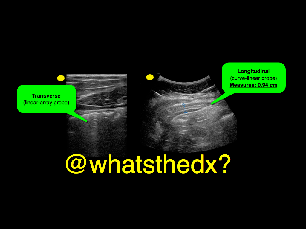
A male patient presents with right lower quadrant pain. The ultrasound is shown at the point of maximal tenderness. The diameter of the structure (image on right) is about 0.94cm. What is this structure and what's the diagnosis?
Show Answer

Appendicitis
  • Ultrasound can be used to diagnose appendicitis; a linear transducer is preferred in thinner / pediatric patients, while a curvilinear probe is may be used larger patients
  • Acute appendicitis causes irritation of the abdominal wall, so when looking for the appendix you need enough depth to visualize the peritoneal wall
  • To perform the scan, start at right upper quadrant in the transverse plane and slide down the abdomen towards the right iliac fossa until bowel gas has disappeared, typically the ileocecal junction. Alternatively, you can start scanning the point of maximal tenderness (i.e. the appendix)
  • Identifying a non-compressible and dilated appendix measuring greater than 6mm in transverse diameter suggesting the diagnosis
  • Color flow can be added to identify hyperemia around the appendix, which increases the specificity of the study.
  • More info on the scan can be found here and two podcasts from the incredible @ultrasoundpodcast guys part I and part II

Show References

Follow me on Twitter (@criticalcarenow) or Google+ (+criticalcarenow)


Title: What's the Diagnosis?

Category: Visual Diagnosis

Posted: 12/30/2014 by Haney Mallemat, MD (Updated: 12/30/2014)

Question

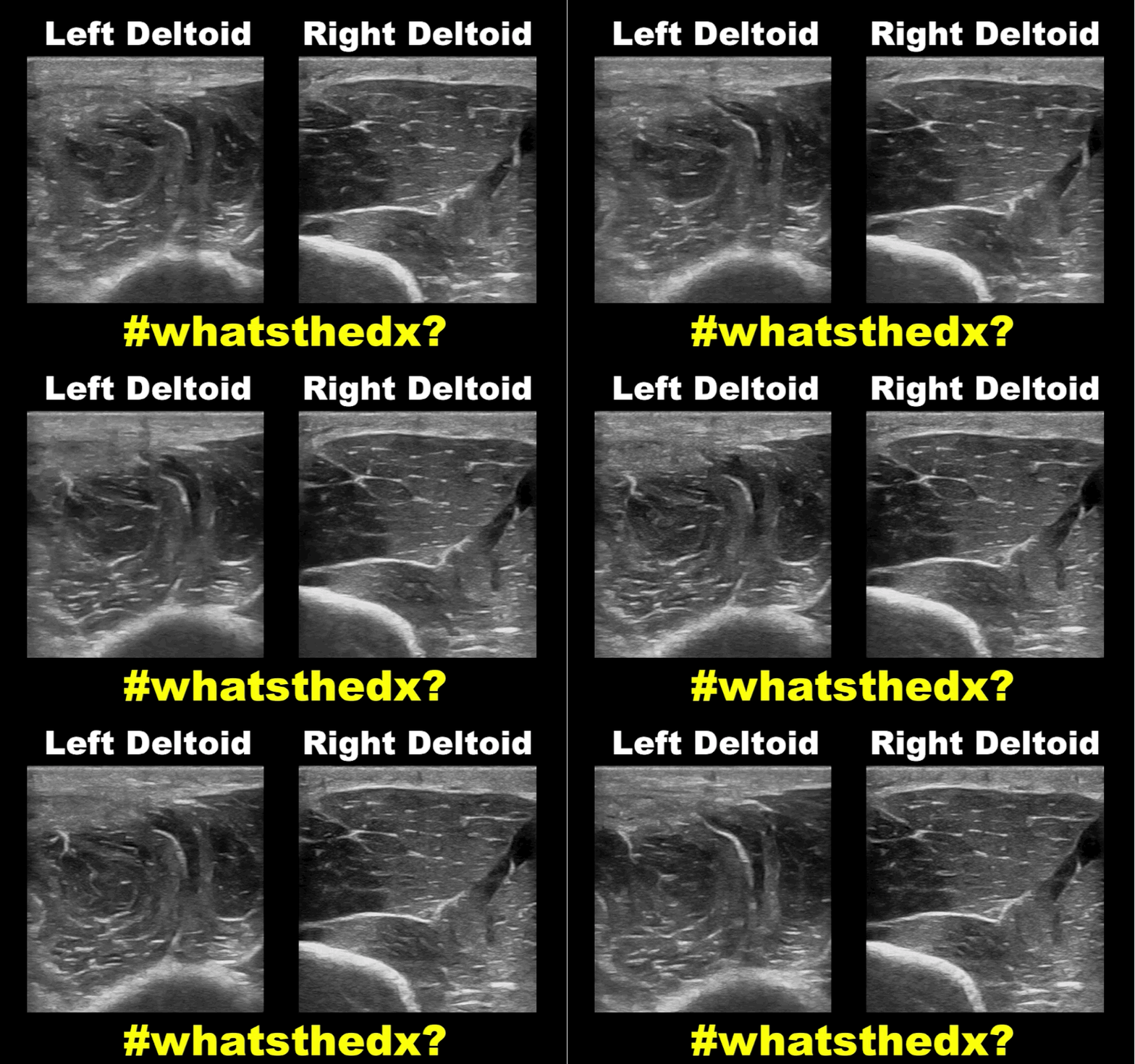
19 year-old male complaining of left arm pain one week after injecting anabolic steroids into his sholder. What's the diagnosis?
Show Answer
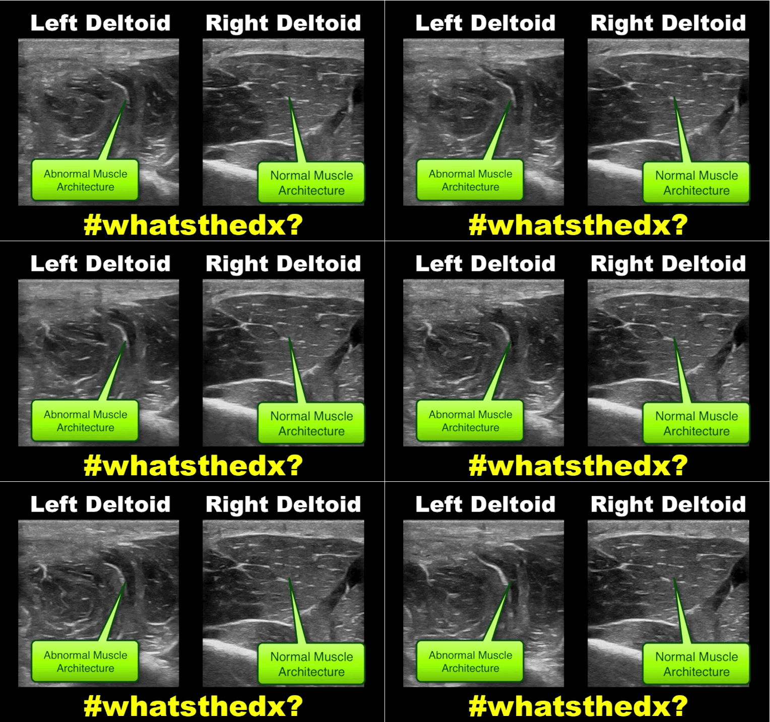
Myositis of the deltoid muscle

Show References

Follow me on Twitter (@criticalcarenow) or Google+ (+criticalcarenow)


Title: What's the Diagnosis?

Category: Visual Diagnosis

Posted: 12/23/2014 by Haney Mallemat, MD (Updated: 12/23/2014)

Question

Hand pain following fist versus face. What's the diagnosis and what nerve block would you use?
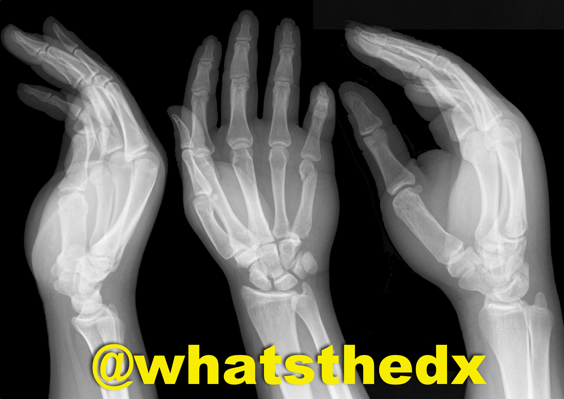
Show Answer
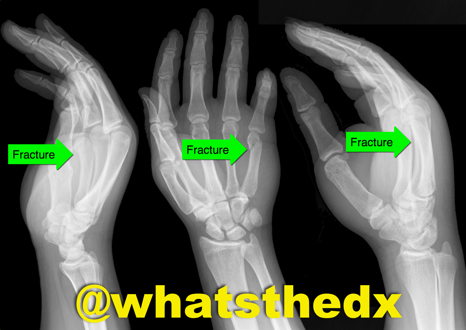
Boxer's fracture. Read more here.

Pain control can be achieved with an ulnar nerve block (e.g., reducation if if angulated). A video for the technique using ultrasound can be found here.

Show References

Follow me on Twitter (@criticalcarenow) or Google+ (+criticalcarenow)


Title: What's the Diagnosis? Dr. Ahmed Alrasheedi

Category: Visual Diagnosis

Posted: 12/15/2014 by Haney Mallemat, MD

Question

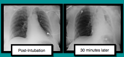
A patient is intubated for respiratory failure and the post-intubation CXR is shown on the left. 30 minutes later the patient desaturates and another CXR is obtained (the one on the right). What’s the diagnosis and what should you do?
Show Answer

Mucus plug

There are several conditions to consider when patients are having difficultly being ventilated. The approach to such a patient can be remembered with a mnemonic found here.

In this case, conditions to consider are:

  • right main-stem intubation (can be seen with ultrasound; here)
  • mucus plug (shift is towards problem)
  • tension pneumothorax (shift is away from problem); use ultrasound

Treating mucus plugs can usually be treated as follows:

  • Deep tracheal suctioning
  • Disconnecting patient from the vent and bagging
  • Chest physiotherapy (i.e., percussing the chest) on the affected area
  • Bronchoscopy, if above does not result in resolution

Show References

Follow me on Twitter (@criticalcarenow) or Google+ (+criticalcarenow)


Title: What's the Diagnosis? Case by Adeleke Oni, MD

Category: Visual Diagnosis

Posted: 12/8/2014 by Haney Mallemat, MD

Question

Patient was found in a house fire and was given a certain medication in the Emergency Room. The patient’s urine turned this color (red), what’s the diagnosis?

Show Answer

Hydroxocobalamin (i.e., Cyanokit)

Cyanide toxicity

  • Occurs secondary to combustion of natural and synthetic products producing cyanide
  • Patients recovered from fires should have a lactate level drawn; levels greater than 10 are concerning for cyanide toxicity.  
  • The Cyanokit contains hydroxocobalamin, a form of vitamin B12 that is used as the antidote.
  • Hydroxocobalmin has many side-effects including self-limited hypertension, flushing of the skin, and red urine for up to 14 days.  
  • See a previous UMEM post by Dr. Bryan Hayes for more information 

Show References

Follow me on Twitter (@criticalcarenow) or Google+ (+criticalcarenow)


Previous  |  1 |  2 |  3 |  4 |  5 |  6 |  7 |  8 |  9 |  ... |  13 |  Next
University of Maryland School of Medicine logo
  • Departments
  • Programs
  • Research Centers
  • Institutes
  • Graduate Education
  • CIBR Cores
  • Offices of the Dean
    • University of Maryland, Baltimore
    • University of Maryland Medical Center
    • Medical Alumni Association of the University of Maryland
    • Health Sciences and Human Services Library
University of Maryland School of Medicine
655 W. Baltimore Street
Baltimore, MD 21201
Contact Us

© University of Maryland School of Medicine

    • Webmaster
    • Web Accessibility
    • Site Index
    • UMB Hotline
    • Employment
    • Map