University of Maryland School of Medicine logo
Emergency Medicine
All Departments
UMSOM Assistant
UMEM Educational Pearls
  • Home
  • Departments
  • Emergency Medicine
  • CME
  • UMEM Educational Pearls
  • CME
  • Courses and Conferences
  • Free Open Access Medical Education
  • UMEM Educational Pearls

Search

21-40 of 245 results with category "Visual Diagnosis"

Previous  |  1 |  2 |  3 |  4 |  5 |  6 |  7 |  ... |  13 |  Next

Title: What's the Diagnosis?

Category: Visual Diagnosis

Posted: 11/7/2016 by Tu Carol Nguyen, DO

Question

8 year-old female with no PMH who presents with concerns for "purple patches" popping up on her arm for 2-3 days. Stated that one appeared and then, the other one appeared 12 hours later. She denied any trauma whatsoever, history of easy bleeding/bruising and did feel safe at home. The rest of the review of systems was negative.

Patient said there was mild pain when the area was touched. The rest of the physical examination was normal.

What's the diagnosis? (Image below)

Show Answer

Superficial Thermal Burn

Upon further questioning, patient stated that she had been making s'mores by roasting marshmallows over an electric stove 3 days prior. The burns showed up the subsequent morning.

  • Thermal injury
    • Types - scald, contact, fire, chemical, electrical, radiation
    • Can still sustain dermal burns from emanating heat without contact
    • Extent of tissue damage based on temperature & duration of exposure
  • Non-accidental burns are common in children
    • 10% of physically abused children have intentional burns/scalds
    • Sites: hand, back, wrist, buttock, feet, legs
    • Type: well-define margins of contact/scald burns in unusual places, glove-and-stocking distribution

Take Home Points:

  • Take a good history
  • Can sustain burns without direct contact (i.e. heat, radiation, etc.)
  • Consider non-accidental burns in children

Previous pearls about burns:

Pediatric Burns:

  • https://umem.org/educational_pearls/1107/
  • https://em.umaryland.edu/educational_pearls/1164/

Show References

Monseau AJ, Reed ZM, Langley KJ, Onks C. Sunburn, Thermal, and Chemical Injuries to the Skin. Prim Care. 2015;42(4):591-605.

"Pathophysiology of Thermal Injury." Civic Plus. 2007.


Title: What is the diagnosis ? (Image by Dr. Tu Nguyen)

Category: Visual Diagnosis

Posted: 10/31/2016 by Hussain Alhashem, MBBS

Question

30 year old female presents with a painful finger for 1 week. Finger exam showed the following. What is the diagnosis ?

Show Answer

Herpetic whitlow

- It is caused by either HSV 1 or 2.

- Infection is usually transmitted by direct unprotected contact.

- Healthcare workers are at risk of contracting the infection if appropriate preventative measures were not taken when dealing with patients who have the infection.

- Diagnosis is mainly clinical. Tzank smears can be done to confirm the diagnosis if in doubt.

- Treatment with Acyclovir is thought to shorten the duration of the infection and prevent recurrence if started within 72 hours of beginning of symptoms.

- Incision of the vesicular lesion is contraindicated and can cause severe bacterial superinfection.

Show References

McDonald, Lucas S., et al. "Hand infections." The Journal of hand surgery36.8 (2011): 1403-1412.


Title: What's the Diagnosis?

Category: Visual Diagnosis

Posted: 10/25/2016 by Tu Carol Nguyen, DO (Updated: 10/26/2016)

Question

20 year-old female presents with sore throat, right throat fullness, difficulty speaking for 2-3 days. A bedside ultrasound and subsequent CT was obtained as seen below. What's the diagnosis?


Show Answer


Peritonsillar Abscess


The ultrasound image is a transcutaneous approach with a linear transducer that is placed at the angle of the mandible of the affected side. This is an alternative approach to an intra-oral ultrasound with the endocavitary transducer if the patient has trismus.

  • Peritonsillar abscess (PTA) is often taught as a clinical diagnosis; however, 36% are often peritonsillar cellulitis (PTC)
    • Sensitivity and specificity of clinical diagnosis of PTA are 78% and 50%, respectively
  • Classically, providers attempt blind needle aspirations (landmark-based)
    • Blind needle aspiration is unreliable for diagnosis of PTA with a false-negative rate of 10-24%
  • Intraoral sonography sensitivity: 90-100%
    • Using ultrasound can avoid unnecessary use of CTs and blind needle asprations


Take Home Points:

  • Consider intra-oral US for diagnosis of PTA vs. PTC and to reduce unecessary radiation with CT
  • Consider transcutaneous US to evaluate for PTA in the setting of trismus
  • Consider intra-oral US-guided needle aspiration of PTA vs. blind needle aspiration


How to do an intra-oral US-guided needle aspiration of PTA, check out:

http://www.ultrasoundpodcast.com/2012/01/episode-21-full-peritonsillar-abscess-podcast/


For a brief video on how to perform a transcutaneous US for PTA:

https://www.youtube.com/watch?v=JkIYOhKCweI&t=28s


Show References

Constantino TG, Satz WA, Dehnkamp W, Goett H. Randomized Trial Comparing Intraoral Ultrasound to Landmark-based Needle Aspiration in Patients with Suspected Peritonsillar Abscess. Academic Emergency Medicine. June 2012; Vol. 19 No. 6: 626-631.

Halm BM, Ng C, Larrabee YC. Diagnosis of a Peritonsillar Abscess by Transcutaneous Point-of-Care Ultrasound in the Pediatric Emergency Department. Pediatr Emerg Care. 2016;32(7):489-92.

Rehrer M, Mantuani D, Nagdev A. Identification of peritonsillar abscess by transcutaneous cervical ultrasound. Am J Emerg Med. 2013;31(1):267.e1-3.


Title: What's the Diagnosis? Image and Case by Dr. Kathleen Stephanos

Category: Visual Diagnosis

Posted: 10/10/2016 by Tu Carol Nguyen, DO

Question

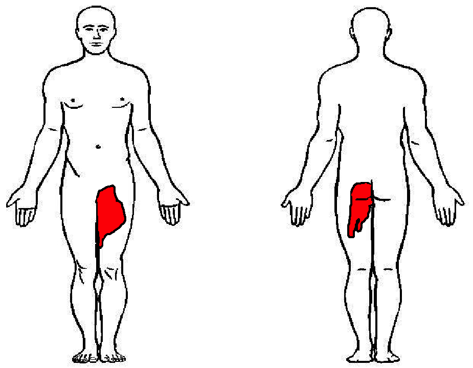
57 year-old female with history of bilateral lung transplants presents with fever, and 2 days of a painful, red, bumpy rash over the left labia and left buttock, but also notes a small tender area on the plantar surface of the left foot.

Below is a figure depicting the location of the rash, as well as a photo of her foot.
Show Answer

This is Herpes Zoster (Shingles).

Presentation:

  • Zoster typically appears along a single dermatome, without crossing midline in immunocompetent hosts, but may involve 2-3 adjacent dermatomes.
  • In immunocompromised patients, systemic involvement may occur.
  • Thoracic and lumbar dermatomes are the most common.
  • Lumbar and sacral dermatomes wrap around the lower extremity and may spare the skin between the perineum and the foot.

Treatment:

  • Immunocompromised patients should be treated with IV Acyclovir 10mg/kg q 8hr for 7 days (Use IBW in obese patients)
  • In immunocompetent individuals, if identified within 48 hours of rash onset, may receive oral therapy with famciclovir, acyclovir or valcyclovir
  • Herpes Zoster alone should not prompt immunosuppression work up
    • However, concerns for underlying immune disorders if:
      • continued development of lesions > 1 week, or
      • involvement of more than 3 dermatomes

Show References

Dworkin RH, Johnson RW, Breuer J, et al. Recommendations for the management of herpes zoster. Clin Infect Dis. 2007;44 Suppl 1:S1.


Title: What is the diagnosis ?

Category: Visual Diagnosis

Posted: 10/3/2016 by Hussain Alhashem, MBBS

Question

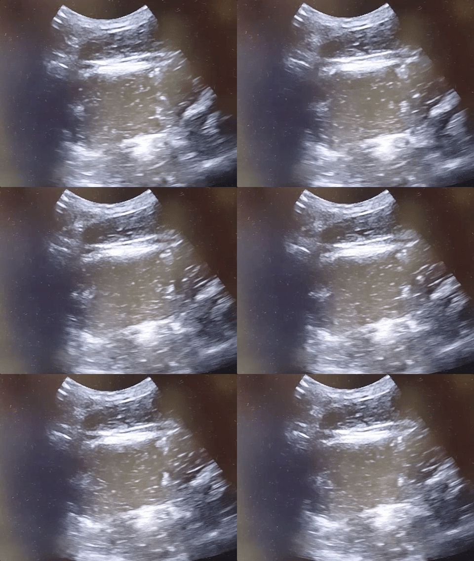
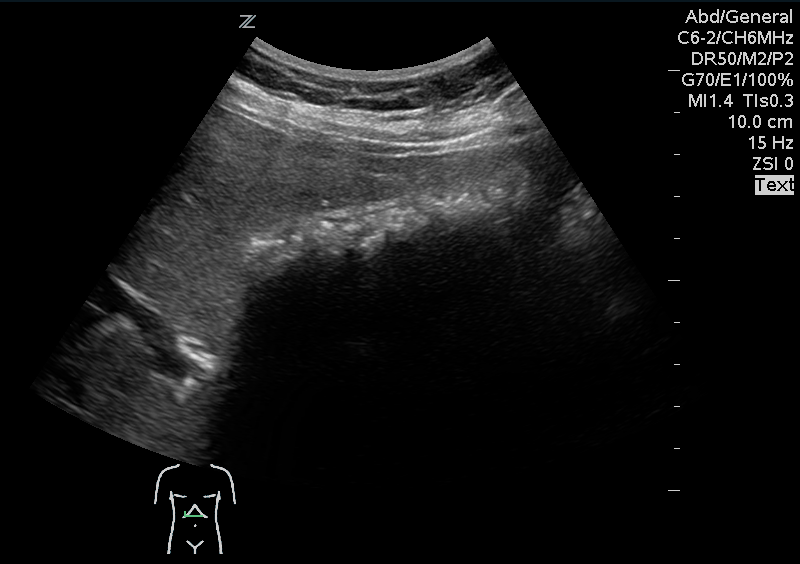
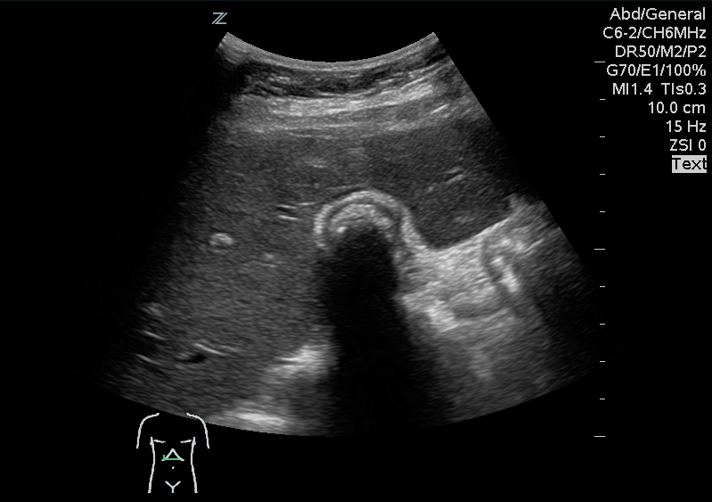
A 41 year old female presenting with intermittent RUQ abdominal pain for 1 week. An ultrasound of the right upper quadrant was performed. What is the diagnosis ?
Show Answer
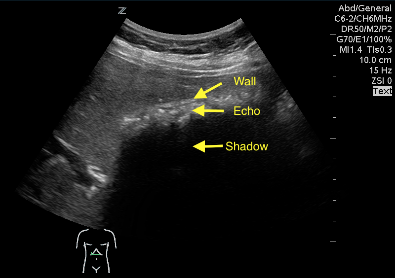
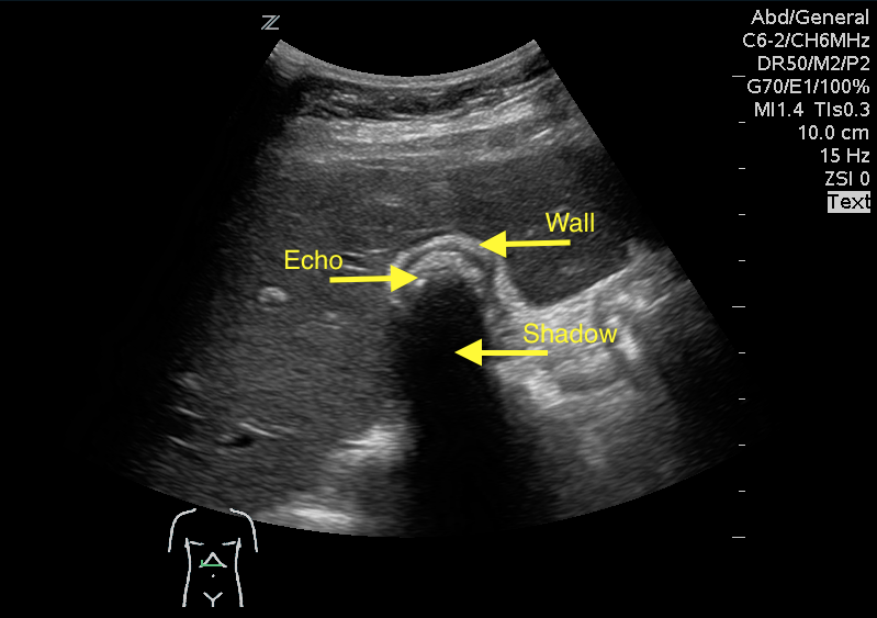
Answer: WES sign

WES sign stands for Wall Echo Shadow sign. It is a triad of:

1- Thick echogenic gall bladder wall (W)

2- Echoes filling the gallbladder (E)

3- A posterior acoustic shadow (S)

  • It is suggestive of either one big stone or multiple stones filling the gallbladder.
  • This sign can be mistaken for either a bowel loop or an empty and contracted gallbladder.
  • To differentiate between WES and a bowel loop, look for visible bowel peristalsis.

Rybicki, F. J. (2000). The WES Sign 1. Radiology, 214(3), 881-882.


Title: What's the Diagnosis? Case by Dr. Lindsay Weiner

Category: Visual Diagnosis

Posted: 9/26/2016 by Tu Carol Nguyen, DO (Updated: 9/26/2016)

Question

22-year-old male with history of autism, mental retardation who is non-verbal presents with abdominal pain and vomiting for one day. Patient was found clutching his abdomen and moaning. What's the diagnosis?


Show Answer

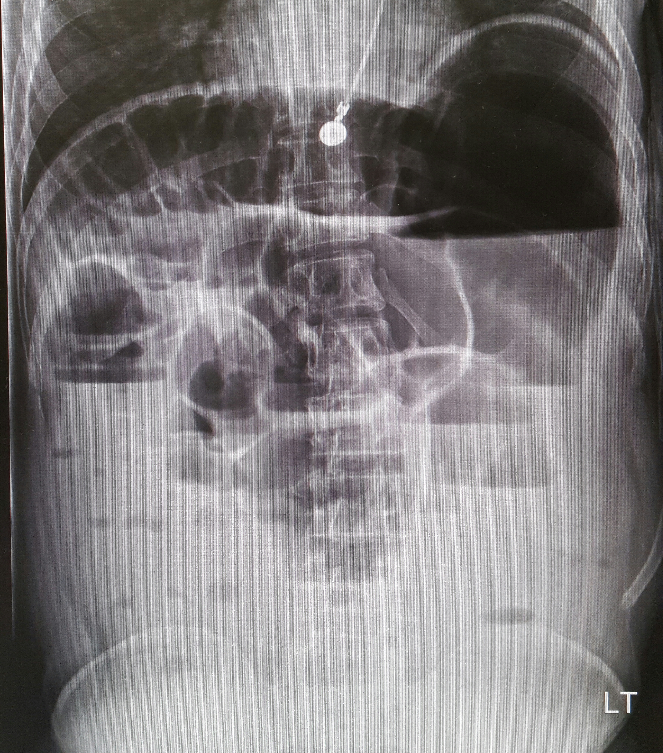
Small Bowel Obstruction


  • Presentation: abdominal pain, nausea, vomiting, early diarrhea progressing to lack of flatus or bowel movements with complete obstruction. Severe obstruction can result in perforation, unstable vital signs, and physical exam signs of peritonitis.
  • Sensitivity and specificity > 90% for diagnosis of SBO with US, with superior test performance compared to X-ray.
  • US findings: dilated small bowel loops > 3 cm; no peristalsis, swirling snowstorm appearance of bowel contents
  • Treatment: bowel rest, IV fluids, and prompt surgery consultation! Consider NG tube for decompression.


See the corresponding upright abdominal x-ray, showing dilated bowel with air fluid levels.


Show References

Kameda T, Taniguchi N. Overview of point-of-care abdominal ultrasound in emergency and critical care. J Intensive Care. 2016 Aug 15;4:53. doi: 10.1186/s40560-016-0175-y. eCollection 2016. Review.

Unl er EE, Yava i O, Ero lu O, Yilmaz C, Akarca FK. Ultrasonography by emergency medicine and radiology residents for the diagnosis of small bowel obstruction. Eur J Emerg Med. 2010 Oct; 17(5):260-4.


Title: What is the diagnosis ? (Case submitted by Dr. Kevin Campos and Dr. Adeleke Oni)

Category: Visual Diagnosis

Posted: 9/19/2016 by Hussain Alhashem, MBBS

Question

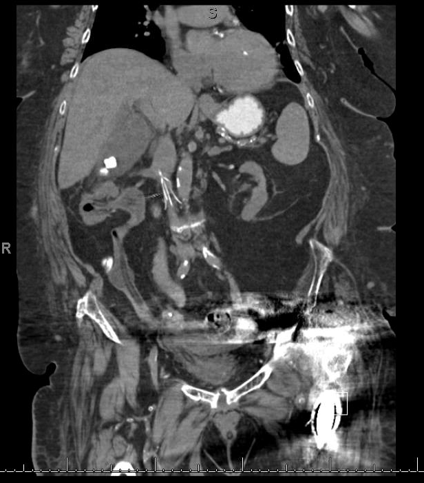
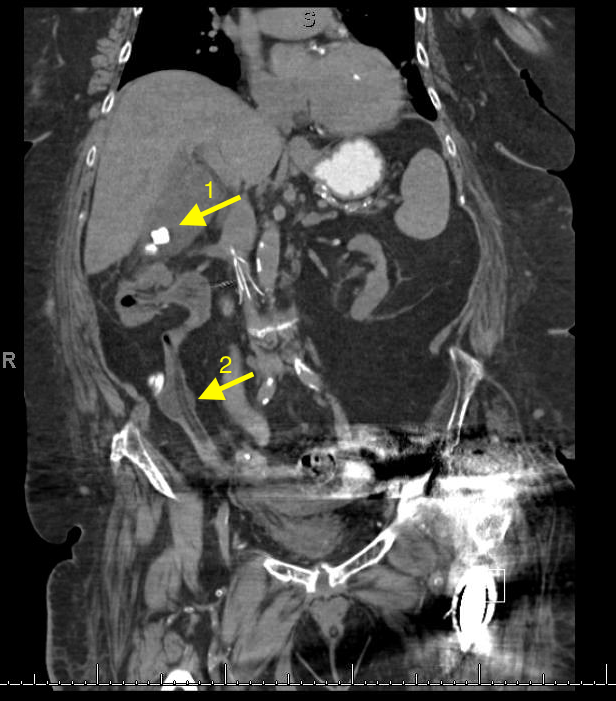
A 67 year old female with history of CVA, presented from a nursing home with RUQ abdominal pain and inablitiy to tolerate PO for 3 days. A CT scan of her abdomen was obtained. What is the diagnosis ?

Show Answer
1- Cholecystitis

Ultrasound remains the best modality to test for cholecystitis. However, CT scans can still be obtained for non-classic presentations. The negative predictive value of CT is still relatively high. CT has a negative predictive value of 89%, compared to 97% to that of ultrasound. The absence of cholecysitis on CT will help the argument against the diagnosis, but if the suspicion is high an ultrasound study should still be obtained.

Things to look for on an abdominal CT that are suggestive of cholecysitis:

  1. Gallbladder distension ( >5 cm width, >8 cm length).

  2. Wall thickening ( >4mm thickness).

  3. Pericholecystic fat stranding.

  4. Presence of gallstones.

2- An inflated foley catheter in the ureter!

Ureteric insertion of foley catheters is a very rare complication of foley catheterization. There are no clear predisposing factors to this complication. However, it is thought that the presence of an underlying anatomical deformity (e.g. abnormal ureteric insertion site) might put the patient at a higher risk for it. Inflating a balloon in the ureter might result in severe ureteric injury. A suggested method to prevent this kind of injury is to perform bladder aspiration to insure balloon positioning prior to inflation.

Show References

References

1- Shakespear, Jonathan S., Akram M. Shaaban, and Maryam Rezvani. "CT findings of acute cholecystitis and its complications." American Journal of Roentgenology 194.6 (2010): 1523-1529.

2- Kim, Myung Ki, and Kwangsung Park. "Unusual complication of urethral catheterization: a case report." Journal of Korean medical science 23.1 (2008): 161.


Title: What's the Diagnosis? Case by Dr. Tejusve Rao

Category: Visual Diagnosis

Posted: 9/12/2016 by Tu Carol Nguyen, DO (Updated: 9/12/2016)

Question


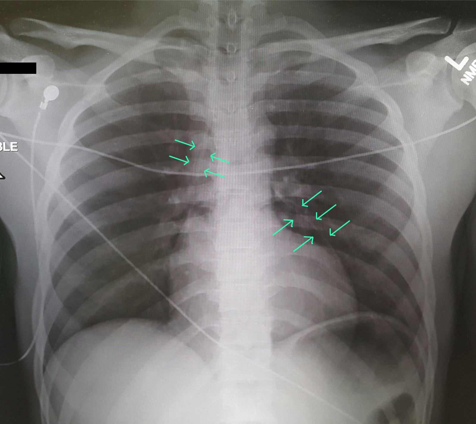
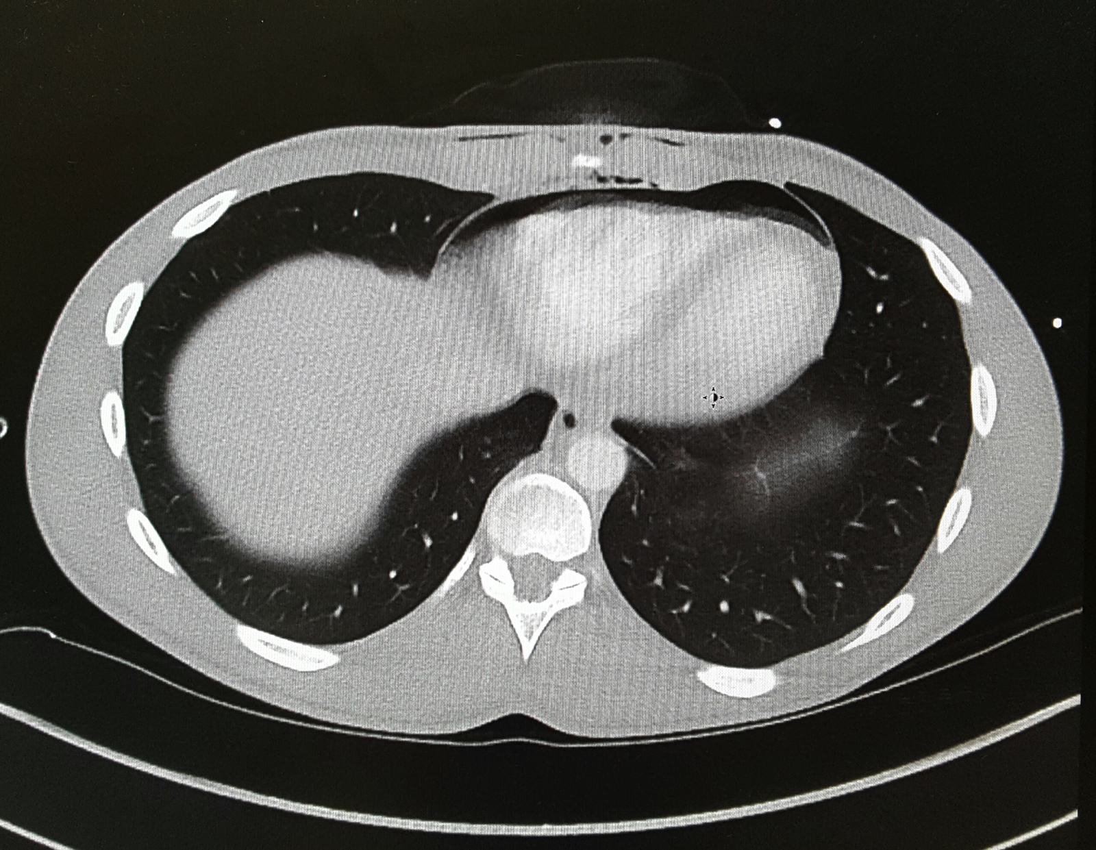
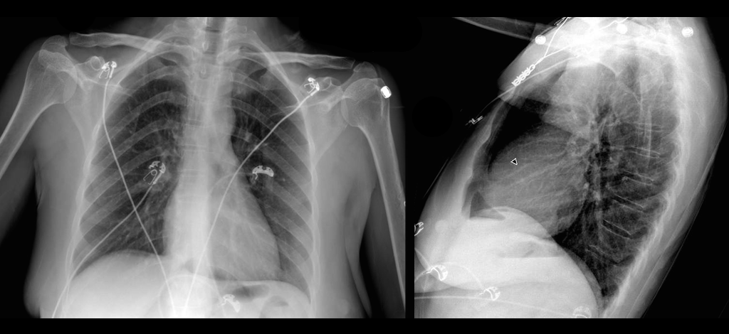
A 25-year-old male was brought in by EMS with a stab wound to the chest. What's the diagnosis?


Show Answer

Pneumopericardium


CT Chest was done with the image below:


Penetrating Cardiac Trauma
  • Initial Assessment of ABC, ATLS primary survey guidelines.
  • Evaluate for tension pneumothorax or cardiac tamponade in all patients presenting with chest trauma and shock.
  • Cardiac Box: Surrounded by sternal notch, xiphoid process and nipples.
  • Order of injury: Right Ventricle --> Left Ventricle --> Right Atrium --> Left Atrium
  • Beck’s Triad: Hypotension, JVD, muffled heart sounds may not be present initially.
  • Conduct FAST exam to examine for cardiac tamponade, hemothorax, pneumothorax.
  • Cardiac Tamponade more common from stab wounds than from gun shot wounds.
  • Hemodynamically unstable patients require immediate operative therapy after quick bedside assessment (physical exam, ultrasound, chest tube as needed)

Differential Diagnosis: Bronchial injury, Diaphragm injury, Hemothorax, Tension Pneumothorax, Aortic Transection, Esophageal injury, Pneumomediastinum

Evaluation: Ultrasound (FAST Exam), CXR, CTA in stable patients, ECG, troponin.

Management: Penetrating cardiac trauma require emergent thoracotomy, pericardial window.


Show References

Clancy K, Velopulos C, Bilaniuk JW, et al. Screening for blunt cardiac injury: an Eastern Association for the Surgery of Trauma practice management guideline. J Trauma Acute Care Surg. 2012;73(5 Suppl 4):S301-6.

El-menyar A, Al thani H, Zarour A, Latifi R. Understanding traumatic blunt cardiac injury. Ann Card Anaesth. 2012;15(4):287-95.

Tintinalli's 7th Edition. Emergency Medicine Manual. Chapter 164: Cardiothoracic Trauma.


Title: What's the Diagnosis?

Category: Visual Diagnosis

Posted: 2/29/2016 by Haney Mallemat, MD

Question

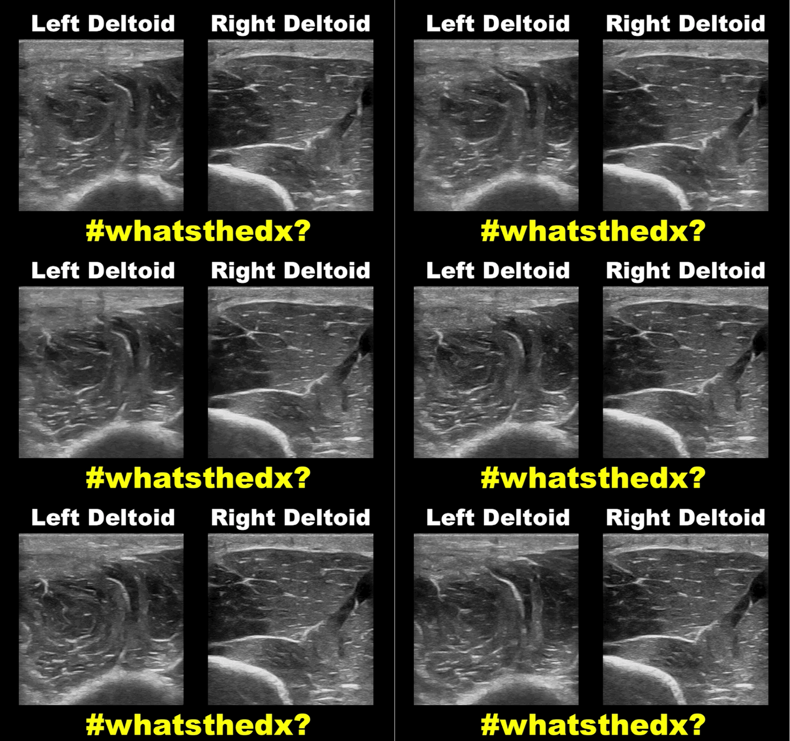
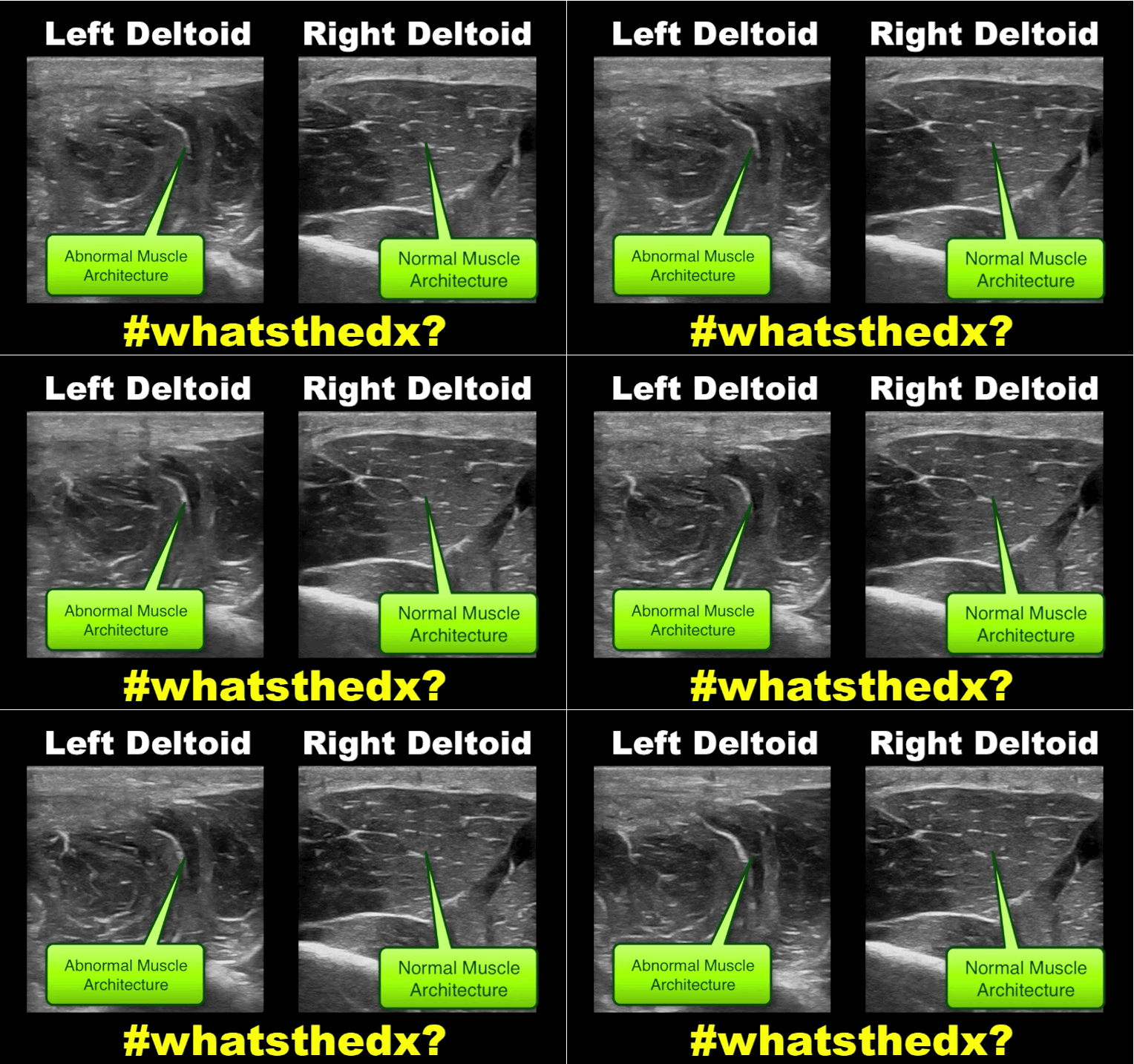
19 year-old male complaining of left arm pain one week after injecting anabolic steroids into his shoulder. What's the diagnosis?

Show Answer

Myositis of the deltoid muscle

Show References

Follow me on Twitter (@criticalcarenow)


Title: What's the Diagnosis? Case by Dr. Ali Farzad

Category: Visual Diagnosis

Posted: 1/18/2016 by Haney Mallemat, MD

Question

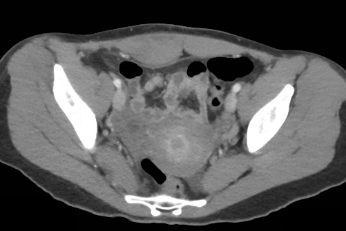
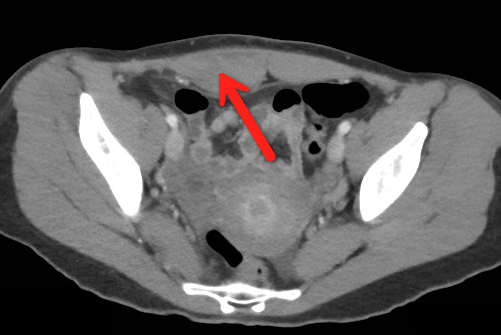
23 year-old female presents complaining of progressive right lower quadrant pain after doing "vigorous" exercise. CT abdomen/pelvis below. What’s the diagnosis? (Hint: it’s not appendicitis)


Show Answer

Rectus sheath hematoma

Rectus Sheath Hematoma (RSH)

Rectus muscle tear causing damage to the superior or inferior epigastric arteries with subsequent bleeding into the rectus sheath; uncommon cause of abdominal pain but mimics almost any abdominal condition.

Diagnose with CT, but try using ultrasound (thanks Dr. Joseph Minardi)

May occur spontaneously, but suspect with the following risk factors:

  • Coagulopathy (#1 cause); acquired (e.g., warfarin) or inherited disorder
  • Rectus muscle trauma
  • Vigorous or sudden contraction of rectus muscle
  • Increased intra-abdominal pressure from vigorous coughing
  • Pregnancy (gestation, labor, or post-partum)

Typically a self-limiting condition, but hypovolemic shock may result from significant hematoma expansion.

Hemodynamically stable (non-expanding hematoma): conservative treatment (rest, analgesia, and ice)

Hemodynamically unstable (expanding hematoma): treat with fluid resuscitation, reversal of coagulopathy, and transfusion of blood products.


Show References

Follow me on Twitter (@criticalcarenow)


Title: What's the Diagnosis? Guest submission by #ChileEM Dr. Pedro Verdugo

Category: Visual Diagnosis

Posted: 1/11/2016 by Haney Mallemat, MD (Updated: 3/10/2016)

Question

What’s the name of this CT finding and name two potential causes?

Show Answer

Pneumobilia (air in the biliary tree). Be careful, this must be distinguished from portal venous gas.

Diagnoses to consider when pneumobilia is present:

  • Infection
    • Cholangitis
    • Liver abscess communicating with biliary tree
    • Emphysematous cholecystitis
  • Biliary instrumentation (e.g., recent ERCP)
  • Incompetent sphincter or Oddi
    • passage of a gallstone
    • sphincterotomy
    • certain medications
  • Biliary-enteric fistula (e.g., Peptic ulcer disease, gallstone ileus, trauma,, etc.)

http://radiopaedia.org/articles/pneumobilia

Show References

Follow me on Twitter (@criticalcarenow)


Title: What's the Diagnosis? Case by UMEM alumni, Dr. Bethany Radin

Category: Visual Diagnosis

Posted: 12/28/2015 by Haney Mallemat, MD

Question

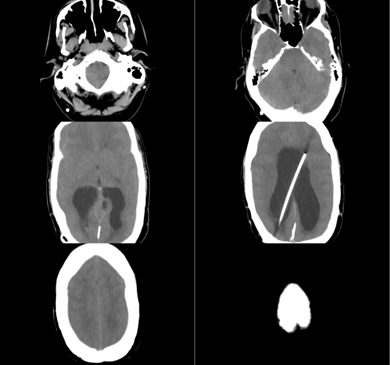
79 year-old male with headaches, ataxia, falls, and difficulty urinating. What's the diagnosis?

Show Answer
Diagnosis: Ventriculomegaly secondary to Normal Pressure Hydrocephalus

An approach to ventriculomegaly

Ventriculomegaly is due to cerebral atrophy (e.g., Parkinson disease) or increased cerebrospinal fluid (CSF) within the ventricles. Increased CSF is due to:

  • Increased CSF production (e.g., choroid-plexus papilloma), or
  • Impaired CSF re-absorption (e.g., subarachnoid-villi inflammation), or
  • Obstruction of CSF flow (e.g., non-communicating hydrocephalus)

Congenital causes of ventriculomegaly:

  • Neuro-tube defect (e.g, myelomeningocele)
  • Aquaductal stenosis
  • CNS malformation (e.g., Dandi-Walker syndrome)
  • Intrauterine infection (e.g., CMV, rubella, etc.)

Acquired causes of ventriculomegaly:

  • Paget's disease (obstruction of CSF flow)
  • Bacterial or viral meningitis (impaired CSF absorption)
  • Tumor (obstruction of CSF flow)
  • Post-hemorrhagic (e.g., trauma, aneurysmal rupture, AVM, coagulopathy) impaired absorption and/or obstruction of flow

Show References

Follow me on Twitter (@criticalcarenow)


Title: What's the Diagnosis? The Critical Care Edition

Category: Visual Diagnosis

Posted: 12/15/2015 by Haney Mallemat, MD

Question

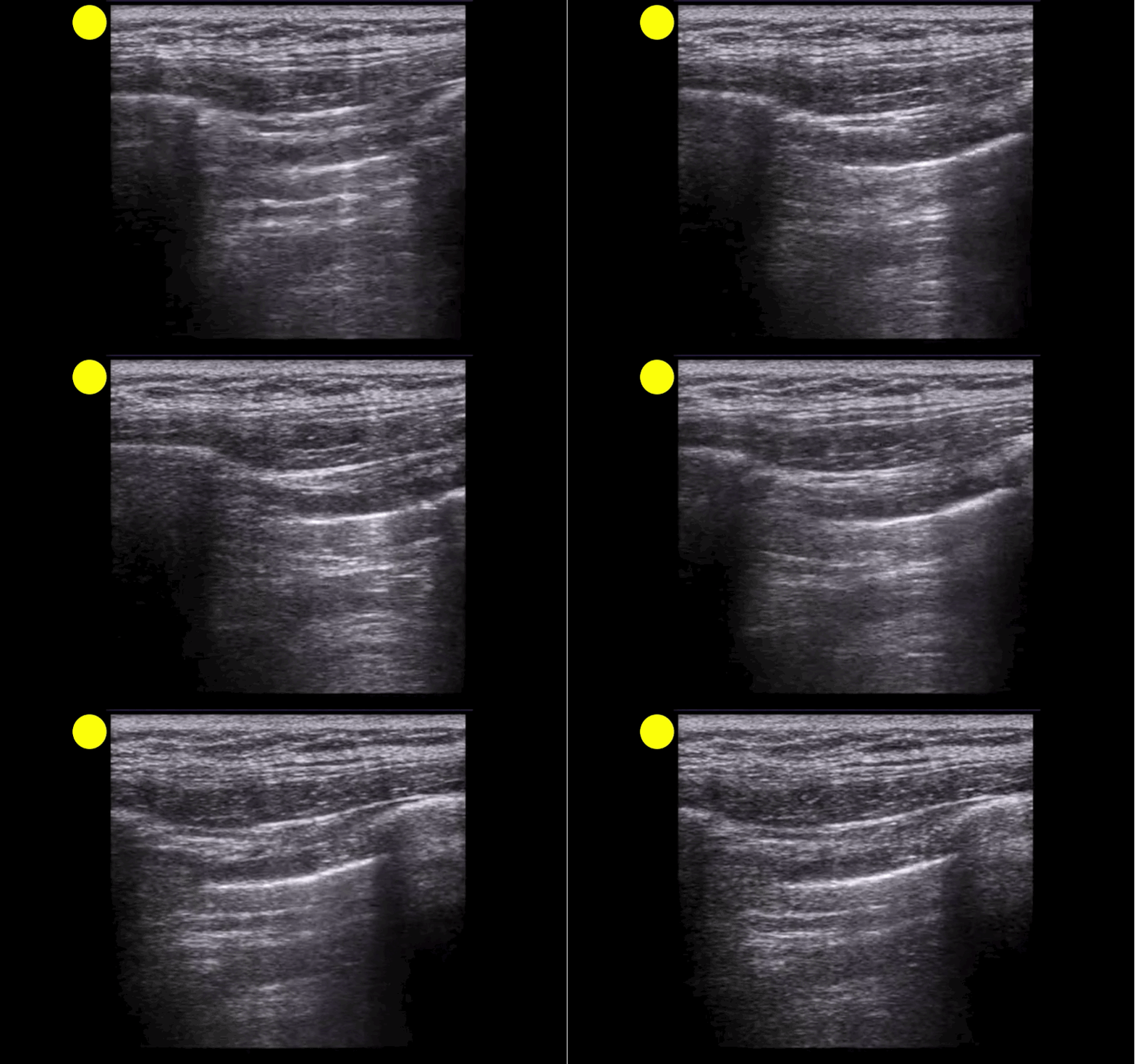
A patient arrives in acute respiratory distress with left sided chest pain. Ultrasound of the left anterior chest is shown; what's the diagnosis and name one false positive?
Show Answer

Lung point indicating pneumothorax (PTX)....see below for the false positives

What's the (Lung) Point

  • Separation of the visceral and parietal pleural secondary to a PTX leads to well recognized loss of lung sliding on ultrasound. 
  • Lung point (LP) on ultrasound is where the transition between pleural sliding and the loss of this sliding is demonstrated. Previous studies have reported that the LP has 100% specificity for a PTX.
  • Although this is a great sign to demonstrate PTX, be aware that there are several LP mimics:
    • lung-heart interface
    • lung-diaphragm interface (lower thorax laterally)
    • pleural effusion
    • blebs
  • Check out these great #FOAM posts by @ultrasoundjelly & @ultrasoundMD for more on the LP (and their mimics). Check them out here & here

Show References

Follow me on Twitter (@criticalcarenow)


Title: What's the Diagnosis? Case by UMEM alumni Dr. Adeleke Oni

Category: Visual Diagnosis

Posted: 12/14/2015 by Haney Mallemat, MD

Question

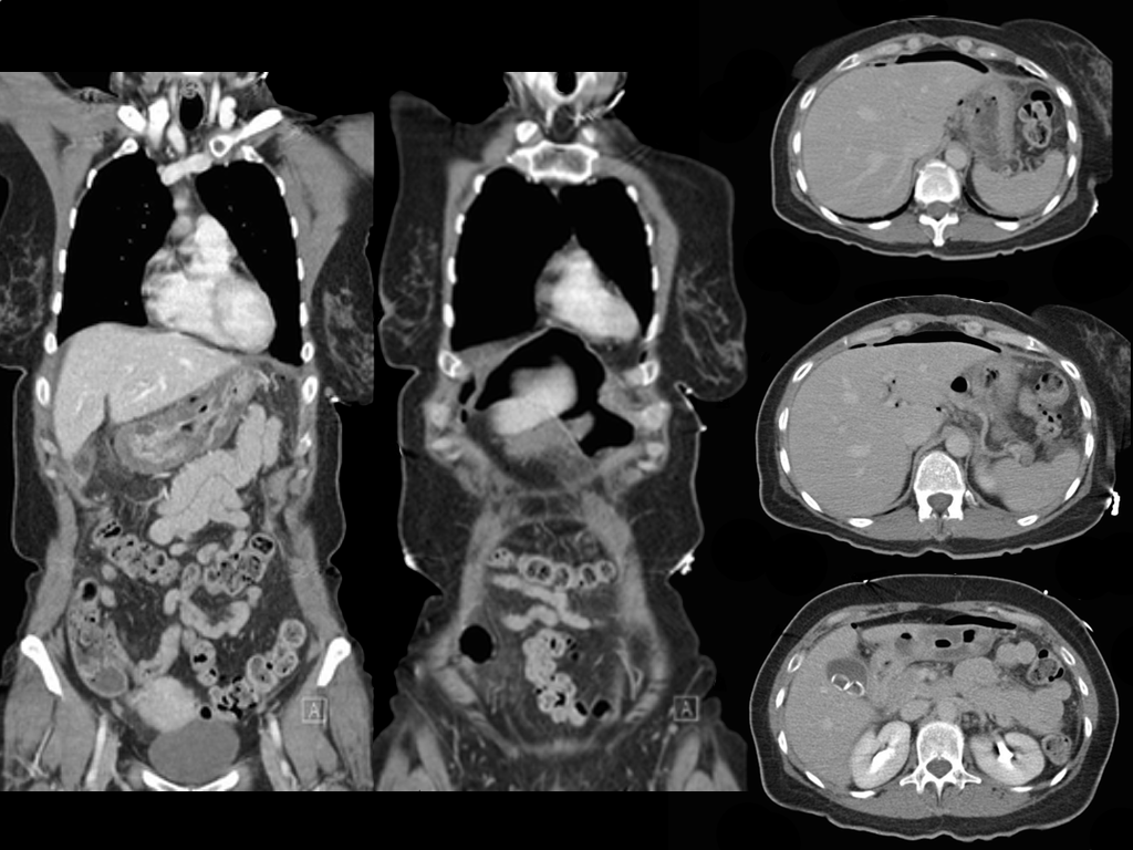
A patient presents with the sudden of onset chest and abdominal pain which woke her up at 2am. She has abdominal tenderness and rebound on exam, what's the diagnosis?

Show Answer

Ruptured gastric ulcer with pneumoperitoneum (CT scan below)

Show References

Follow me on Twitter (@criticalcarenow)


Title: What's the Diagnosis? By Dr. Samira Bhattacharya and Dr. Girish Sethuraman

Category: Visual Diagnosis

Posted: 12/8/2015 by Haney Mallemat, MD (Updated: 12/8/2015)

Question

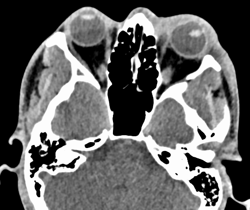
27 year-old presents after being punched in the face. Decreased vision in left eye, what's the diagnosis?

Show Answer

Lens disclocation

Show References

Follow me on Twitter (@criticalcarenow)


Title: What's the Diagnosis? Image by Dr. Jeffrey Leiter

Category: Visual Diagnosis

Posted: 11/30/2015 by Haney Mallemat, MD

Question

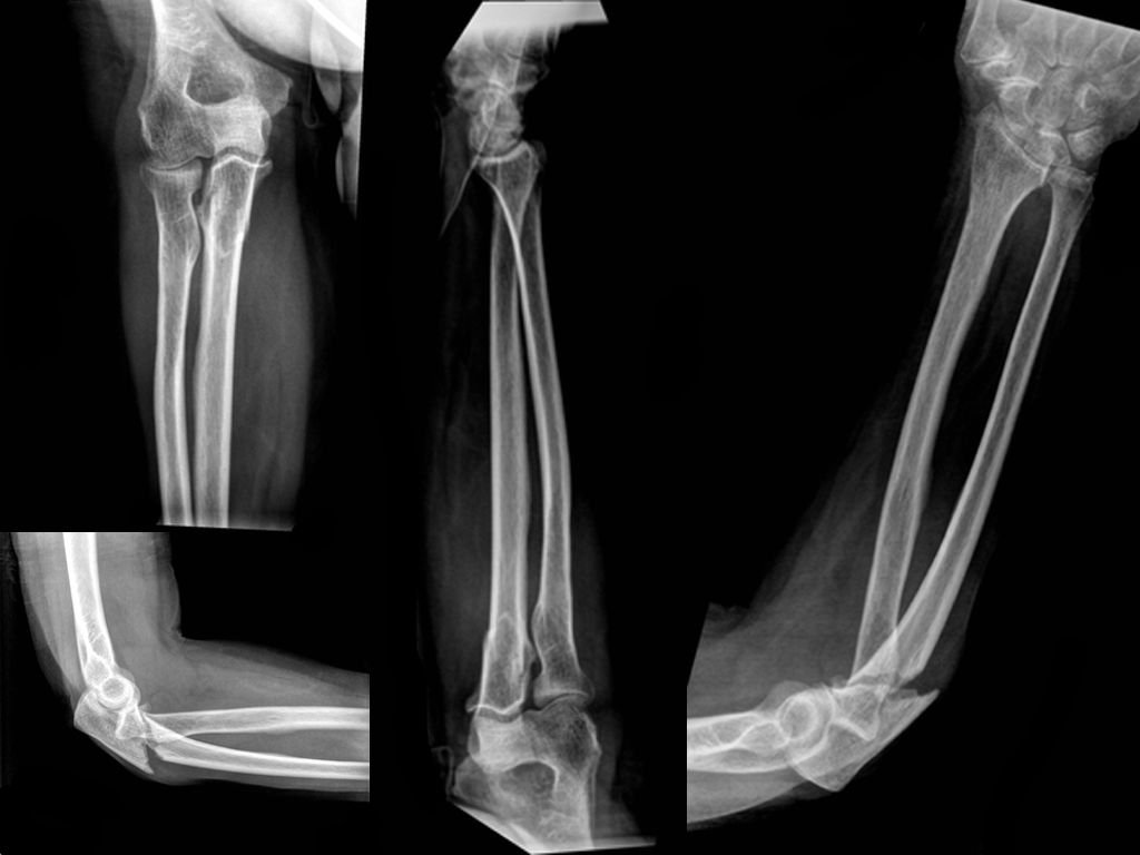
Patient presents with right elbow pain after a fall. What's the diagnosis and what other injury should you look for?
Show Answer

Displaced fracture of the proximal ulna.

Pearls

  • Always look for injury at the radial head when the proximal ulna is fractured, specifically looking for a radial head dislocation; this pattern is known as a Monteggia fracture.
  • Remember the mnemonic MUGR (MU-GR)
    • Monteggia fracture-dislocation = Ulnar fracture with a proximal radial dislocation
    • Galeazzi fracture-dislocation = Radial fracture with a distal radioulnar dislocation

Show References

Follow me on Twitter (@criticalcarenow)


Title: What's the Diagnosis? Case by Dr. Mak Moayedi

Category: Visual Diagnosis

Posted: 11/23/2015 by Haney Mallemat, MD (Updated: 12/5/2015)

Question

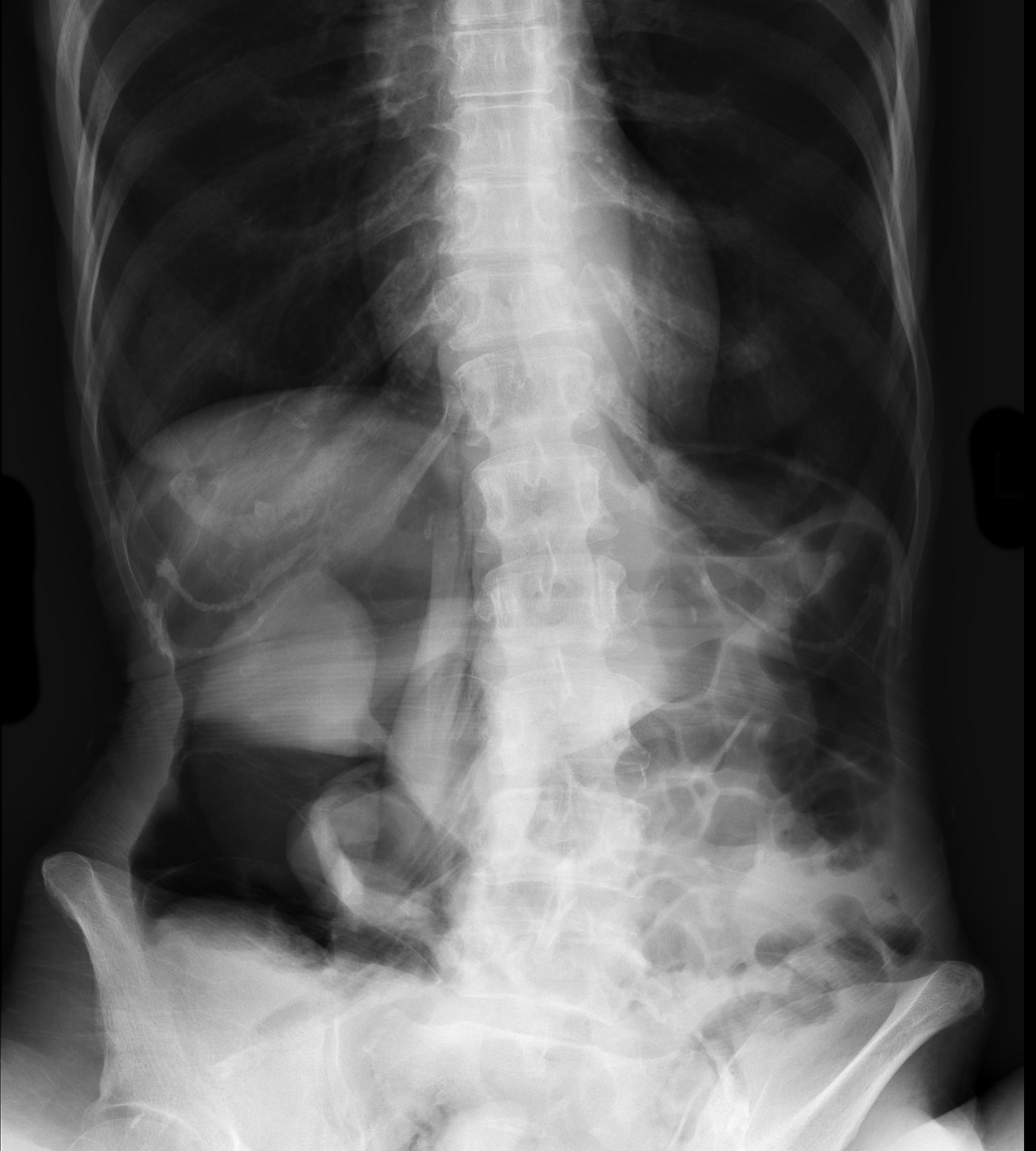
An elderly patient presents with a history of weight loss and chronic constipation. The abdominal Xray is shown below. What's the diagnosis?

This one is tricky so here's a hint: why is the right kidney and psoas muscle so well defined?
 

Show Answer
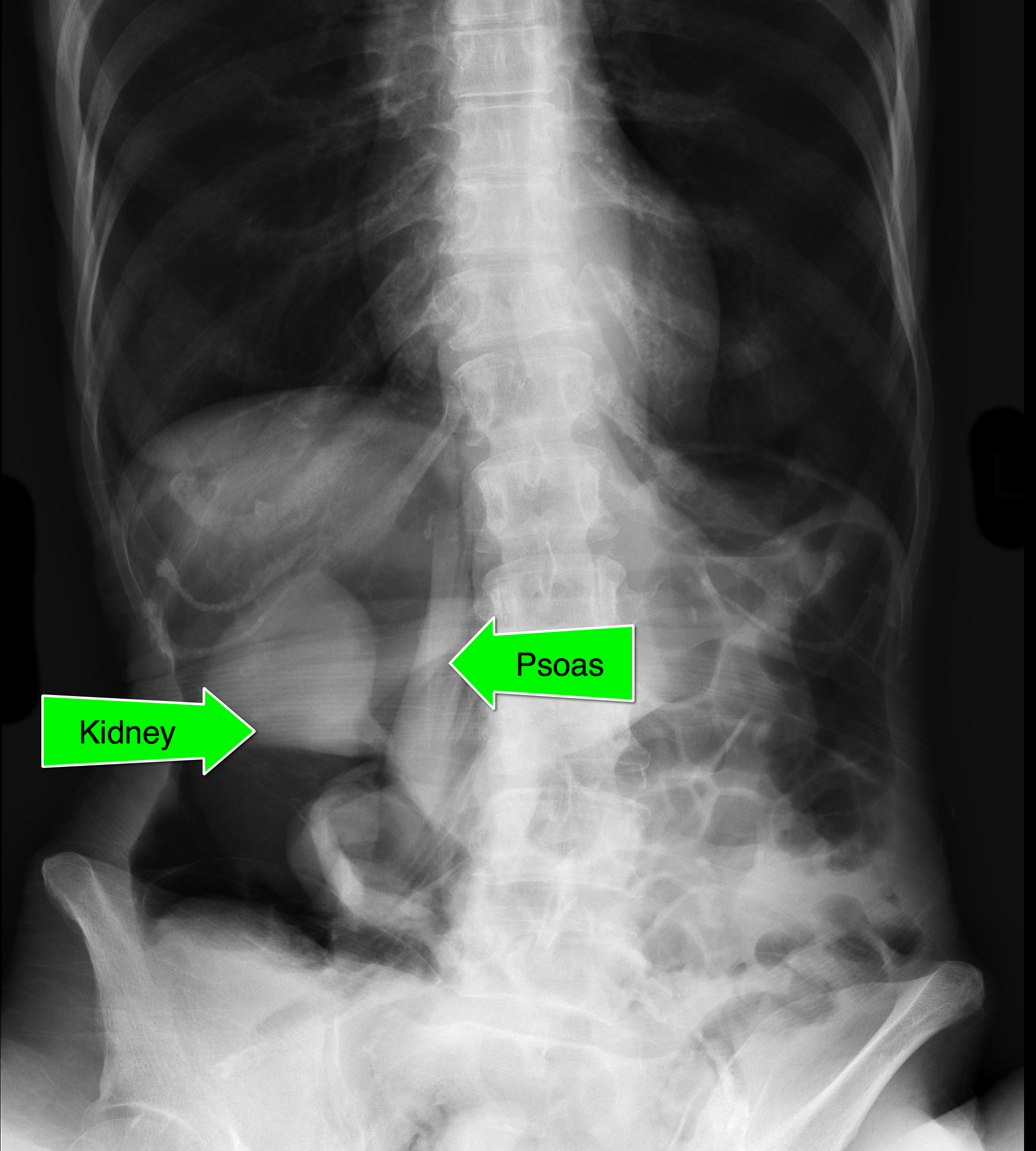
  • Massive retroperitoneal and peritoneal free-air from ascending colon rupture
  • Colon cancer was eventually diagnosed in the operating room.
  • Notice that the kidney and psoas muscle outlined by the free-air below.

Show References

Follow me on Twitter (@criticalcarenow)


Title: What's the Diagnosis?

Category: Visual Diagnosis

Posted: 11/2/2015 by Haney Mallemat, MD

Question

Patient complains of facial and neck swelling, what's the diagnosis?

Show Answer

Subcutaneous emphysema

Show References

Follow me on Twitter (@criticalcarenow)


Title: What's the Diagnosis? Case by Dr. Samuel Cordeiro

Category: Visual Diagnosis

Posted: 10/19/2015 by Haney Mallemat, MD

Question

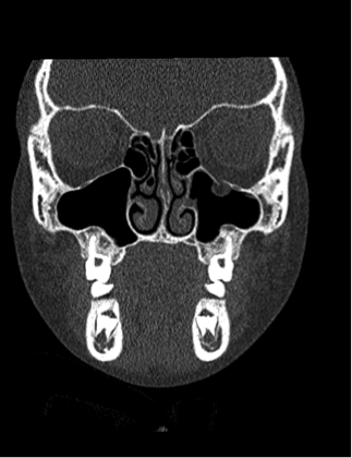
8 year-old female presents with nausea, vomiting, double-vision and inability to move her left eye upwards after being kicked in the face at school. What's the diagnosis?
Show Answer

Orbital floor fracture with entrapment of the inferior rectus muscle.

  • Orbital floor fractures are the most commonly fractured part of the pediatric orbit. Although treated conservatively in adults, pediatric patients can benefit from early repair.
  • Children are at increased risk for a “trap-door” type fracture, which can entrap the extra-ocular muscles because their orbital floor is more flexible.
  • Signs of entrapment include abnormal extra-ocular movements, diplopia, nausea, and vomiting requiring urgent subspecialty evaluation.
  • Bradycardia may occur secondary to the oculo-cardiac reflex when muscle entrapment occurs and is a helpful clue when present


Show References

Follow me on Twitter (@criticalcarenow)


Title: What's the Diagnosis? Case by Dr. Kathleen Stephanos

Category: Visual Diagnosis

Posted: 10/12/2015 by Haney Mallemat, MD

Question

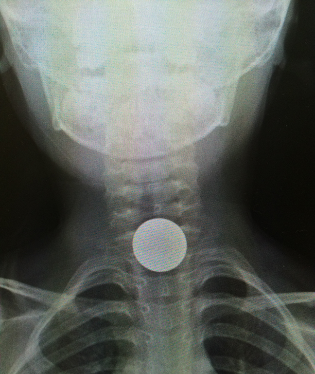
5 year-old boy who presents with sudden onset hoarse voice, and drooling without a fever. 
Show Answer

Coin lodged in the esophagus

Coin ingestions

  • More than 100,000 foreign body ingestions are reported in children each year; coins are #1 cause
  • Although not always 100% true, coins typically appear circular on an AP Xray of the neck when in the esophagus and linear when in the trachea
  • Coins usually pass without issue, but warning signs are drooling, dysphagia, hoarse voice, wheezing, or stridor
  • 10-20% require endoscopy for removal and 1% require surgery
  • Objects lodged in the middle esophagus should raise concern for underlying pathology such as strictures, masses, or webs

Show References

Follow me on Twitter (@criticalcarenow)

Waltzman ML, Baskin M, Wypij D, Mooney D, Jones D, Fleisher G. A randomized clinical trial of the management of esophageal coins in children. Pediatrics. 2005;116(3):614.

Cevik M, Gókdemir MT, Boleken ME, Sogut O, Kurkcuoglu C. The characteristics and outcomes of foreign body ingestion and aspiration in children due to lodged foreign body in the aerodigestive tract. Pediatr Emerg Care. 2013;29(1):53.


Previous  |  1 |  2 |  3 |  4 |  5 |  6 |  7 |  ... |  13 |  Next
University of Maryland School of Medicine logo
  • Departments
  • Programs
  • Research Centers
  • Institutes
  • Graduate Education
  • CIBR Cores
  • Offices of the Dean
    • University of Maryland, Baltimore
    • University of Maryland Medical Center
    • Medical Alumni Association of the University of Maryland
    • Health Sciences and Human Services Library
University of Maryland School of Medicine
655 W. Baltimore Street
Baltimore, MD 21201
Contact Us

© University of Maryland School of Medicine

    • Webmaster
    • Web Accessibility
    • Site Index
    • UMB Hotline
    • Employment
    • Map