University of Maryland School of Medicine logo
Emergency Medicine
All Departments
UMSOM Assistant
UMEM Educational Pearls
  • Home
  • Departments
  • Emergency Medicine
  • CME
  • UMEM Educational Pearls
  • CME
  • Courses and Conferences
  • Free Open Access Medical Education
  • UMEM Educational Pearls

Search

1-13 of 13 results by Tu Carol Nguyen


Title: What's the Diagnosis? Case by Dr. Eric Friedman

Category: Visual Diagnosis

Posted: 11/26/2019 by Tu Carol Nguyen, DO

Question

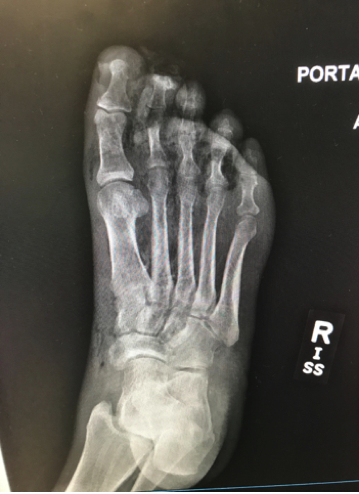
A ~55 year-old female with a history of ESRD and diabetes who presented to the ED with progressively worsening foot odor. An x-ray was performed. The picture below shows the right foot.

What is the diagnosis?


Show Answer

Necrotizing infection of the foot

  • Labs will show increased lactate, increased CK, increased WBC, and decreased sodium
    • Admission serum lactate (>6mmol/L) and sodium levels (<135mEq/L) are predictors of mortality
  • On x-ray, the characteristic finding of gas in the soft tissues is seen in only a minority of cases.
  • The presence of gas in the facial planes is highly specific, but not very sensitive.
  • Diagnosis of necrotizing fasciitis or myonecrosis can only be established by surgical exploration.
  • Treatment includes a Carbapenem or Zosyn plus Clindamycin and Vancomycin.


Show References

https://radiopaedia.org/articles/necrotising-fasciitis

Yaghoubian et al. Use of admission serum lactate and sodium levels to predict mortality in necrotizing soft-tissue infections. Archives of surgery. 2007.

Anaya DA and Dellinger EP.  Necrotizing soft-tissue infection: diagnosis and management. Clinical infectious diseases. 2007.


Title: Trouble treating your gastroparetic? Consider an antipsychotic! (Submitted by Dr. Bradford Schwartz)

Category: Pharmacology & Therapeutics

Posted: 4/27/2017 by Tu Carol Nguyen, DO

Haloperidol has a higher D2 receptor antagonist effect than standard antiemetic treatment agents such as metoclopramide. In addition, newer antipsychotic agents such as Olanzapine have a high affinity at multiple antiemetic sites such as the dopamine and serotinergic receptors.

While formal RCT's are still in the works, multiple sources including palliative care, emergency medicine, and pain journals support their use in refractory emesis.


Consider Haloperidol 3-5 mg IV. 
Check an EKG for long QTc prior to use. Consider dose reduction of haloperidol in those with hepatic impairment. Also consider dose reduction in patients taking carbamazepine, phenytoin, phenobarbital, rifampicin, or quinidine due to that pesky CYP3A4 inhibition. 

Consider Olanzapine 2-5 mg IV.

Several case reports have shown a higher rate of success with olanzapine for refractory emesis. Olanzapine has similar precautions as those to haloperidol (EKG, hepatic impairment), although it's CYP drug interactions are less common. Additionally, use olanzapine cautiously in hyperglycemic patients as there are several case reports of olanzapine prompting episodes of DKA. Consider frequent blood sugar checks or small doses of insulin in hyperglycemic patients. 


Take Home Points:

Consider the antipsychotic agents Haloperidol or Olanzapine for patients with refractory emesis, they may be more effective than traditional antiemetics. 

Get an EKG prior to administration to check for QTc prolongation. As the classical and atypical antipsychotic agents are sedating, use caution in conjunction with other sedating medications (such as benzodiazepines).  


Show Additional Information

Show References

Glare P, Miller J, Nikolova T, Tickoo R. Treating nausea and vomiting in palliative care: a review. Clin Interv Aging. 2011;6:243-59.

Prommer E. Olanzapine: palliative medicine update. Am J Hosp Palliat Care. 2013 Feb;30(1):75-82

Pommer E. Role of Haloperidol in Palliative Medicine: An Update. Am J Hosp Palliat Care. 2012 Jun;29(4):295-301


Navari RM, Nagy CK, Gray SE. The use of olanzapine versus metoclopramide for the treatment of breakthrough nausea and vomiting in patients receiving highly emetogenic chemotherapy. Support Care Cancer. 2013 Jun;21(6):1655-63.

Jackson WC, Tavernier L (2003) Olanzapine for intractable nausea in palliative care patients. J Palliat Med 6:251–255 
Hasse Abrahamsson. Treatment options for patients with severe gastroparesis. Gut. 2007 Jun; 56(6): 877–883.
 
Bradford MV, Glode A. Olanzapine: An antiemetic option for chemotherapy-induced nausea and vomiting. J Adv Pract Onc. 2014 Jan;5(1):24-9.
 
Chan EW, Knott, JC, Taylor DM, Phillips GA, Kong DC. Intravenous olanzapine- another option for the acutely agitated patient. Emery Med Australas. 2009 Jun; 21 (3) 241-2
 
Cole JB et al. A prospective observational study of patients receiving intravenous and intramuscular olanzapine in the emergency department. Ann Emerg Med 2016 Nov 4; [e-pub]. 
 
C. Roldan, Y. Chathampally. Haloperidol vs. placebo in addition to conventional therapy to treat pain secondary to gastroparesis in the emergency department. Journal Of Pain. April 2015 Volume 16, Issue 4, Supplement, Page S34

P. Stalcup, B. Croft, R. Ramirez, M. Darracq. Research Forum Abstract: 204 Haloperidol Undermining Gastroparesis Symptoms in the Emergency Department. Annals Of Emergency Medicine. October 2016. Volume 68, Issue 4, Supplement, Page S80


Ramirez R et al. Haloperidol Undermining Gastroparesis Symptoms (HUGS) in the Emergency Department. AJEM 2017


Lindenmayer JP, Patel R. Olazapine-induced ketoacidosis with diabetes mellitus (letter) Am J Psychiatry. 1999;156:1471

Roefaro J, Mukherjee SM. Olanzapine-lnduced hyperglycemic nonketonic coma. Ann Pharmacother. 2001;35:300–2.

 Lee JS, Kim JY, Ahn JH, Kim CY. Diabetic ketoacidosis in a schizophrenic patient treated with olanzapine: a case report. J Korean Neuropsychiatr Assoc. 2005;44:116–119.

Ragucci KR, Wells BJ. Olanzapine-induced diabetic ketoacidosis. Ann Pharmocother. 2001; 35: (12) 1556-8


Title: What is the diagnosis ? (Case by Hussain Alhashem)

Category: Visual Diagnosis

Keywords: Pleural effusion; POCUS (PubMed Search)

Posted: 4/17/2017 by Tu Carol Nguyen, DO

Question

A 50 years old male with a history of CHF, presenting to the ED with progressively worsening shortness of breath. POCUS was performed. The picture shows the left lower part of the chest. What is the diagnosis?

Show Answer

Answer: Pleural effusion

  • POCUS is faster than X-rays and better at quantifying the amount of fluid in a pleural effusion.
  • It is also useful in guiding thoracentesis. 
  • The diagnosis of pleural effusion by ultrasound is made by visualizing fluid above the diaphragm. 
  • One sign that can help with detecting fluid is the presence of V lines (not shown in this image). V lines are the spine shadows visualized through a pleural effusion. The presence of V lines strongly supports the presence of fluid in the pleural space. 

Show References

Eibenberger, K. L., Dock, W. I., Ammann, M. E., Dorffner, R., Hörmann, M. F., & Grabenwöger, F. (1994). Quantification of pleural effusions: sonography versus radiography. Radiology, 191(3), 681-684.

Atkinson, P., Milne, J., Loubani, O., & Verheul, G. (2012). The V-line: a sonographic aid for the confirmation of pleural fluid. Critical ultrasound journal, 4(1), 19.


Title: What's the Diagnosis?

Category: Visual Diagnosis

Posted: 2/13/2017 by Tu Carol Nguyen, DO

Question

56 year-old male with history of hypertension presents with complaints of right scrotal swelling and pain. Denies any urinary symptoms, abdominal pain, nausea/vomiting or change in bowel habits or prior episodes. Temp was 99.0.

A scrotal ultrasound was done and an image of the right testis was seen (below). What's the diagnosis?


Show Answer

Answer: Right Epididymitis (and Hydrocele)


Take Home Points:

  • Epididymitis is heterogeneous on grayscale US
    • 100% sensitivity of acute inflammation if hypervascular on Color Doppler
  • Treatment:
    • Age < 35 years and sexually active - more commonly caused by C. trachomatis or N. gonorrhoeae
      • Ceftriaxone 250mg IM once + Doxycycline 100mg BID x 10 days
    • Age > 35 years - more comonly caused by obstructive urinary disease (enteric organisms)
      • Levaquin 500mg daily x 10 days OR Ofloxacin 300mg BID x 10 days

Show References

Kühn AL, Scortegagna E, Nowitzki KM, Kim YH. Ultrasonography of the scrotum in adults. Ultrasonography. 2016;35(3):180-97.

Centers for Disease Control and Prevention. Sexually Transmitted Diseases Treatment Guidelines, 2015. MMWR Recomm Rep 2015;64(No. RR-3): 1-137.


Title: What's the Diagnosis? Case by Dr. Phillip Magidson

Category: Visual Diagnosis

Posted: 1/30/2017 by Tu Carol Nguyen, DO

Question

25 year-old female with hx of cerebral palsy with significant developmental delay, s/p G-tube who presented with acute hypoxic respiratory failure, hypotension and a distended, tense abdomen. A CT was done with the scout film below. What's the diagnosis?


Show Answer

Answer: Cecal Volvulus

Patient was subsequently intubated, had an NG tube placed with over 500cc of fluid returned. Patient had multiorgan failure and received fluids, antibiotics, pressors, blood products and went to the OR, and had a partial bowel resection. 

One way to differentiate cecal volvulus from sigmoid volvulus is that sigmoid volvulus generally do not have haustra. 

Show References

Tonerini M, Pancrazi F, Lorenzi S, Pacciardi F, Ruschi F, et al. (2015) Cecal volvulus: what the radiologist needs to know. Glob Surg, 1: DOI: 10.15761/GOS.1000106.


Title: What's the Diagnosis?

Category: Visual Diagnosis

Posted: 1/9/2017 by Tu Carol Nguyen, DO

Question

A 60 year-old man with history of atrial fibrillation, CAD presents with left lower leg/foot pain for a few days. His foot is seen below. What's the diagnosis?


Show Answer

Mottling with concerns for Acute Limb Ischemia


Key Points:

  • Assess appearance, temperature, sensation, strength and pulses
  • Remember 6 P's (paresthesia, pain, pallor, pulselessness, poikilothermia, paralysis)
  • Irreversible tissue necrosis can occur by 6 hours from onset
  • Start IV Heparin bolus (100 units/kg or 5000u) and continuous IV infusion (1000 units/hr) with considerations for any contraindications
  • Immediate vascular consult

Show References

Creager MA, Kaufman JA, Conte MS. Clinical practice. Acute limb ischemia. N Engl J Med. 2012;366(23):2198-206.

http://www.emdocs.net/acute-limb-ischemia-pearls-pitfalls/


Title: What's the Diagnosis? Image by Dr. Karen Baker

Category: Visual Diagnosis

Posted: 12/27/2016 by Tu Carol Nguyen, DO

Question

68 year-old male presents with 3-4 days of hedache, anorexia for 2 weeks and "balance trouble." His blood pressure was 226/140 and he states he has not been on his medications for 6 months.
Physical examination revealed a shuffled gait with his walker and the rest of his physical exam and neurologic exam was unremarkable. 
 
His CT is seen below. What's the diagnosis?


Show Answer

Answer: Large left cerebellar intraparenchymal hemorrhage with surrounding vasogenic edema with mild infratentorial midline shift of ~4mm


Take Home Points:

  • Ambulate every patient when doing a neurologic exam (as they can tolerate)
  • Reverse anticoagulation, if necessary
  • BP control in intracerebral hemorrhage
    • Still exist different opinions with BP thresholds
    • 2015 AHA/ASA guidelines: reduction of SBP to 140 is safe
    • Refer to a previous pearl for more information

Show References

Hemphill JC, Greenberg SM, Anderson CS, et al. Guidelines for the Management of Spontaneous Intracerebral Hemorrhage: A Guideline for Healthcare Professionals From the American Heart Association/American Stroke Association. Stroke. 2015;46(7):2032-60.


Title: What's the Diagnosis? Case by Dr. Harry Achterberg

Category: Visual Diagnosis

Posted: 12/5/2016 by Tu Carol Nguyen, DO

Question

27 year-old G2P1 presents with 3 days of abdominal pain that is mostly suprapubic. Denies any urinary symptoms and vaginal bleeding. Physical examination reveals slight rebound in the right lower quadrant.

An ultrasound revealed the following. What's the diagnosis?


Show Answer

Pregnancy with Appendicitis


Take Home Points:

  • Ultrasound: Outer appendiceal diameter > 6mm and noncompressible supports diagnosis of acute appendicitis
    • 100% sensitivity, 68% specificity
  • There is no difference in ability for US to diagnosis acute appendicitis between pregnant and non-pregnant women
  • Consider MRI for nondiagnostic US
    • varying literature shows 92-100% for both sensitivity & specificity


See previous pearl for how to conduct an ultrasound to evaluate for appendicitis.
 

Show References

Dewhurst C, Beddy P, Pedrosa I. MRI evaluation of acute appendicitis in pregnancy. J Magn Reson Imaging. 2013;37(3):566-75.

Israel GM, Malguria N, Mccarthy S, Copel J, Weinreb J. MRI vs. ultrasound for suspected appendicitis during pregnancy. J Magn Reson Imaging. 2008;28(2):428-33.

Segev L, Segev Y, Rayman S, Nissan A, Sadot E. The diagnostic performance of ultrasound for acute appendicitis in pregnant and young nonpregnant women: A case-control study. Int J Surg. 2016;34:81-85.

Segev L, Segev Y, Rayman S, Nissan A, Sadot E. Acute Appendicitis During Pregnancy: Different from the Nonpregnant State?. World J Surg. 2016.

Theilen LH, Mellnick VM, Longman RE, et al. Utility of magnetic resonance imaging for suspected appendicitis in pregnant women. Am J Obstet Gynecol. 2015;212(3):345.e1-6.


Title: What's the Diagnosis?

Category: Visual Diagnosis

Posted: 11/7/2016 by Tu Carol Nguyen, DO

Question

8 year-old female with no PMH who presents with concerns for "purple patches" popping up on her arm for 2-3 days. Stated that one appeared and then, the other one appeared 12 hours later. She denied any trauma whatsoever, history of easy bleeding/bruising and did feel safe at home. The rest of the review of systems was negative.

Patient said there was mild pain when the area was touched. The rest of the physical examination was normal.

What's the diagnosis? (Image below)

Show Answer

Superficial Thermal Burn

Upon further questioning, patient stated that she had been making s'mores by roasting marshmallows over an electric stove 3 days prior. The burns showed up the subsequent morning.

  • Thermal injury
    • Types - scald, contact, fire, chemical, electrical, radiation
    • Can still sustain dermal burns from emanating heat without contact
    • Extent of tissue damage based on temperature & duration of exposure
  • Non-accidental burns are common in children
    • 10% of physically abused children have intentional burns/scalds
    • Sites: hand, back, wrist, buttock, feet, legs
    • Type: well-define margins of contact/scald burns in unusual places, glove-and-stocking distribution

Take Home Points:

  • Take a good history
  • Can sustain burns without direct contact (i.e. heat, radiation, etc.)
  • Consider non-accidental burns in children

Previous pearls about burns:

Pediatric Burns:

  • https://umem.org/educational_pearls/1107/
  • https://em.umaryland.edu/educational_pearls/1164/

Show References

Monseau AJ, Reed ZM, Langley KJ, Onks C. Sunburn, Thermal, and Chemical Injuries to the Skin. Prim Care. 2015;42(4):591-605.

"Pathophysiology of Thermal Injury." Civic Plus. 2007.


Title: What's the Diagnosis?

Category: Visual Diagnosis

Posted: 10/25/2016 by Tu Carol Nguyen, DO (Updated: 10/26/2016)

Question

20 year-old female presents with sore throat, right throat fullness, difficulty speaking for 2-3 days. A bedside ultrasound and subsequent CT was obtained as seen below. What's the diagnosis?


Show Answer


Peritonsillar Abscess


The ultrasound image is a transcutaneous approach with a linear transducer that is placed at the angle of the mandible of the affected side. This is an alternative approach to an intra-oral ultrasound with the endocavitary transducer if the patient has trismus.

  • Peritonsillar abscess (PTA) is often taught as a clinical diagnosis; however, 36% are often peritonsillar cellulitis (PTC)
    • Sensitivity and specificity of clinical diagnosis of PTA are 78% and 50%, respectively
  • Classically, providers attempt blind needle aspirations (landmark-based)
    • Blind needle aspiration is unreliable for diagnosis of PTA with a false-negative rate of 10-24%
  • Intraoral sonography sensitivity: 90-100%
    • Using ultrasound can avoid unnecessary use of CTs and blind needle asprations


Take Home Points:

  • Consider intra-oral US for diagnosis of PTA vs. PTC and to reduce unecessary radiation with CT
  • Consider transcutaneous US to evaluate for PTA in the setting of trismus
  • Consider intra-oral US-guided needle aspiration of PTA vs. blind needle aspiration


How to do an intra-oral US-guided needle aspiration of PTA, check out:

http://www.ultrasoundpodcast.com/2012/01/episode-21-full-peritonsillar-abscess-podcast/


For a brief video on how to perform a transcutaneous US for PTA:

https://www.youtube.com/watch?v=JkIYOhKCweI&t=28s


Show References

Constantino TG, Satz WA, Dehnkamp W, Goett H. Randomized Trial Comparing Intraoral Ultrasound to Landmark-based Needle Aspiration in Patients with Suspected Peritonsillar Abscess. Academic Emergency Medicine. June 2012; Vol. 19 No. 6: 626-631.

Halm BM, Ng C, Larrabee YC. Diagnosis of a Peritonsillar Abscess by Transcutaneous Point-of-Care Ultrasound in the Pediatric Emergency Department. Pediatr Emerg Care. 2016;32(7):489-92.

Rehrer M, Mantuani D, Nagdev A. Identification of peritonsillar abscess by transcutaneous cervical ultrasound. Am J Emerg Med. 2013;31(1):267.e1-3.


Title: What's the Diagnosis? Image and Case by Dr. Kathleen Stephanos

Category: Visual Diagnosis

Posted: 10/10/2016 by Tu Carol Nguyen, DO

Question

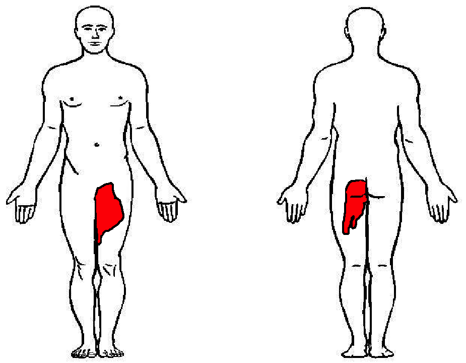
57 year-old female with history of bilateral lung transplants presents with fever, and 2 days of a painful, red, bumpy rash over the left labia and left buttock, but also notes a small tender area on the plantar surface of the left foot.

Below is a figure depicting the location of the rash, as well as a photo of her foot.
Show Answer

This is Herpes Zoster (Shingles).

Presentation:

  • Zoster typically appears along a single dermatome, without crossing midline in immunocompetent hosts, but may involve 2-3 adjacent dermatomes.
  • In immunocompromised patients, systemic involvement may occur.
  • Thoracic and lumbar dermatomes are the most common.
  • Lumbar and sacral dermatomes wrap around the lower extremity and may spare the skin between the perineum and the foot.

Treatment:

  • Immunocompromised patients should be treated with IV Acyclovir 10mg/kg q 8hr for 7 days (Use IBW in obese patients)
  • In immunocompetent individuals, if identified within 48 hours of rash onset, may receive oral therapy with famciclovir, acyclovir or valcyclovir
  • Herpes Zoster alone should not prompt immunosuppression work up
    • However, concerns for underlying immune disorders if:
      • continued development of lesions > 1 week, or
      • involvement of more than 3 dermatomes

Show References

Dworkin RH, Johnson RW, Breuer J, et al. Recommendations for the management of herpes zoster. Clin Infect Dis. 2007;44 Suppl 1:S1.


Title: What's the Diagnosis? Case by Dr. Lindsay Weiner

Category: Visual Diagnosis

Posted: 9/26/2016 by Tu Carol Nguyen, DO (Updated: 9/26/2016)

Question

22-year-old male with history of autism, mental retardation who is non-verbal presents with abdominal pain and vomiting for one day. Patient was found clutching his abdomen and moaning. What's the diagnosis?
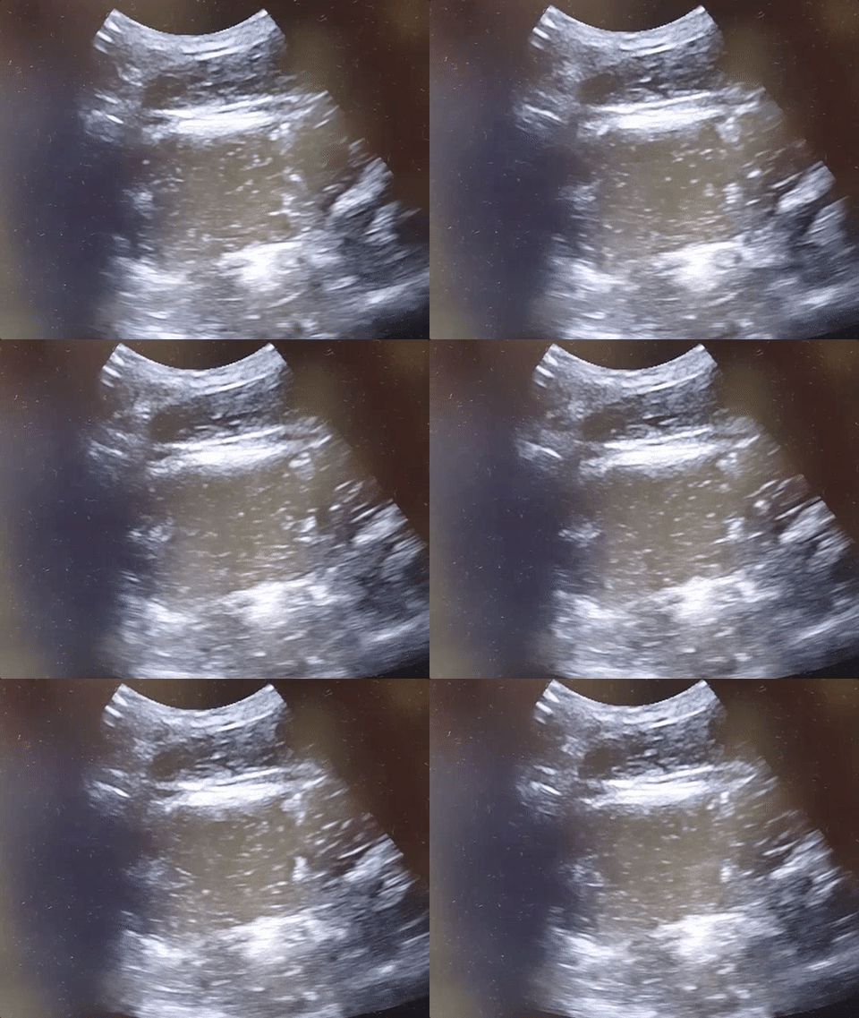


Show Answer
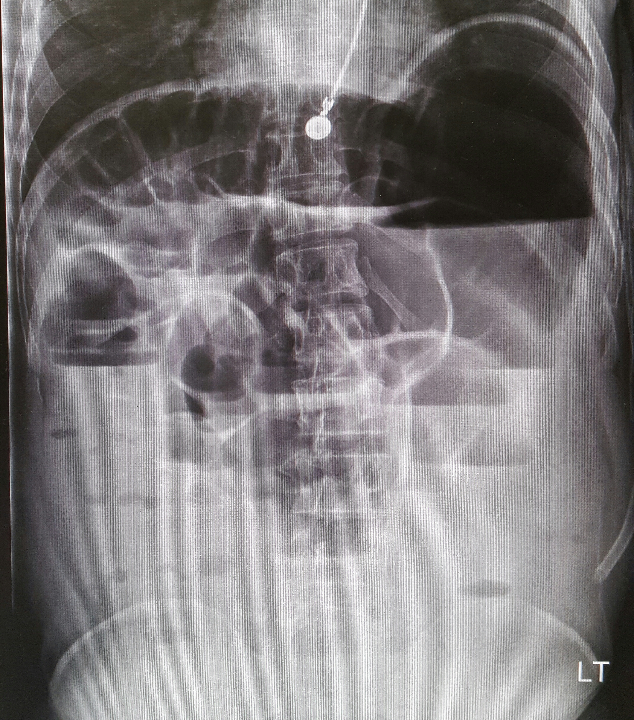
Small Bowel Obstruction


  • Presentation: abdominal pain, nausea, vomiting, early diarrhea progressing to lack of flatus or bowel movements with complete obstruction. Severe obstruction can result in perforation, unstable vital signs, and physical exam signs of peritonitis.
  • Sensitivity and specificity > 90% for diagnosis of SBO with US, with superior test performance compared to X-ray.
  • US findings: dilated small bowel loops > 3 cm; no peristalsis, swirling snowstorm appearance of bowel contents
  • Treatment: bowel rest, IV fluids, and prompt surgery consultation! Consider NG tube for decompression.


See the corresponding upright abdominal x-ray, showing dilated bowel with air fluid levels.


Show References

Kameda T, Taniguchi N. Overview of point-of-care abdominal ultrasound in emergency and critical care. J Intensive Care. 2016 Aug 15;4:53. doi: 10.1186/s40560-016-0175-y. eCollection 2016. Review.

Unl er EE, Yava i O, Ero lu O, Yilmaz C, Akarca FK. Ultrasonography by emergency medicine and radiology residents for the diagnosis of small bowel obstruction. Eur J Emerg Med. 2010 Oct; 17(5):260-4.


Title: What's the Diagnosis? Case by Dr. Tejusve Rao

Category: Visual Diagnosis

Posted: 9/12/2016 by Tu Carol Nguyen, DO (Updated: 9/12/2016)

Question


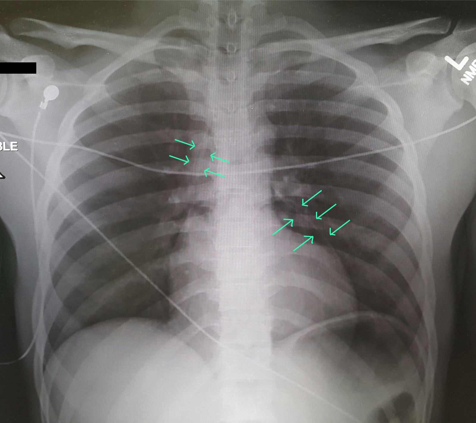
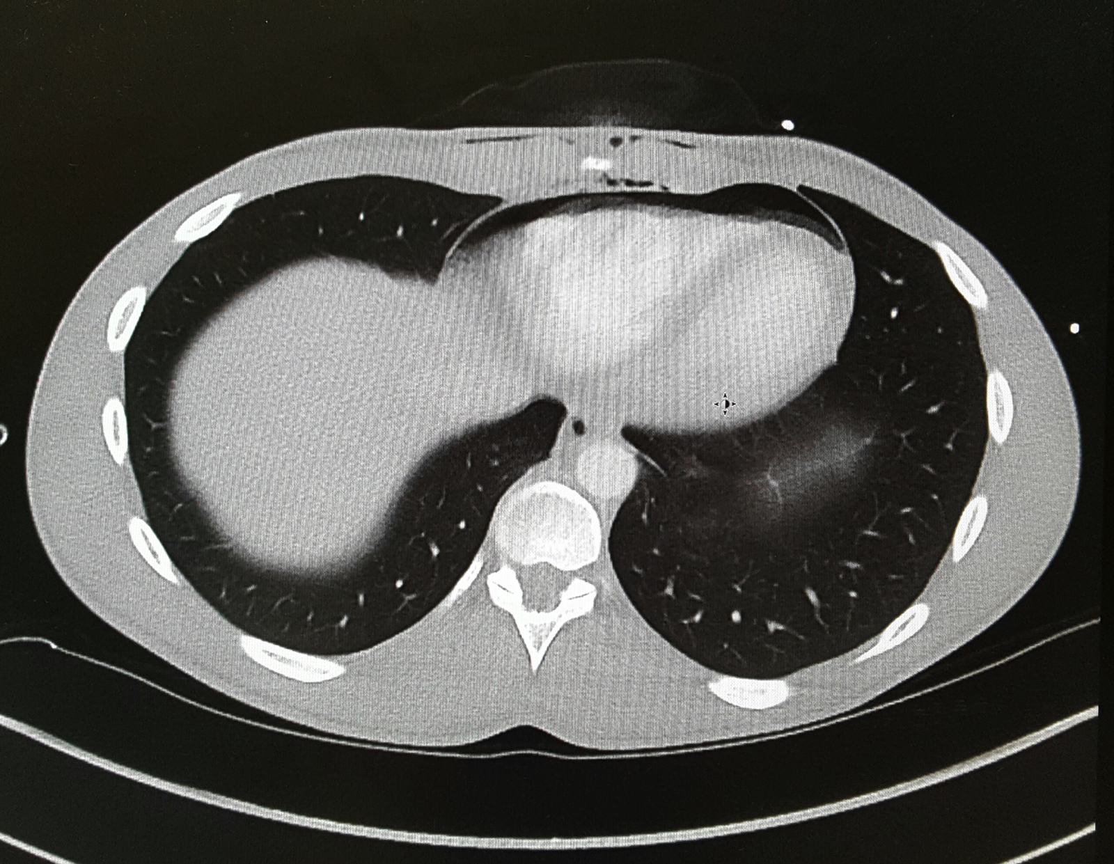
A 25-year-old male was brought in by EMS with a stab wound to the chest. What's the diagnosis?
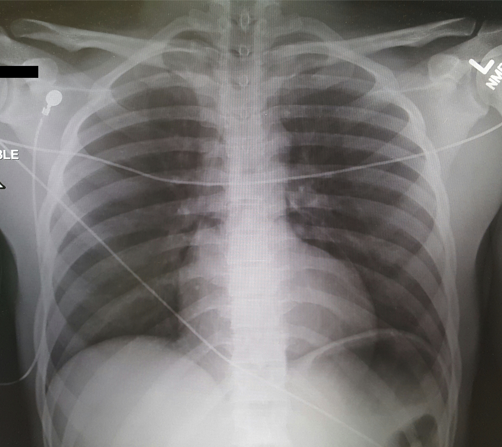


Show Answer

Pneumopericardium


CT Chest was done with the image below:


Penetrating Cardiac Trauma
  • Initial Assessment of ABC, ATLS primary survey guidelines.
  • Evaluate for tension pneumothorax or cardiac tamponade in all patients presenting with chest trauma and shock.
  • Cardiac Box: Surrounded by sternal notch, xiphoid process and nipples.
  • Order of injury: Right Ventricle --> Left Ventricle --> Right Atrium --> Left Atrium
  • Beck’s Triad: Hypotension, JVD, muffled heart sounds may not be present initially.
  • Conduct FAST exam to examine for cardiac tamponade, hemothorax, pneumothorax.
  • Cardiac Tamponade more common from stab wounds than from gun shot wounds.
  • Hemodynamically unstable patients require immediate operative therapy after quick bedside assessment (physical exam, ultrasound, chest tube as needed)

Differential Diagnosis: Bronchial injury, Diaphragm injury, Hemothorax, Tension Pneumothorax, Aortic Transection, Esophageal injury, Pneumomediastinum

Evaluation: Ultrasound (FAST Exam), CXR, CTA in stable patients, ECG, troponin.

Management: Penetrating cardiac trauma require emergent thoracotomy, pericardial window.


Show References

Clancy K, Velopulos C, Bilaniuk JW, et al. Screening for blunt cardiac injury: an Eastern Association for the Surgery of Trauma practice management guideline. J Trauma Acute Care Surg. 2012;73(5 Suppl 4):S301-6.

El-menyar A, Al thani H, Zarour A, Latifi R. Understanding traumatic blunt cardiac injury. Ann Card Anaesth. 2012;15(4):287-95.

Tintinalli's 7th Edition. Emergency Medicine Manual. Chapter 164: Cardiothoracic Trauma.


University of Maryland School of Medicine logo
  • Departments
  • Programs
  • Research Centers
  • Institutes
  • Graduate Education
  • CIBR Cores
  • Offices of the Dean
    • University of Maryland, Baltimore
    • University of Maryland Medical Center
    • Medical Alumni Association of the University of Maryland
    • Health Sciences and Human Services Library
University of Maryland School of Medicine
655 W. Baltimore Street
Baltimore, MD 21201
Contact Us

© University of Maryland School of Medicine

    • Webmaster
    • Web Accessibility
    • Site Index
    • UMB Hotline
    • Employment
    • Map