University of Maryland School of Medicine logo
Emergency Medicine
All Departments
UMSOM Assistant
UMEM Educational Pearls
  • Home
  • Departments
  • Emergency Medicine
  • CME
  • UMEM Educational Pearls
  • CME
  • Courses and Conferences
  • Free Open Access Medical Education
  • UMEM Educational Pearls

Search

221-240 of 245 results with category "Visual Diagnosis"

Previous  |  1 |  ... |  7 |  8 |  9 |  10 |  11 |  12 |  13 |  Next

Title: What's the Diagnosis? Case submitted by Dr. Zachary Dezman

Category: Visual Diagnosis

Posted: 11/28/2011 by Haney Mallemat, MD (Updated: 11/28/2011)

Question

9 year-old boy with sudden onset of unilateral facial swelling. What’s the diagnosis?

Show Answer

Answer: Acute Parotitis

  • Viral parotitis
    • Usually bilateral swelling without warmth or erythema; typically a benign course
    • 85% of cases occur in children <15 years old
    • Common causes: Influenza, Para-influenza, and Coxsackie; Rarely Paromyxovirus (Mumps) due to vaccination programs
    • Treat conservatively; sialagogues to stimulate gland drainage (lemon drops or orange juice), warm compresses, or local massage.
  • Bacterial parotitis
    • Commonly presents with unilateral pain, induration, erythema, and tenderness; 20-50% mortality!
    • Risk factors: chronic illness, age > 60, recent surgery, and dehydration
    • Treatment: Antibiotics (covering oral flora) and certain cases may require surgical intervention
    • Lack of improvement within 48 hours requires CT scan to exclude abscess and IV antibiotics

Bonus Trivia: U.S. President Garfield died from parotitis after becoming dehydrated following abdominal surgery

Shelly J. McQuone MD, Acute Viral and Bacterial Infections of the Salivary Glands, Otolaryngologic Clinics of North America, Volume 32, Issue 5 (October 1999)

Show References

Follow me on Twitter (@criticalcarenow) or Google+ (+haney mallemat)


Title: What's the diagnosis? Pearl submitted and written by Dr. Zachary Dezman

Category: Visual Diagnosis

Posted: 10/31/2011 by Haney Mallemat, MD (Updated: 10/31/2011)

Question

72 year-old man, one-week post right fem-pop bypass presents with painful blue and black toe. Diagnosis?


Show Answer

Answer: Blue-Toe Syndrome

  • Blue-toe syndrome is acute digital-ischemia, presenting as one or more cold, painful, and cyanotic toes (or other digits).
  • Multiple causes:
    • Emboli (most common)
    • Cholesterol emboli from recent endovascular procedure or atherosclerotic plaque rupture
    • Vascular aneurysm
    • Infectious (e.g., endocarditis)
    • Medications (e.g., warfarin)
    • Drugs (e.g., cocaine, methamphetamine, etc.)
  • Emergency Department evaluation should focus on determining the underlying cause to prevent limb loss.
  • Patients should have emergent vascular consult and admission for potential revascularization.

Show References

Hirschman, J. et al. Blue (or purple) toe syndrome. J Am Acad Dermatol.2009 Jan;60(1):1-20

O'Keeffe S, et al. Blue toe syndrome: Causes and management.Arch Intern Med. 1992 Nov;152(11):2197-202.

Follow me on Twitter (@criticalcarenow) or Google+ (+haney mallemat)


Title: What s the Diagnosis? (Case submitted and written by Dr. Rich Lichenstein)

Category: Visual Diagnosis

Posted: 10/17/2011 by Haney Mallemat, MD

Question

5 year-old male with developmental delay presents with intractable non-bloody and non-bilious vomiting over 10 days; bowel movements are normal. Four weeks ago he was placed in a hip-spica cast following a motor vehicle crash. Abdominal x-ray is below. Diagnosis?

Show Answer

Answer: CAST syndrome (also known as Superior Mesenteric Artery Syndrome)

  • Caused by extrinsic compression of the SMA with ensuing partial gastric outlet obstruction
  • Commonly seen in the second decade of life when there is increased spinal flexibility and truncal casting increases lumbar lordosis. This changes the take-off of the SMA which crosses the horizontal part of the duodenum.
  • Those at risk include those with patients with spinal disease, hip and pelvic fractures.  Children with disabilities who have poor fat mass, hypotonicity, and skeletal distortion may also be affected.
  • Clinical symptoms include nausea, vomiting, abdominal distention which may lead to dehydration, metabolic alkalosis and shock and sometimes death.
  • Treatment includes IV replacement therapy, NG tube to decompress the stomach and duodenum and replacement of the cast.

Show References

Wheeless Textbook of Orthopedics. Updated August 29,2011

Lichenstein, R. Radiology Cases in Pediatric Emergency Medicine, Volume 5, Number 16

Follow me on Twitter (@criticalcarenow) or Google+ (+haney mallemat)


Title: What's the Daignosis? (submitted and written by Dr. Katherine Baugher)

Category: Visual Diagnosis

Posted: 10/3/2011 by Haney Mallemat, MD

Question

Question: 50-year-old diabetic female s/p foot burn several weeks ago, now presenting with pain and discharge from a poorly healing wound. Diagnosis?

Show Answer

Answer:  Osteomyelitis

Osteomyelitis 

· Acute or chronic bone infection

· Risk factors: Immunosuppression (diabetes, chronic steroid use, AIDS, and sickle-cell dz.)

· Secondary to direct trauma, contiguous spread from local infection, or hematogenous spread (in children)

· Common bacteria: S. Aureus, Pseudomonas, Salmonellae (classically in Sickle cell dz.)

· X-ray (limited sensitivity):

- 3-5 days post-infection: Soft-tissue swelling

- 14-21 days: Some patients demonstrate bony changes (e.g., periosteal elevation, bone lucencies, etc.)

- >28 days: >90% with Xray findings

· MRI is the imaging gold standard

· Two of the following needed for diagnosis:

- Purulent aspiration

- Positive blood or tissue culture

- Positive imaging

- Tenderness + erythema / edema

· Antibiotic coverage based on culture results. When immediate empiric therapy required (sepsis), cover most likely pathogen plus MRSA.


References

Carek PJ, Dickerson LM, Sack JL. Diagnosis and management of osteomyelitis. Am Fam Physician. 2001 Jun 15;63(12):2413-20.

Pruthi S, Thapa MM. Infectious and inflammatory disorders. Radiol Clin North Am. Nov 2009;47(6):911-26.

Zink BJ, Raukar NP. Bone and Joint Infections. In: Marx JA, Hockberger RS, Walls RM, Adams JG, Barsan WG, Biros MH, Danzl DF, Gausche-Hill M, Ling LJ, Newton EJ, eds. 7th ed. Emergency Medicine: Concepts and Clinical Practice.Volume 2. Philadelphia, PA: Mosby; 2010:1821-1830.

Follow me on Twitter (@criticalcarenow) or Google+ (+haney mallemat)


Title: What s the diagnosis? Case submitted by Dr. Ari Kestler

Category: Visual Diagnosis

Posted: 9/19/2011 by Haney Mallemat, MD (Updated: 8/28/2014)

Question

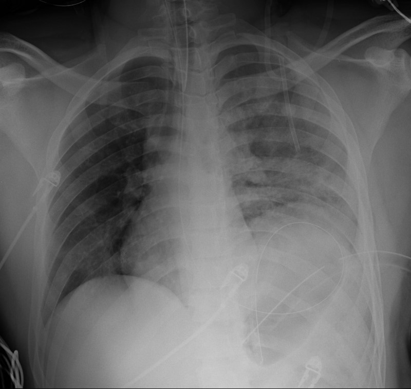
19 year-old male s/p high-speed MVC with hypotension and diminished breath sounds on left. Diagnosis?
Show Answer
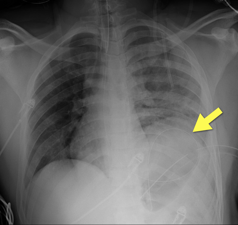
Answer: Diaphragmatic rupture (Note position of NG tube on image below).

·Uncommon; less than 1% of all traumatic injuries

·Diagnosis may be obvious on CXR (as in example) or subtle (e.g., diaphragmatic elevation, basilar atelectasis, etc.)

·Requires a high-index of suspicion because delayed diagnosis increases the risk of abdominal organ herniation or strangulation.

·Mortality depends on the mechanism and presence of associated injuries; mortality is highest for blunt injuries.

Show References

Bhavnagri S, et al. When and how to image a suspected broken rib. Cleveland Clinic J Med. 2009;76(5):309.

Williams M, et al. Predictors of mortality in patients with traumatic diaphragmatic rupture and associated thoracic and/or abdominal injuries. Am Surg. 2004;70(2):157.

National Trauma Data Base. American College of Surgeons 2000-2004.

Follow me on Twitter (@criticalcarenow) or Google+ (+haney mallemat)


Title: Interesting Case

Category: Visual Diagnosis

Posted: 9/12/2011 by Rob Rogers, MD (Updated: 7/22/2026)

Question

A 50 year-old patient presents after a self-inflicted eye injury. The patient had taken some type of needle and inserted it into their eye.

What is the diagnosis and what complications might result?


Show Answer

The image shows air bubbles in the anterior chamber. Since air has been introduced into the eye, the patient is at risk for elevated intraocular pressures. In addition, you would need to consider endophthalmitis as a complication.


Title: What's the diagnosis? Images by Dr. Mak Moayedi

Category: Visual Diagnosis

Posted: 9/5/2011 by Haney Mallemat, MD (Updated: 8/28/2014)

Question

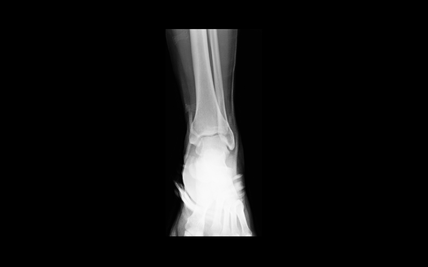
26 year old male presents s/p basketball dunk. Diagnosis?

Show Answer

Answer: Talar dislocation

Below, a post-reduction Xray, as well as a link to a video demonstrating a few ankle reductions (the video's background music makes all the difference): http://www.youtube.com/watch?v=AzqCsLX1K1E

Show References

Follow me on Twitter (@criticalcarenow) or Google+ (+haney mallemat)


Title: What's the Diagnosis?

Category: Visual Diagnosis

Posted: 8/29/2011 by Rob Rogers, MD (Updated: 7/22/2026)

Question


Patient presents with right-sided chest and shoulder pain....

What's the diagnosis?


Show Answer

Herpes Zoster

Show References


Title: What's the diagnosis? Dr. Michael Santiago

Category: Visual Diagnosis

Posted: 8/22/2011 by Haney Mallemat, MD

Question

79 y.o. male lung cancer patient with tachypnea, tachycardia, and normal blood pressure. Click here: http://vimeo.com/27973006

Possible diagnosis?

Show Answer

Answer: Right ventricular (RV) dysfunction secondary to submassive pulmonary embolism (PE).


Ultrasound for suspected PE

Consider bedside echo with PE and elevated troponin or BNP.

Recall the classes of PE:

  1. Non-massive PE: Normal RV function with normotension
  2. Submassive PE: RV dysfunction (see below) with normotension
  3. Massive PE: RV dysfunction with hypotension
  4. Cardiac arrest (Pulseless electrical activity)

Ultrasound “clues” of submassive / massive PE:

  1. Right ventricular enlargement: RV diastolic volume is >60% of the LV; normally  RV is 60% the size of the LV.
  2. Interventricular septal shift ("D-sign"): The normally concentric LV cavity will now look like the letter “D” from diastolic septal flattening; "D" sign indicates elevated pulmonary pressure (e.g., pulmonary embolism)
  3. McConnell’s sign: Hypokinesis in RV free wall with normal apical motion; originally though to be specific for PE, but sensitivity and specificity have been questioned because other disorders also have a "McConnel'-type" appearance (severe pulmonary hypertension, RV infarction); can still help add to pre-test probability.
  4. Free-floating thrombus: clot visualized in pulmonary artery and/or in right-side of heart (rare)

Show References

Lodato JA, Parker Ward RP, Lang RM. Echocardiographic Predictors of Pulmonary Embolism in Patients Referred for Helical CT. Echocardiography 2008;25:584-590.

McConnell MV, Solomon SD, Rayan ME, et. al. Regional Right Ventricular Dysfunction Detected by Echocardiography in Acute Pulmonary Embolism. Am J Cardiol. 1996; 78: 469-473.

ACEP. Critical Issues in the Evaluation and Management of Adult Patients Presenting to the Emergency Department With Suspected Pulmonary Embolism. http://www.acep.org/content.aspx?id=30060. [July 24, 2011].

John Griffiths. Respiratory: Management of small, submassive and massive pulmonary embolism. http://www.frca.co.uk/article.aspx?articleid=100750. [July 24, 2011].


Follow me on Twitter (@criticalcarenow) or Google+ (+haney mallemat)


Title: What's the Diagnosis? Images submitted by Dr. Joy Kay

Category: Visual Diagnosis

Posted: 8/8/2011 by Haney Mallemat, MD (Updated: 8/8/2011)

Question

13 year-old right-hand dominant male following assault with blunt object. Diagnosis?


Show Answer

Answer: Monteggia fracture


Monteggia Fracture

  • Ulnar fracture with dislocation of proximal radioulnar joint (often subtle); do not confuse with Galeazzi fracture (radial-shaft fracture with distal radioulnar dislocation)
  • Due to fall on outstretched hand with arm in hyper-pronation or with direct trauma to forearm as in defensive wounds (e.g., nightstick injury).
  • Ulnar fracture typically the proximal third (although any portion can be involved).
  • Relatively uncommon; 1-2% forearm fractures
  • Interosseous membrane (between radius and ulna) transmits forces to radioulnar joints and causes associated dislocations.
  • Suspected forearm fractures should always include Xray of the wrist, forearm, and elbow  
  • Bado classification system (Type I-IV) used; based on displacement of radial head.
  • Children may be treated with closed reduction and immobilization; adults usually require open reduction and internal fixation.
  • Radial head dislocations should be reduced within 6-8 hours as can lead to articular damage and/or nerve injury.
  • Radial, ulnar and/or median nerve neuropraxias (motor or sensory deficits) may complicate injury with resolution over several weeks.

Show References

Bruce H.E., Harvey J.P., Wilson J.C. Monteggia Fractures. J Bone Joint Surg Am. 1974;56:1563.

Reckling F.W. Unstable fracture-dislocation of the forearm (Monteggia and Galeazzi lesions). J Bone Joint Surg Am. 1982;64:857.

http://emedicine.medscape.com/article/1231438-overview
 

Follow me on Twitter (@criticalcarenow) or Google+ (+haney mallemat)


Title: What's the Daignosis?

Category: Visual Diagnosis

Posted: 7/25/2011 by Haney Mallemat, MD (Updated: 8/28/2014)

Question

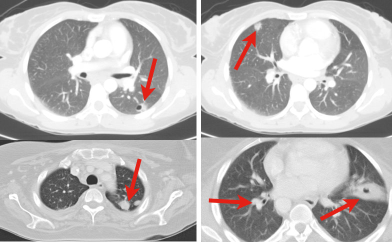
34 y.o. male with history of IVDA (intravenous drug abuse) complains of fever, chills and cough. Diagnosis?


Show Answer
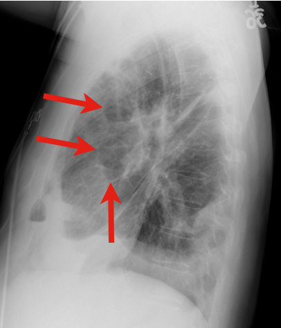
Answer: Lung Abscess (from septic pulmonary emboli)

Lung Abscess

  • Necrosis of lung parenchyma with pus and debris-filled cavities

  • Caused by direct injury (e.g., aspiration pneumonia) or secondary causes (e.g., tricuspid  endocarditis, bacteremia, etc.)

  • Suspect with:

    • Loss of airway reflexes (e.g., CVA, seizures, alcohol / narcotic abuse, etc)

    • Poor dentition

    • Immunosuppression

    • IVDA

  • Gram positives, negatives and anaerobic bacteria have all been implicated.

  • CXR may suggest diagnosis, but CT scan better identifies abscess, necrotic tissue, empyema, or other pathology (see image below).

  • After drawing blood cultures, broad-spectrum antibiotics should be started and narrowed once culture data is available; address underlying cause (e.g., valve replacement for endocarditis).

  • Prognosis is generally good with normal immune function and antibiotics, but mortality sharply increases with immunocompromise and treatment delay.

Show References

Mansharamani N, et al. Lung abscess in adults: clinical comparison of immunocompromised to non-immunocompromised patients. Respiratory Medicine. Mar 2002;96(3):178-85

Follow me on Twitter (@criticalcarenow) or Google+ (+haney mallemat)


Title: What's the Diagnosis? Written by Dr. Ari Kestler

Category: Visual Diagnosis

Posted: 7/11/2011 by Haney Mallemat, MD (Updated: 8/28/2014)

Question

48 year old male following 15 foot fall onto both feet. What is the diagnosis?
…and why is it called the “Lover’s Fracture”?
 

Show Answer
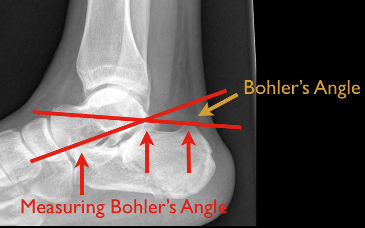
Answer: Calcaneus fracture; historically called the “Lover’s Fracture” for “lovers” jumping out of bedroom windows to evade suspicious spouses and landing directly on their feet.

Calcaneus fractures

  • Most commonly fractured tarsal bone
  • 2 types:
    • Extra-articular fracture from direct blow, twisting force and repetitive forces (causing stress fractures)
    • Intra-articular fracture from axial loading secondary to fall >6 feet, motor vehicle crash, etc; this is the classic “lover’s fracture”
  • 10% of axial loaded intra-articular fractures associated with:
    • Bilateral calcaneus fractures and/or,
    • Thoracic or lumbar compression fractures and/or,
    • Proximal femur or tibial plateau fractures
  • Ankle Xray is diagnostic for fractures and to measure Bohler’s angle (angle formed by intersection of lines connecting apex of anterior process with the apex of posterior facets and apex of posterior facet with the posterior tuberosity; see figure below)
    • Normally 20-40 degrees; <20 degrees increases suspicion for intra-articular fracture
  • Ankle CT in select cases; Xray may underestimate some injuries
  • Typically, extra-articular fractures treated with closed reduction and casting, while intra-articular fractures by open reduction and internal fixation (closed reduction in select cases)

Show References

Rosen's Emergency Medicine: Online Edition

Follow me on Twitter @criticalcarenow


Title: What's the diagnosis? Written by Dr. Ari Kestler

Category: Visual Diagnosis

Posted: 6/27/2011 by Haney Mallemat, MD

Question

49 y.o. female on Trimethoprim/sulfamethoxazole presents with rash and oral mucus membrane lesions. Diagnosis?

Show Answer

Answer: Steven-Johnson Syndrome

  • Malaise and fever prodrome, followed by erythematous or purpuric macules and plaques
    • <10% body-surface area (BSA) and mucosal membrane involvement (e.g., ocular, oral, genital)
    • Toxic Epidermal Necrolysis defined as >30% BSA involvement
  • Immune-complex hypersensitivity reaction from viral, bacterial, chemical, or drug exposure.
    • Common medications:
      • Antibiotics; Sulfonamides > PCN > Cephalosporins
      • NSAIDs
      • Anti-psychotics
  • Management:
    • Remove possible trigger(s)
    • Treat like burns; supportive care (hemodynamic support, electrolyte management, analgesia)
    • Strongly consider Burn ICU.

Show References

French LE. Toxic epidermal necrolysis and Stevens Johnson syndrome: our current understanding. Allergol Int. Mar 2006;55(1):9-16

Schöpf E. Toxic epidermal necrolysis and Stevens-Johnson syndrome. An epidemiologic study from West Germany. Arch Dermatol. 1991;127(6):839.

Roujeau JC. Severe adverse cutaneous reactions to drugs. N Engl J Med. 1994;331(19):1272.


Follow me on Twitter @criticalcarenow


Title: What's the Diagnosis? Written by Dr. Katherine Baugher

Category: Visual Diagnosis

Posted: 6/13/2011 by Haney Mallemat, MD (Updated: 6/13/2011)

Question

13 y.o. with shoulder trauma (during basketball game). Arm held in adduction and exquisite scapular tenderness. Diagnosis?

Show Answer

Answer: Scapular Fracture


Scapular Fracture

  • Uncommon (1% of all fractures)

  • Extensive force required; high probability of associated injuries (pneumothorax, shoulder dislocation).

  • 5 types: body/spine, acromion, neck, glenoid, and coracoid

  • Arm held in adduction and pain with shoulder movement; may mimic rotator cuff tear.

  • Obtain X-rays (AP shoulder and lateral scapula) or CT (if displaced).

  • Conservative management for non-displaced fractures; Orthopedic reduction for displaced fractures.

  • Complications include post-traumatic arthritis or bursitis.

  • NSAIDs are first-line analgesics.

Show References

Rosen P, Barkin R. Emergency Medicine: Concepts and Clinical Practice. Mosby Year Book; 2010:573-574.

Zlowodzki M, Bhandari M, Zelle BA, Kregor PJ, Cole PA. Treatment of scapula fractures: systematic review of 520 fractures in 22 case series. J Orthop Trauma. Mar 2006;20(3):230-3.


Title: Mystery Case

Category: Visual Diagnosis

Posted: 6/6/2011 by Rob Rogers, MD

Question

A 20 year-old female presents with bilateral neck pain that occurred at rest. No other complaints. See if you can find the subtle clue on the x-ray...


Show Answer

The x-ray shows air in the soft tissue of the right neck and supraclavicular area. Close inspection of the mediastinum reveals subtle air along the heart border. Diagnosis: pneumomediastinum.

Often the first clue of pneumomediastinum is air in the neck and supraclavicular area on plain film. So, always look at the soft tissues on chest x-ray.


Title: What's the diagnosis?

Category: Visual Diagnosis

Posted: 5/30/2011 by Haney Mallemat, MD

Question

13 y.o. female with ankle pain following fall down escalator. What's the diagnosis? (Hint: Look very closely)

Show Answer

Answer: Tri-plane ankle fracture

Tri-plane Fractures (Submitted and written by Dr. Michael Santiago)

  • Multi-planar ankle fracture in older children and adolescents during an 18 month window prior to distal tibial-physis closure.
  • 3 planes (see Xray below): coronal (A: tibal metaphysis), sagital (B: epiphysis), and transverse (C: growth plate)
  • Associated fibular spiral fracture (50% cases)
  • Commonly due to external rotational or "twisting" forces.
  • Xrays are helpfully, but CT scan may be indicated as fractures may be more displaced than radiographs suggest.
  • Non-operative treatment for non-displaced fractures:
    • Closed reduction with long-leg splint/cast
    • Reduction may slip once swelling has subsided
  • Indications for operative repair:
    • >2mm displacement of fracture segments
    • Intra-articular fracture


Title: What's the Diagnosis?

Category: Visual Diagnosis

Posted: 5/23/2011 by Haney Mallemat, MD

Question

50 yo female s/p motor vehicle crash. Diagnosis?

Show Answer

Answer: Pneumomediastinum from pneumothorax


Pneumomediastinum


Mediastinal air has multiple causes:

  • Intrathoracic injury (e.g., Pneumothorax)
  • Upper respiratory tract (e.g., Injury during intubation) 
  • Digestive tract perforation (e.g., Boerhaave’s syndrome)
  • Intra-abdominal perforation (e.g., EGD complication)
  • Anaerobic infection at sites communicating with mediastinum
    • Ludwig’s angina
    • Pneumoperitoneum
    • Bacterial mediastinitis

Listen for Hamman's sign ("crunching" with cardiac auscultation) and feel for crepitus (distinct feeling on palpation).

Treat the precipitating cause.

Usually resolves without mediastinal decompression; air travels along tissue planes decompressing increased mediastinal pressure.


Title: What is the Diagnosis?

Category: Visual Diagnosis

Posted: 5/9/2011 by Haney Mallemat, MD

Question

70 yo female from nursing home with fever. RUQ ultrasound is shown below. Diagnosis?

Show Answer

Acalculous Cholecystitis

Acalculous cholecystitis is due to gallbladder (GB) wall ischemia and GB stasis (reduced GB contraction from reduced PO intake)

which all leads to “sludging” (Figure 1; A), thickened GB walls (B), and pericholecystic fluid (C).


Risk factors include critical illness (especially sepsis), acute cholecystitis, total parenteral nutrition, diabetes, and GB dysmotility.


Treatment includes antibiotics covering enteric / biliary pathogens and cholecystectomy (if a surgical candidate) or percutaneous

cholecystostomy if unsuitable for the OR. 

Complications include perforation, GB gangrene, and extrabiliary abscess.


Title: What's the Diagnosis?

Category: Visual Diagnosis

Posted: 4/25/2011 by Haney Mallemat, MD (Updated: 7/22/2026)

Question

Patient presents with the following X ray after yawning. Diagnosis?

Show Answer

Diagnosis: Mandibular Dislocation

The link below demonstrates an interesting alternative to the traditional reduction method; prospective randomized trails are pending.

http://www.youtube.com/watch?v=Kp8AzHIC0hM

Show References

Thanks to Dr. George Kochman for the image and Dr. Christopher Doty (EM Program Director at SUNY Downstate / Kings County Hospital Center) for the video.


Title: What's the diagnosis? Written by John Greenwood, MD

Category: Visual Diagnosis

Posted: 4/11/2011 by Haney Mallemat, MD (Updated: 4/11/2011)

Question

60 y/o male transferred from local rehab facility c/o abdominal pain.


Show Answer

Ogilvie's syndrome / Acute colonic pseudo-obstruction (ACPO)

Syndrome of decreased GI motility believed to be an autonomic imbalance (specifically, parasympathetic suppression) predominantly in the cecum and right colon. ACPO is also commonly due to narcotic overuse / abuse. 

Differential diagnosis:

  • Mechanical large bowel obstruction
  • Toxic megacolon / colitis
  • Ileus 

Abdominal XR:

  • Gaseous distention without fluid levels (UNLIKE obstruction)
  • Small bowel dilation is typically absent (UNLIKE ileus)
  • Preserved haustral markings and smooth inner contour (UNLIKE colitis)

Treatment:

  • Treat underlying cause (e.g., discontinue narcotics) 
  • Neostigmine
  • Therapeutic Colonoscopy
  • Surgical decompression (if severe)

Show References

Fazel A, Verne GN.  New solutions to an old problem: Acute colonic pseudo-obstruction. JClin Gastroenterol 2005; Vol 39(1): 17-20.

Ozkurt H, Yilmaz F, et al.  Acute colonic pseudo-obstruction (Ogilvie's syndrome): radiologic diagnosis and medical treatment with neostigmine.  Report of 4 cases.  Am J of Emer Med 2009; Vol 27: 757.e1 - 757e4.


Previous  |  1 |  ... |  7 |  8 |  9 |  10 |  11 |  12 |  13 |  Next
University of Maryland School of Medicine logo
  • Departments
  • Programs
  • Research Centers
  • Institutes
  • Graduate Education
  • CIBR Cores
  • Offices of the Dean
    • University of Maryland, Baltimore
    • University of Maryland Medical Center
    • Medical Alumni Association of the University of Maryland
    • Health Sciences and Human Services Library
University of Maryland School of Medicine
655 W. Baltimore Street
Baltimore, MD 21201
Contact Us

© University of Maryland School of Medicine

    • Webmaster
    • Web Accessibility
    • Site Index
    • UMB Hotline
    • Employment
    • Map