University of Maryland School of Medicine logo
Emergency Medicine
All Departments
UMSOM Assistant
UMEM Educational Pearls
  • Home
  • Departments
  • Emergency Medicine
  • CME
  • UMEM Educational Pearls
  • CME
  • Courses and Conferences
  • Free Open Access Medical Education
  • UMEM Educational Pearls

Search

201-220 of 245 results with category "Visual Diagnosis"

Previous  |  1 |  ... |  6 |  7 |  8 |  9 |  10 |  11 |  12 |  13 |  Next

Title: What's the Diagnosis? Written by Ali Farzad, MD

Category: Visual Diagnosis

Posted: 7/16/2012 by Haney Mallemat, MD (Updated: 7/16/2012)

Question

23 year-old female presents complaining of progressive right lower quadrant pain after doing "vigorous" pushups. CT abdomen/pelvis below. What’s the diagnosis? (Hint: it’s not appendicitis)


Show Answer

Answer: Rectus Sheath Hematoma


Rectus Sheath Hematoma (RSH)

Rectus muscle tear causing damage to the superior or inferior epigastric arteries with subsequent bleeding into the rectus sheath; uncommon cause of abdominal pain but mimics almost any abdominal condition.

May occur spontaneously, but suspect with these risk factors:

  • Coagulopathy (#1 cause); acquired (e.g., warfarin) or inherited disorder
  • Rectus muscle trauma
  • Vigorous or sudden contraction of rectus muscle
  • Increased intra-abdominal pressure from vigorous coughing
  • Pregnancy (gestation, labor, or post-partum)

Typically a self-limiting condition, but hypovolemic shock may result from significant hematoma expansion.

  • Hemodynamically stable (non-expanding hematoma): conservative treatment (rest, analgesia, and ice)
  • Hemodynamically unstable (expanding hematoma): treat with fluid resuscitation, reversal of coagulopathy, and transfusion of blood products.


Show References

Follow me on Twitter (@criticalcarenow) or Google+ (+haney mallemat)


Title: What's the diagnosis? Written by Danya Khoujah, MD

Category: Visual Diagnosis

Posted: 7/9/2012 by Haney Mallemat, MD (Updated: 7/9/2012)

Question

A previously healthy 3 year-old male presents with a one-day history of fever, drooling, and refusal to move his neck. The lateral neck x-ray is shown. What's the diagnosis?


Show Answer

Answer: Retropharyngeal abscess


Retropharyngeal Abscess (RPA)

  • Commonly diagnosed in young children because the retropharyngeal space contains lymph nodes that atrophy before puberty; there is an increasing incidence in adults
  • Etiologic bacteria: Aerobic (e.g., Beta-hemolytic Strep and Staph), anaerobic (e.g., Bacteroides), or gram-negative species (e.g., Haemophillus); 50% of cases preceded by a URI
  • Signs / Symptoms: fever, drooling, sore throat, dysphagia, stiff neck, vocal changes (e.g., "hot-potato" voice), stridor, or respiratory distress.
  • Radiologic studies
    • ​Lateral X-ray with soft tissue swelling, gas, or air-fluid levels.
    • Neck CT w/ IV contrast (definitive) defines the presence of an abscess, extent of disease, and presence of cellulitis.
  • Treatment: Antibiotics with or without surgical drainage
  • Complications: mediastinitis, epidural abscess, sepsis, airway obstruction, and jugular venous thrombosis.

Show References

Follow me on Twitter (@criticalcarenow) or Google+ (+haney mallemat)


Title: Got (Ultrasound) Clips?

Category: Visual Diagnosis

Posted: 7/2/2012 by Haney Mallemat, MD

Have you ever had to give a national presentation, but can't find that one ultrasound clip or image that you really need? You could "borrow" it from someone on the internet, but you are secretly afraid that the "owner" of the clip is lurking somewhere in the audience. Well, the guys at the Ultrasound Podcast (www.ultrasoundpodcast.com) have come to your rescue by creating SonoCloud, a free access ultrasound library. At Sonocloud, you will find several categories of ultrasound clips and images for you to view and share,...and again it's FREE. In fact, the only thing you are expected to do is upload some of your own ultrasounds to share. 

So head over to www.sonocloud.org, create your free account, and begin exploring...and while you're there, why don't you upload a clip or two?

Show References

Follow me on Twitter (@criticalcarenow) or Google+ (+haney mallemat)


Title: What's the Diagnosis? Image submitted by Dr. Zachary Dezman

Category: Visual Diagnosis

Posted: 6/25/2012 by Haney Mallemat, MD (Updated: 6/25/2012)

Question

77 year old male presents to the Emergency Department one week after a motor vehicle crash in which he suffered minor facial injuries. He is now concerned because his eye looks like this. Diagnosis? 

Show Answer

Answer: Subconjunctival Hemorrhage

Subconjunctival hemorrhage is the accumulation of blood between the conjunctiva and sclera (i.e., subconjunctival space) secondary to bleeding from conjunctival or episcleral blood vessels.

Subconjunctival hemorrhage may be caused by:

  • Trauma
  • Excessive coughing or valsalva
  • High-blood pressure
  • Coagulopathy: acquired (e.g., aspirin use) or inherited (e.g., Factor VIII deficiency)
  • Post-occular surgery (normal complication)
  • Febrile systemic-infections (e.g., meningococcal septicemia)
  • Following CPR-associated chest compressions
  • Post-cardiac angiography or open-heart surgery

Treatment should be directed towards reversing the underlying cause and providing symptomatic relief (e.g., artificial tears)

Prognosis is typically good. Occasionally the hemorrhage becomes yellowish-green (similar to a bruise) during the healing phase; this eventually resolves.

Bonus Pearl: As July 1rst approaches a new class of doctors will begin their journey to become Emergency Medicine specialists. The EM Basic website, created by Dr. Steve Carroll, was developed as a boot camp guide for Emergency Medicine. At the EM Basic website, you can find podcasts and discussions of must know topics within Emergency Medicine. Check it out today at http://embasic.org or on iTunes

Show References

Follow me on Twitter (@criticalcarenow) or Google+ (+haney mallemat)


Title: What's the Diagnosis? Case submitted by Bethany Radin

Category: Visual Diagnosis

Posted: 6/18/2012 by Haney Mallemat, MD

Question

79 year old male with headaches, ataxia, falls, and difficulty urinating. What's the diagnosis?

Show Answer

Diagnosis: Ventriculomegaly secondary to Normal Pressure Hydrocephalus


An approach to ventriculomegaly

Ventriculomegaly is due to cerebral atrophy (e.g., Parkinson disease) or increased cerebrospinal fluid (CSF) within the ventricles. Increased CSF is due to:

  • Increased CSF production (e.g., choroid-plexus papilloma), or
  • Impaired CSF re-absorption (e.g., subarachnoid-villi inflammation), or
  • Obstruction of CSF flow (e.g., non-communicating hydrocephalus)

Congenital causes of ventriculomegaly:

  • Neuro-tube defect (e.g, myelomeningocele)
  • Aquaductal stenosis
  • CNS malformation (e.g., Dandi-Walker syndrome)
  • Intrauterine infection (e.g., CMV, rubella, etc.)

Acquired causes of ventriculomegaly:

  • Paget's disease (obstruction of CSF flow)
  • Bacterial or viral meningitis (impaired CSF absorption)
  • Tumor (obstruction of CSF flow)
  • Post-hemorrhagic (e.g., trauma, aneurysmal rupture, AVM, coagulopathy) impaired absorption and/or obstruction of flow


Title: What's the diagnosis? Case written by Dr. Nadia Eltaki

Category: Visual Diagnosis

Posted: 6/11/2012 by Haney Mallemat, MD

Question

19 year-old male presents with L ankle pain and obvious deformity after jumping out of a window and landing on his inverted foot. What's the diagnosis?

Show Answer

Sub-talar dislocation

  • Dislocation of distal talar articulation at talocalcaneal and talonavicular joints (ankle joint is undisturbed)
  • Also known as "basketball foot"; a common mechanism of injury
  • Medial dislocation is more common than lateral (85% vs 15%) and it is usually caused by injury while foot is in inversion
  • Closed reduction is facilitated with knee in flexion to relax the gastrocnemius
  • Approximately 20% of lateral dislocations require open reduction
  • CT scan is recommended following closed reduction to rule out osteochondral lesions and better assess the reduction

Show References

Wheeless' Textbook of Orthopedics

Follow me on Twitter (@criticalcarenow) or Google+ (+haney mallemat)


Title: Ultrasound for Confirmation of Endotracheal Intubation

Category: Visual Diagnosis

Posted: 5/28/2012 by Haney Mallemat, MD

Ultrasound is useful during intubation; here is a video explaining how: http://ultrarounds.com/ultrarounds.com/Visual_Pearl_May_28,_2012.html


Today's Bonus Pearl:

EMRA has developed a great antibiotic guide for the iphone (http://itunes.apple.com/us/app/2011-emra-antibiotic-guide/id393020737?mt=8) or android (https://play.google.com/store/apps/developer?id=Emergency+Medicine+Residents'+Association). This app is a bit pricey ($15.99), but is easy to use and well organized. Enjoy!  

Show References

Chou, H. et al. Tracheal rapid ultrasound exam (T.R.U.E.) for confirming endotracheal tube placement during emergency intubation. Resuscitation. Jun 2011

Werner SL,et al. Pilot study to evaluate the accuracy of ultrasonography in confirming endotracheal tube placement. Ann Emerg Med 2007;49:75–80.

Follow me on Twitter (@criticalcarenow) or Google+ (+haney mallemat)


Title: Psoas Abscess

Category: Visual Diagnosis

Posted: 5/14/2012 by Haney Mallemat, MD

This week's visual pearl is an interesting ultrasound of a psoas abscess submitted by Dr. Sa'ad Lahri. He is an Attending physician in the Emergency Department of the Khayelitsha Hospital in Cape Town, South Africa. The video quality is grainy, but it automatically replays so you can watch it a few times.

http://ultrarounds.com/ultrarounds.com/Visual_Pearl_May_14,_2012.html

Show References

Follow me on Twitter (@criticalcarenow) or Google+ (+haney mallemat)


Title: An alcoholic with fever and cough

Category: Visual Diagnosis

Keywords: fever, cough, alcoholic (PubMed Search)

Posted: 5/7/2012 by Rob Rogers, MD (Updated: 7/22/2026)

Question

An alcoholic patient presents with a cough, fever, and very foul smelling breath (worse than usual)

What's the diagnosis? And what are the risk factors?


Show Answer

Lung abcess


Risk factors for a lung abscess include:

  • Airway foreign body
  • Alcoholism
  • Cystic fibrosis
  • Periodontal disease
  • Diabetes
  • Drug Abuse
  • Esophageal surgery
  • Gastric surgery
  • Loss of consciousness
  • Lung cancer
  • Seizures
  • Sinusitis
  • Stroke


Show References

case compliments of our very own Zac Dezman


Title: What's the Diagnosis?

Category: Visual Diagnosis

Posted: 4/30/2012 by Haney Mallemat, MD (Updated: 4/30/2012)

Question

68 yo man presents with new-onset seizures; his CT is shown below. What is your differential diagnosis?

Show Answer

Cerebral Ring-Enhancing Lesions

Neoplasm

  • Primary brain tumor
  • Primary CNS lymphoma
  • Metastatic disease

Infectious

  • Pyogenic abscess
  • Tuberculoma
  • Syphilis
  • Neurocysticercosis  
  • Toxoplasmosis
  • Nocardiosis
  • Cryptococcosis
  • Aspergillosis

Neurologic

  • Multiple sclerosis (lesions within the white matter)
  • Sacroidosis
  • Vasculitis
  • Aneurysm
  • Resolving hematoma
  • Cerebral infarct
  • Postoperative change

Bonus pearl: Do you like Emergency Ultrasound and want a quick review before you scan your next patient? Well, check out the "One-minute Ultrasound App". It's provides a quick review for many essential ultrasound studies...and yes, it's FREE for both iphone and android.

Iphone: http://itunes.apple.com/us/app/one-minute-ultrasound/id512301845?mt=8&ls=1

Adroid: https://play.google.com/store/apps/details?id=com.v1_4.oneminuteus.com&feature=search_result#?t=W251bGwsMSwxLDEsImNvbS52MV80Lm9uZW1pbnV0ZXVzLmNvbSJd


Garg, R., Sinha, M. Multiple ring-enhancing lesions of the brain. J Postgrad Med. 2010 Oct-Dec; 56(4):307-16

Follow me on Twitter (@criticalcarenow) or Google+ (+haney mallemat)


Title: What s the Diagnosis? Case submitted and written by Dr. Zachary Dezman

Category: Visual Diagnosis

Posted: 4/16/2012 by Haney Mallemat, MD (Updated: 4/16/2012)

Question

67 yo male presents with burning substernal chest pain; worse with meals and when supine. What's the diagnosis?


Show Answer

Answer: Hiatal hernia

  • Hiatal hernias are commonly retrosternal and present with GERD-like symptoms
  • A barium swallow is the diagnostic test of choice.

The differential diagnosis for circumscribed air-fluid levels on chest X-ray includes:

  • Hiatal hernia
  • Cavitary disease (e.g., tuberculosis)
  • Empyema
  • Lung abscess

Bonus Pearl

Do you like listening to pre-recorded lectures, especially when they’re free? Then check out Free Emergency Medicine Talks (http://freeemergencytalks.net) where you can listen to millions (ok, more like 1,384) of free lectures recorded at major conferences around the world.

Follow me on Twitter (@criticalcarenow) or Google+ (+haney mallemat)


Title: What's the Diagnosis? Case submitted and written by Dr. Zachary Dezman

Category: Visual Diagnosis

Posted: 3/26/2012 by Haney Mallemat, MD (Updated: 3/26/2012)

Question

26 year old male with pain when he extends his 4th finger as well as swelling of that digit. Diagnosis?

Show Answer

Answer: Infectious Flexor Tenosynovitis

Infectious Flexor Tenosynovitis

- Closed space infection of the flexor tendon sheath; an orthopedic emergency

- Typically an infection with skin flora secondary to penetrating trauma

- Diagnosed by Kanavel's Signs:

           - Finger held in slight flexion
           - Fusiform swelling
           - Pain on passive extension
           - Pain while palpating the tendon sheath

- If early and not severe a course of IV antibiotics (covering skin flora) may be tried, however, surgical intervention is often necessary; early consultation with a hand surgeon is highly recommended

Follow me on Twitter (@criticalcarenow) or Google+ (+haney mallemat)


Title: What's the Diagnosis? Case submitted and written by Dr. Zachary Dezman

Category: Visual Diagnosis

Posted: 3/12/2012 by Haney Mallemat, MD (Updated: 8/12/2014)

Question

14 year-old male presents with right-sided testicular pain. What's the diagnosis?


Show Answer

Answer: Testicular torsion

  • Most common pediatric cause of testicular pain; Occurs in 1 in 4,000 males less than 25 years-old
  • Sudden and severe pain with diffuse testicular swelling and tenderness; right side is affected more than the left.
  • Most sensitive sign (90-100%) is absent or decreased cremaster reflex.
  • Diagnosis is made by ultrasound demonstrating an enlarged testicle, increased echogenicity, and little to no Doppler flow (as seen above) compared to a testicle with normal flow (see below)
  • Requires emergent urology consult for surgical detorsion and fixation of testis to prevent recurrence; testicular salvage rate is 90% within 6 hours of presentation as compared to 20% at 24-48 hours
  • Manual detorsion should be attempted if urology consult is delayed.

References

Shan Yin, MD, MPH, Jennifer L. Trainor, MD, Diagnosis and Management of Testicular Torsion, Torsion of the Appendix Testis, and Epididymitis, Clin Ped Emerg Med, 10:38-44, 2009

Follow me on Twitter (@criticalcarenow) or Google+ (+haney mallemat)


Title: What's the Diagnosis? Case submitted by Dr. Bethany Radin

Category: Visual Diagnosis

Posted: 2/27/2012 by Haney Mallemat, MD (Updated: 8/28/2014)

Question

24 year-old male presents following fall from a scaffolding and complains of wrist pain. Diagnosis?

Show Answer
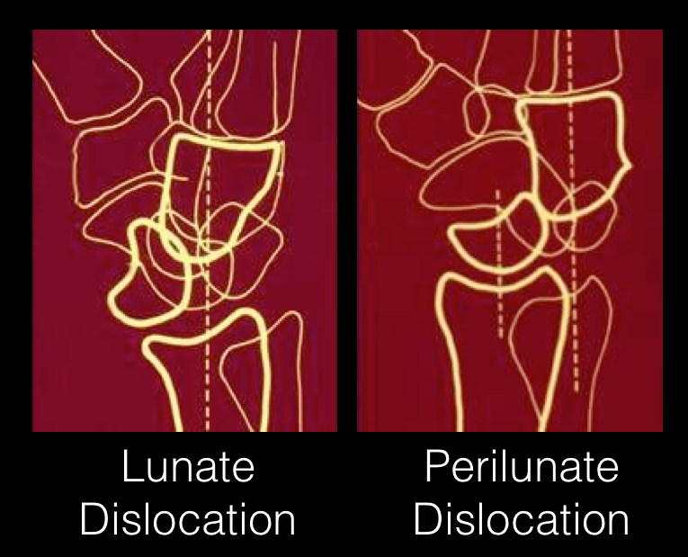
Answer: Perilunate dislocation

Perilunate dislocations usually occur following high-energy trauma (e.g., fall from a height); the mechanism is usually wrist hyperextension, ulnar deviation, and carpal supination.

Tenderness is palpated along the dorsum of the wrist; specifically distal to the lister tubercle along the scapholunate ligament; injuries may be associated with scaphoid fracture

Paresthesias may also occur along the median nerve distribution with up to 25% of cases developing carpal tunnel syndrome.

Treatment options include closed reduction and casting or open reduction and ligamentous repair with internal fixation.

The figure below illustrates the differences between lunate and perilunate dislocations.

References

Lunate and Perilunate Dislocations. Kannikeswaran, N & Sethuraman, U.

http://www.radiologyassistant.nl/en/42a29ec06b9e8 (diagram)

Follow me on Twitter (@criticalcarenow) or Google+ (+haney mallemat)


Title: What's the Diagnosis? Case submitted by Dr. Joeseph Martinez

Category: Visual Diagnosis

Posted: 2/13/2012 by Haney Mallemat, MD (Updated: 8/28/2014)

Question

35 year old male with sudden onset of abdominal pain. Diagnosis?

Show Answer
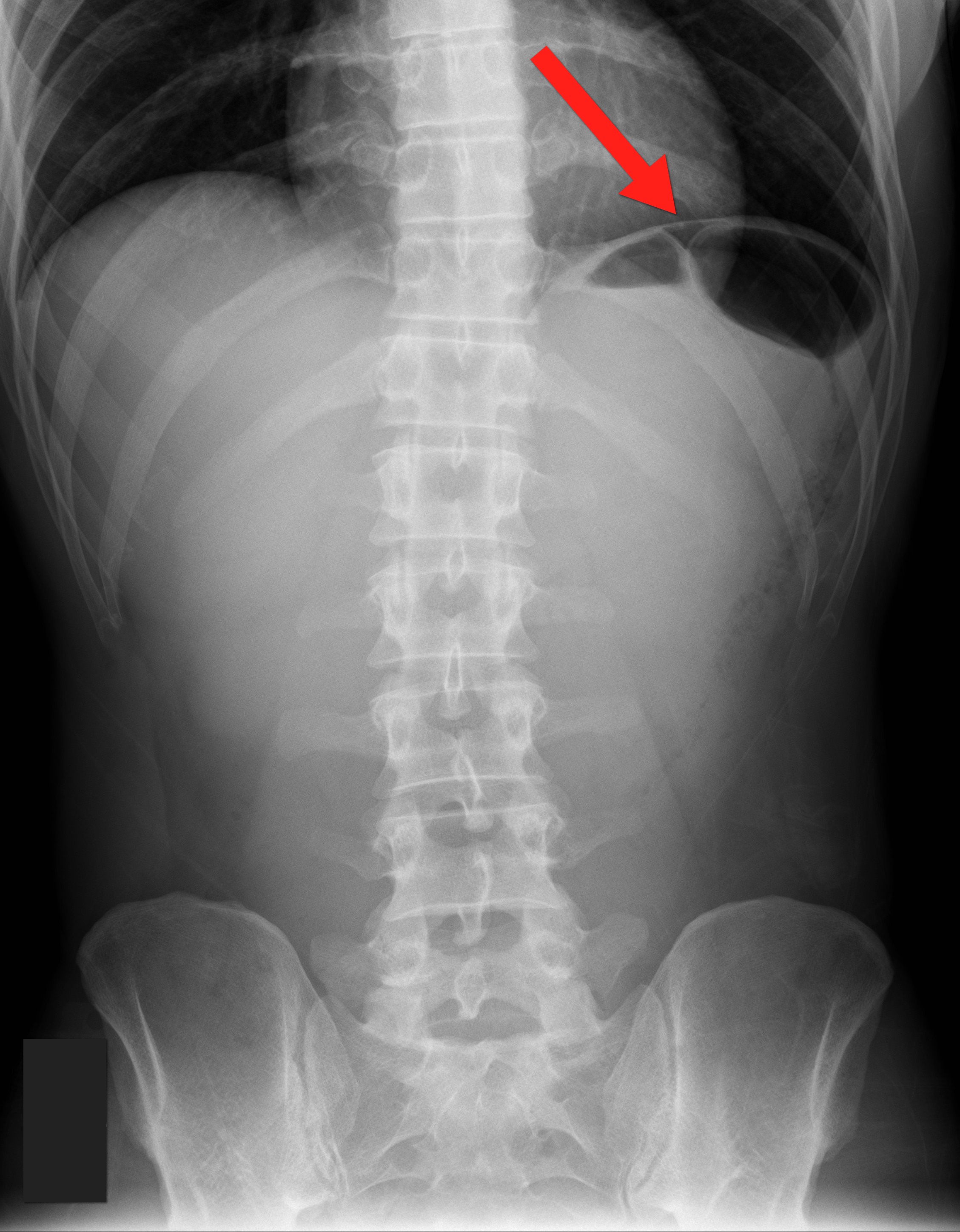
Answer: Pneumoperitoneum

Show References

Follow me on Twitter (@criticalcarenow) or Google+ (+haney mallemat)


Title: What's the Diagnosis? By Michael Allison

Category: Visual Diagnosis

Posted: 2/6/2012 by Haney Mallemat, MD (Updated: 8/28/2014)

Question

28 y.o. male felt his left knee "pop" after landing from a jump while playing basketball. Knee exam revealed limited knee extension. X-ray is shown. Diagnosis?

Show Answer
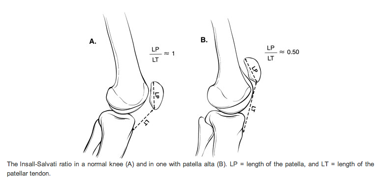
Answer: Patella Alta secondary to Patellar tendon rupture

  • Mechanism is knee flexion combined with forceful contraction of the quadriceps
  • Swelling, patellar tenderness, and limited extension of knee
  • Radiographs are helpful in diagnosis when using the Insall-Salvatti ratio (see below)
    • This ratio is the patellar length (PL) (in cm) divided by the distance between inferior pole of the patella and tibial tuberosity (PT)
    • Insall-Salvatti ratio PL/PT =1 is normal; <0.8 is predictive of tendon rupture

References

  • Insall J, Salvati E. Patella position in the normal knee joint. Radiology 1971;101:101
  • Sibley T, Algren D, Ellison S. Bilateral patellar tendon ruptures without predisposing systemic disease or steroid use: a case report and review of the literature. American Journal of Emergency Medicine 2012; 30: 261.e3-261.e

Follow me on Twitter (@criticalcarenow) or Google+ (+haney mallemat)


Title: What's the diagnosis? Case submitted by Emilie Calvello

Category: Visual Diagnosis

Posted: 1/23/2012 by Haney Mallemat, MD (Updated: 1/23/2012)

Question

20 year old female complains of “itchy” rash to her foot x 1 week and recently the rash has spread to her other other foot and both hands (shown below). No past medical history, no fever or chills, no mucus membranes involvement, no new medications, no tick bites, no travel. She is also 16 weeks pregnant. What’s the diagnosis?

Show Answer

Answer: Secondary syphilis

  • The differential diagnosis for rash on the palms and soles is small and should always include syphilis.
  • The rash may be maculopapular, papular, annular, or pustular.
  • Syphilis easily crosses the placenta and fetal damage can occur; the extent of the damage depends upon its stage of development at the time of infection and the duration of untreated infection.
  • Without therapy, fetal infections  may result in miscarriage, stillbirth, growth restriction, hydrops fetalis, premature delivery, and neonatal death.
  • "The physician who knows syphilis knows medicine." -Sir William Osler

Show References

Follow me on Twitter (@criticalcarenow) or Google+ (+haney mallemat)


Title: What's the diagnosis? Image submitted by Dr. Michael Abraham

Category: Visual Diagnosis

Posted: 1/9/2012 by Haney Mallemat, MD (Updated: 8/28/2014)

Question

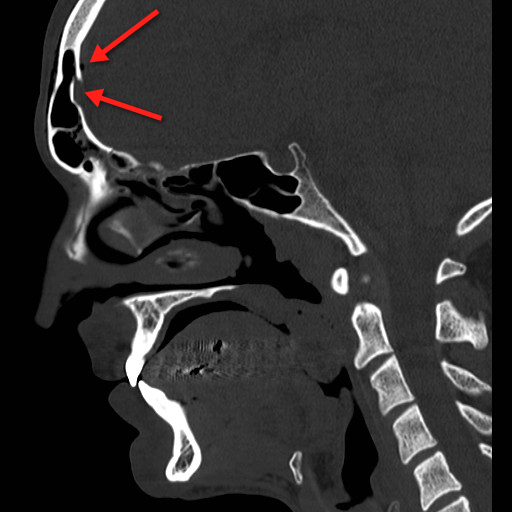
23 year-old male fell off porch while intoxicated. The head CT is shown below. Diagnosis?

Show Answer
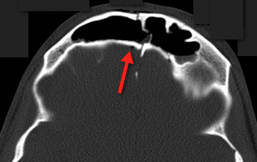
Answer: Frontal sinus fracture (inner and outer table) with pneumocephalus.

A few quick pearls when managing skull fractures:

Medical management:

  • Little evidence for prophylactic antibiotics; even with a laceration over fracture (i.e., an open fracture)
  • Little evidence for prophylactic anti-convulsants

Surgical management, if:

  • Skull depression >5mm relative to adjacent segment(s)
  • Dural tear; suggested by CSF or pneumocephalus
  • Debridement and washout for suspected or confirmed wound contamination

Show References

Follow me on Twitter (@criticalcarenow) or Google+ (+haney mallemat)


Title: What's the diagnosis? Case submitted by Dr. Bethany Radin

Category: Visual Diagnosis

Posted: 12/26/2011 by Haney Mallemat, MD

Question

64 year old male with emphysema and stage 4 lung cancer presents in respiratory distress. What's the diagnosis?

Show Answer

Answer: Pneumothorax (left chest) and bullous disease (right chest).

In questionable cases like this, bedside ultrasound can help distinguish between bullae and pneumothorax. Bullae should have a positive lung sliding sign whereas pneumothorax does not.

See the referenced case report for more information:

Simon, B. et al. Two cases where bedside ultrasound was able to distinguish pulmonary bleb from pneumothorax. J Emerg Med. 2005 Aug;29(2):201-5.

Show References

Follow me on Twitter (@criticalcarenow) or Google+ (+haney mallemat)


Title: What's the Diagnosis?

Category: Visual Diagnosis

Posted: 12/12/2011 by Haney Mallemat, MD (Updated: 8/28/2014)

Question

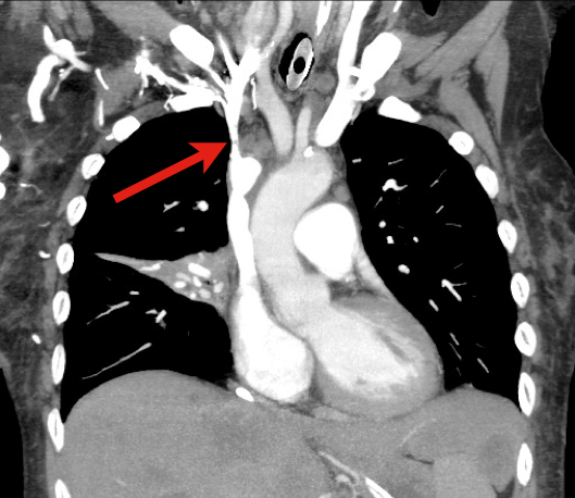
60 year old male with 6 months of weight loss and recent epistaxis. Diagnosis?

Show Answer

Answer: Partial obstruction of the Superior vena cava (SVC)


SVC Syndrome

SVC syndrome is caused by compression or obstruction of the superior vena cava blocking anterograde flow.

Most cases are secondary to extrinsic compression by malignancy. Other causes are secondary to thrombosis and internal obstruction (e.g., central venous catheter placement).

Symptoms present sub-acutely, worsen with bending over, and are secondary to increased venous pressure in the head and neck (e.g., epistaxis, headache, tinnitus, conjunctival injection, neck swelling, etc.).

Treatment focuses on reversing the underlying cause (e.g., radiation or chemotherapy if due a sensitive tumor) and treatment of symptoms:

  • Steroids, diuretics, and hyperosmolar agents to reduce edema
  • Elevate the head of bed to reduce brain edema
  • Anti-emetics to prevent vomiting-associated increases in intracranial pressure

Rice TW, Rodriguez RM, Light RW. The superior vena cava syndrome: clinical characteristics and evolving etiology. Medicine (Baltimore). Jan 2006;85(1):37-42.

Nunnelee JD. Superior vena cava syndrome. J Vasc Nurs. Mar 2007;25(1):2-5; quiz 6.

Follow me on Twitter (@criticalcarenow) or Google+ (+haney mallemat)


Previous  |  1 |  ... |  6 |  7 |  8 |  9 |  10 |  11 |  12 |  13 |  Next
University of Maryland School of Medicine logo
  • Departments
  • Programs
  • Research Centers
  • Institutes
  • Graduate Education
  • CIBR Cores
  • Offices of the Dean
    • University of Maryland, Baltimore
    • University of Maryland Medical Center
    • Medical Alumni Association of the University of Maryland
    • Health Sciences and Human Services Library
University of Maryland School of Medicine
655 W. Baltimore Street
Baltimore, MD 21201
Contact Us

© University of Maryland School of Medicine

    • Webmaster
    • Web Accessibility
    • Site Index
    • UMB Hotline
    • Employment
    • Map