
Search
Subscribe to daily UMEM Pearls (FREE!) via:
461-480 of 889 results with category "Critical Care"
- What type of fluid we use in critical care resuscitation has been hotly debated for some time
- The most recent battles have been played out between NS and plasmalyte or buffered solutions
- There has been some evidence that high chloride solutions can lead to renal injury requiring renal replacement therapy (RRT)
- Does a buffered crystalloid reduce renal complications compared with normal saline in patients admitted to the ICU?
- The SPLIT Trial (Saline vs Plasma-Lyte) from New Zealand ICU's adds more to our knowledge about this topic while enrolling over 2,000 patients
- Summary:
- Primary outcome was a rise in creatinine
- There was no difference in the primary outcome or incidence of AKI
- There was no difference in use of RRT or mortality
- Suggesting that is doesnt make too much of a difference
- There were some limitations: 90% of patients were given fluid before enrollment that was buffered crystalloid and patients were only given around 2 liters on average of fluid in the ICU
The Bottom Line: This was a nicely designed study to evaluate the safety of both fluids. It does suggest that either fluid type is for the most part OK. But in patients requiring hefty fluid boluses, we should be cautious in what type of fluid we choose.
Show References
Mechanical Ventilation for Septic Patients in Resource-Limited Settings
- An international team of physicians just published a series of recommendations for ventilatory support of septic patients in resource-limited settings.
- Pearls from these recommendations include:
- Elevate the head of the bed to 30o - 45o
- Consider tidal volumes of 5 - 7 ml/kg PBW in all patients
- Use minimum levels of PEEP ( 5 cm H2O) in all patients with sepsis and acute respiratory failure (unless the patient has moderate to severe ARDS)
- Lower FiO2 to target SpO2 > 88% or PaO2 > 60 mm Hg
- Use lung ultrasound to evaluate pulmonary edema when CXR is not available
- Consider using SpO2 to FiO2 (S/F) as an alternative to P/F when blood gas analyzers are not available
Show References
--The role of antibiotics in acute exacerbations of COPD remains controversial in many settings. However, a recent Cochrane review concludes that antibiotics have "large and consistent" benefit in ICU admissions [1]:
- decreased length of hospital stay
- decreased treatment failure
- decreased mortality
--However, patients on antibiotics had increased side effects, are at risk for increased drug-drug interaction (think azithromycin/levofloxacin), and the effect on multi-drug resistance is unclear.
--GOLD Guidelines are a bit more liberal with their recommendations for antibiotics [2], recommending antibiotics based on symptoms or in patients needing mechanical support.
--TAKEAWAY -- if your patient needs BiPAP or ICU, they should also get antibiotics!
Show References
- Invasive candidal infections can carry a high mortality (up to 40%) and can hard to diagnose
- In the ICU it is important to know which patients are at risk for developing invasive candidal infections
Risk factors for invasive candidal infections
- Critical illness (long ICU stays)
- Abdominal surgery (anastomotic leaks, repeat laporatomies)
- Necrotizing pancreatitis
- Hematologic malignencies
- Solid organ transplant
- Solid organ tumors
- Neonates (low birth wt, preterm)
- Use of broad spectrum antibiotics
- Central lines/PICC lines
- TPN
- Hemodialysis
- Steroid use
- Candidal colinization (urine, sputum)
Show References
Pain Management in the Critically Ill Patient
- Pain is common, often underappreciated, and routinely undertreated in our critically ill patients.
- Poorly treated pain has been shown to adversely affect both short- and long-term outcomes.
- Key pearls when treating pain in the critically ill:
- Vital signs should not be used in isolation to assess pain
- Use a validated assessment tool to objectively quantify pain (i.e., Critical Care Pain Observation Tool)
- An analgosedation strategy (analgesics before sedative medications) has been shown to decrease duration of mechanical ventilation and decrease ICU LOS
- Opioids have no maximum or ceiling dose. The appropriate dose is that which controls pain with the fewest side effects.
Show References
There is more than the standard preparations of plasma, platelets, and PRBCs in the blood bank. Certain patients will require these specialized preparations when a transfusion is required. Here are three to know:
- Leukoreduced (PRBCs are run through a filter to reduce the total WBC burden)
- Most of the blood in USA is leukoreduced
- Should be requested for pre-transplant patients and patients who previously experienced febrile non-hemolytic reactions
- Irradiated PRBCs (radiation incapacitates donor WBCs)
- Irradiation prevents the fatal transfusion-associated graft versus host disease, which occurs in patients who are severely immunosuppressed or who are closely related to the blood product donors.
- Washed RBCs/platelets (washing removes plasma, cell fragments and excess potassium)
- Washed cells are used for neonates/pediatric patients due to sensitivity to potassium in normal products; in adults, it is used for patients with prior allergic reactions to blood products or IgA deficiency
Show References
- A recent trial looked at the three common sites for central venous catheters in 3471 catheter insertions
- The primary outcome was a composite of catheter-related bloodstream infection and DVT
- The femoral line group had a higher risk of DVT and infections although the risk from both is still very low
- Pneumothorax occurred in 1.5% of subclavian lines and 0.5% of jugular lines
- Subclavian lines are thought to have lower infection rates due to a longer subcutaneous courses before entry. They also have the lowest bacterial bioburden and tend to be protected against disruption
Show References
Classically, aortic dissection presents as tearing or ripping chest pain that radiates to the back in a HYPERtensive patient.
However, type A aortic dissections can quickly become HYPOtensive due to any the primary cardiac complications from retrograde dissection into:
- The pericardium causing cardiac tamponade
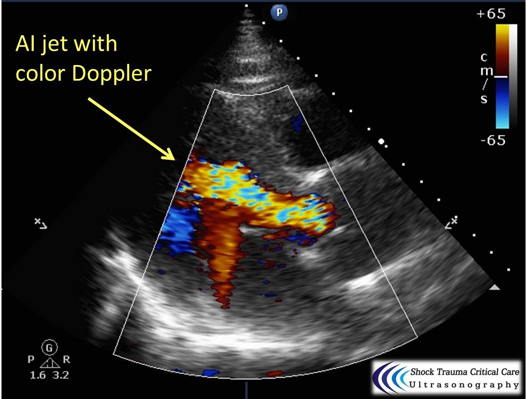
- The aortic valve causing wide-open aortic insufficiency
- One of the coronary arteries (typically the RCA presenting as inferior STEMI)
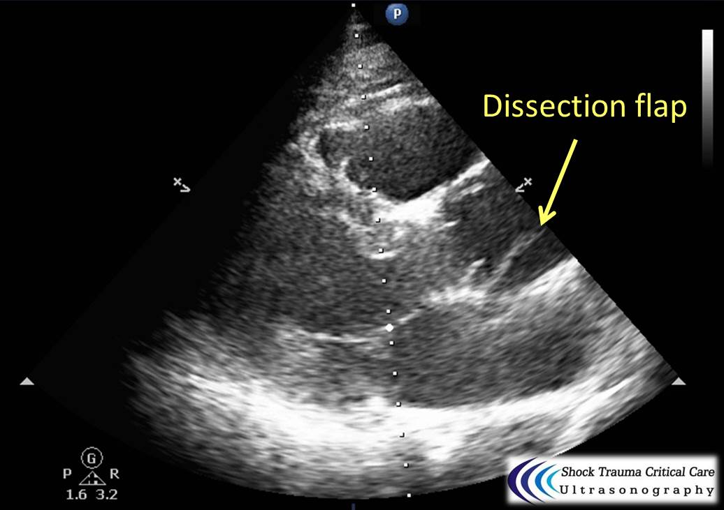
Bedside echo can't rule out aortic dissection, but it can help rule in the diagnosis (figure 1) or complications (figure 2) at times.
Attachments
- Evaluating the systolic function of the RV is an important skill and there are described methods.
- One of the simplest method is using the tricuspid annular plane of systolic excursion (or T.A.P.S.E.)
- This is how far the tricuspid annulus travels from diastole to systole because the RV contracts in a longitudinal fashion from the base (diastole) to the apex (systole)
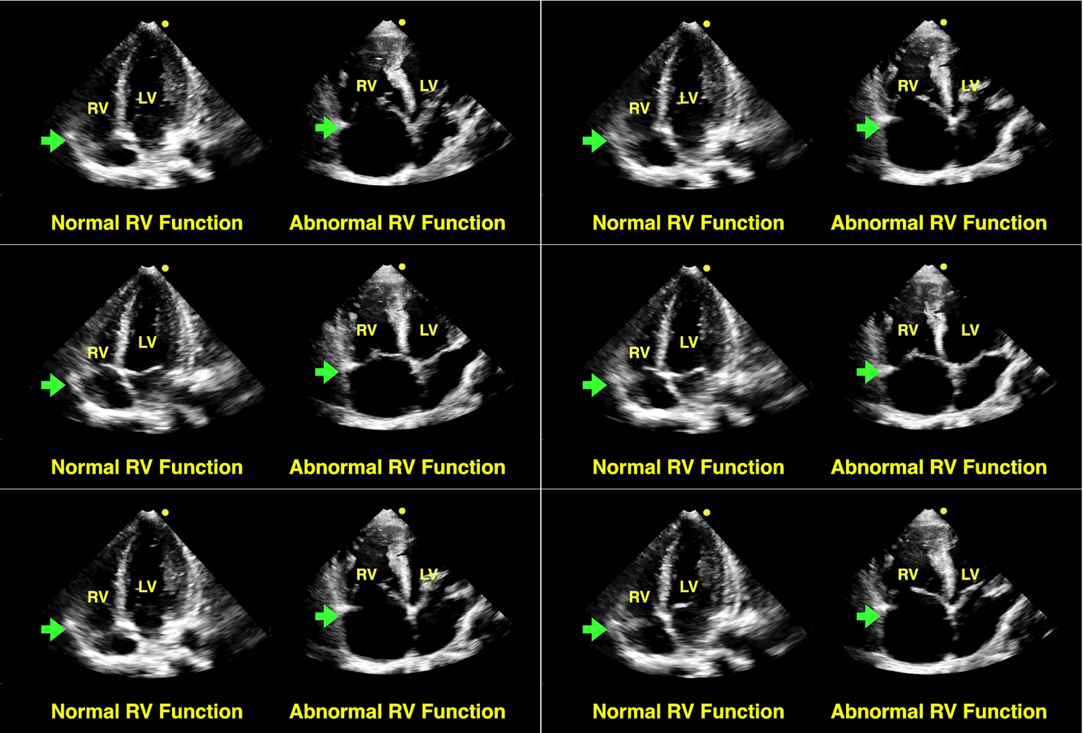
- A TAPSE of <17mm is consistent with abnormal function and >17mm is normal. An eyeball method of assessment can be done when grossly obvious or M-mode can be used when an accurate assessment is required.
- The clip below demonstrates the technique, which should always be performed from an apical four-chamber view.
- Want more info on the RV, then click here for a whole podcast on it.
Show References
SIMV (Synchronized intermittent mandatory ventilation)
- A common mode of ventilation that all pratitioners should be familiar with
- It provides a minimum number of fully assisted breaths synchronized with patient respiratory effort
- Patient or time triggered
- Flow limited
- Volume cycled
- Any additional breaths are unassisted and determined by patient effort
- SIMV=AC when heavily sedated
- The idea is exercise the patients lungs but this can lead to increased work of breathing and fatigue, and prolong extubation when used
Hyperoxia in the Critically Ill
- Oxygen is liberally administered to many critically ill patients, thereby exposing them to supranormal arterial oxygen levels.
- Hyperoxia results in the formation of reactive oxygen species, which adversely affect the pulmonary, vascular, cnetral nervous, and immune systems.
- Though the optimal PaO2 remains unknown, recent evidence indicates that hyperoxia is associated with increased mortality in post-cardiac arrest, CVA, acute coronary syndrome, and traumatic brain injury patients.
- Take Home Point: Carefully titrate oxygen to the lowest tolerable level to meet the patient's needs.
Show References
Your ESLD patient is hypotensive with a tense abdomen, and he needs a paracentesis!
--ALWAYS use ultrasound to localize a fluid pocket [Fig 1]! Take the time to use color Doppler to look for underlying abdominal wall varices [Fig 2]. Cirrhotic patients frequently have abnormal abdominal wall vasculature [1-2].
--Hemorrhage from paracentesis is exceedingly rare, and reversal of mild coagulopathy probably isn't that important [3-4].
--In hypotensive patients, consider placement of a small pigtail catheter for slow, continuous drainage (e.g. 8.3F pericardiocentesis catheter) instead of large-volume paracentesis. Non-tunneled catheter infection risk goes up after 72h [5].
--Albumin replacement improves mortality and incidence of renal failure in patients with SBP or other infection [6-7].
Show References
Attachments
- 1509011640_Figure_1_--_Ascites_pocket.jpg (78 Kb)
- 1509011640_Figure_2_--_Color_over_abd_wall_varices.jpg (89 Kb)
The RV is a low-pressure chamber that doesn’t tolerate acute increases in pulmonary pressures (e.g., ARDS, pulmonary embolism, etc.); acute increases can lead to RV dysfunction / failure
Managing RV dysfunction requires a three-pronged approach:
- Optimize preload – give small fluid boluses (e.g., 250cc) but not too much, because too much can worsen RV function. Use ultrasound to determine volume status
- Optimize RV function – Consider starting inotropes (e.g., dobutamine) for better RV contractility and concurrently start pulmonary vasodilators (e.g., inhaled nitric oxide); also minimize hypoxemia and hypercarbia
- Prevent systemic hypotension – hypotension reduces coronary perfusion that leads to RV ischemia and dysfunction; use norepinephrine to keep blood pressure >65
- Bottom-line: Don't under-estimate the importance of the RV when resuscitating your patients
Show References
Pressure Regulated Volume Control (PRVC)
Here are some basic pearls about PRVC Ventilation
- Form of Assist Control (AC) ventilation: patient initiated or ventilator intiated
- Constant pressure through inspiration
- Decelerating inspiratory flow pattern
- Ventilator adjusts pressure breath to breath based on patient’s airway resistance and compliance
- Not recommended for asthma or COPD
- Set: RR, tidal volume, upper pressure limit, oxygen level, I:E ratio (can start at 1:2), PEEP
Benefits: minimum PIP, guaranteed tidal volume, patient can trigger more breaths, improved oxygenation, breath by breath changes
Is It Really ARDS?
- Recent literature suggests that the incidence of ARDS in intubated ED patients may be as high as 10%.
- The Berlin Definition of ARDS includes the acute onset of bliateral opacities (CXR or chest CT) that is not fully explained by pulmonary edema or fluid overload.
- Emergency physicians and Intensivists are well versed in lung-protective ventilator settings for patients with ARDS.
- However, several diseases can appear simliar to ARDS and may require different ventilator strategies and treatments.
- In the absence of clinical risk factors for ARDS (e.g., sepsis, trauma), consider the following in your differential:
- Idiopathic pulmonary fibrosis
- Interstitial pneumonitis
- Granulomatosis with polyangitis (Wegener's)
- Diffuse alveolar hemorrhage
- Goodpasture's syndrome
Show References
Ever forget all the things that make up MUDPILES in your AG acidosis differential?
Instead, consider the less-complicated mnemonic "KILR"!
K Ketoacidosis (diabetic, alcoholic, starvation)
I Ingestion (salicylate, acetaminophen, methanol, ethylene glycol, CO, CN, iron, INH)
L Lactic acidosis (infection, hemorrhage, hypoperfusion, alcohol, metformin)
R Renal (uremia)
Once you rule out the KLR causes, begin to consider ingestion or a tox source as your source. Remember that many of the listed ingestions can also cause a lactic acidosis.
Show References
It's July, that means new doctors are learning to do central-lines...here's a quick video with some quick pearls on how to do that. Enjoy!
Show References
Care of Drowning Patients in the ED
- 500,000 worldwide deaths per year/10 per day in US on average
- Main goal of resuscitation is to reverse hypoxemia: endotracheal intubation, mouth-to-mouth, BVM depending on setting
- In water resuscitation can be considered (mouth-to-mouth only) while the patient is being actively rescued
- CPR with Airway emphasis- five rescue breaths, 30 compressions, then 2 breaths/30 compressions until the airway can be secured
- Turning the patient over and performing abdominal thrusts or back blows is not helpful
- ARDSnet protocol is generally used when intubated
- No steroids or prophylactic antibiotics are indicated
- Consider trauma (CT head and C-spine precautions based on history/exam)
- Warm up your patient as needed--assume hypothermia on presentation
- Can consider therapeutic hypothermia after ROSC and when rewarmed---no clear benefit here
Show References
Blood Pressure Management in Severe Preeclampsia
- Severe preeclampsia (preeclampsia + at least one severe complication) accounts for almost 40% of deaths in obstetrical ICU admissions.
- Systolic arterial hypertension is the most important predictor of morbidity in patients with severe preeclampsia.
- First-line agents to reduce blood pressure in severe preeclampsia are nicardipine and labetalol.
- Hydralazine is no longer recommended as first-line therapy.
- Magnesium is used as an anticonvulsant and should not be considered an antihypertensive.
Show References
With a new academic year starting, it is important to review some details on central lines
Complications of central lines (TLC-Triple lumen catheter)
- Pneumothorax (more common with subclavian)
- Arterial puncture (more common with femoral)
- Catheter malposition
- Subcutaneous hematoma
- Hemothorax
- Catheter related infection (historically more with femoral)
- Catheter induced thrombosis
- Arrhythmia (usually from guidewire insertion)
- Venous air embolism (avoid with Trendelenburg position)
- Bleeding
Avoiding infections: hand hygiene, chlorhexidine skin antisepsis, maximal barrier precautions, remove unnecessary lines, full gown and glove w/ mask and sterile technique.
Catheter position: 16-18cm for Right sided and 18-20 cm for Left sided. But can vary based on height, neck length, and catheter insertion site. Approximate length based on these factors.
Flow rates: Remember that putting in a central line does not necessarily improve your flow rates in resuscitation
16 G IV: 220 ml/min
Cordis/introducer sheath: 126 ml/min
18 G IV: 105 ml/min
16G distal port TLC: 69 ml/min
Ports (Can vary with type of catheter)
1. Distal exit port (16G)
2. Middle port (18G)
3. Proximal port (18G)
Arterial puncture: hold pressure for 5 mins and evaluate for hematoma formation (harder for subclavian approach)
Arterial cannulation: Has decreased due to ultrasound use but if you do cannulate an arterial site, don’t panic. Don’t remove the line. You can check a blood gas or arterial pulse waveform to confirm placement. Call vascular surgery for open removal and repair or endovascular repair. You could potentially remove a femoral arterial line and hold pressure but seek vascular advice regarding possible closure devices to use after removal.