University of Maryland School of Medicine logo
Emergency Medicine
All Departments
UMSOM Assistant
UMEM Educational Pearls
  • Home
  • Departments
  • Emergency Medicine
  • CME
  • UMEM Educational Pearls
  • CME
  • Courses and Conferences
  • Free Open Access Medical Education
  • UMEM Educational Pearls

Search

61-80 of 364 results with category "Cardiology"

Previous  |  1 |  2 |  3 |  4 |  5 |  6 |  7 |  8 |  9 |  ... |  19 |  Next

Title: Recent Negative Stress Tests in Chest Pain Bouncebacks

Category: Cardiology

Keywords: ACS, Stress Test (PubMed Search)

Posted: 2/23/2014 by Ali Farzad, MD (Updated: 3/23/2014)

Over-reliance on stress tests is a common reason for misdiagnosis or delays in diagnosis in patients with ACS.
 
The utility of a recent negative stress test is limited when it is used to determine the risk for an ACS in patients presenting to the emergency department with symptoms of cardiac ischemia. 
 
Several studies, including a meta analysis, show that while a positive stress test can be useful in determining the next appropriate step of a patient's care, a negative stress test may not be as useful.
 
ED patients who bounceback after a negative stress test, represent a much higher risk population that may be at the same risk for ACS as those without previous testing.
 
Bottom Line:
No test is capable of reliably stratifying a patient’s risk to zero. If you are concerned about an ED chest pain patient with a HPI suggestive of ACS, treat conservatively and do not be misled by a recent negative stress test.
 
Bonus:
Working in an observation unit and don't know what stress test to order? Check out Dr. Mattu's lecture Non-invasive cardiac stress testing: What every emergency physician needs to know (Need EmedHome subscription for link to work).


Show References

1.      Banerjee A, Newman DR, Van den Bruel A, Heneghan C. Diagnostic accuracy of exercise stress testing for coronary artery disease: a systematic review and meta-analysis of prospective studies. International Journal of Clinical Practice. 2012;66(5):477–492.

2.      Walker J, Galuska M, Vega D. Coronary disease in emergency department chest pain patients with recent negative stress testing. West J Emerg Med. 2010;11(4):384–388.

3.      Nerenberg RH, Shofer FS, Robey JL, Brown AM, Hollander JE. Impact of a negative prior stress test on emergency physician disposition decision in ED patients with chest pain syndromes. The American journal of emergency medicine. 2007;25(1):39–44. 

Want more emergency cardiology pearls?
Follow me on Twitter @alifarzadmd


Title: Early Atherosclerosis Detection

Category: Cardiology

Posted: 2/16/2014 by Semhar Tewelde, MD (Updated: 7/21/2026)

Early Atherosclerosis Detection

50 middle-aged asymptomatic subjects free of vascular disease underwent carotid ultrasound (CUS) for risk stratification were also invited to undergo coronary computed tomography angiography (CCTA) or coronary artery calcium score (CAC) to identify which of the 3 imaging modalities was best at identification of early atherosclerosis

Atherosclerosis was observed in 28%, 78%, and 90% of subjects using CAC, CCTA, and CUS, respectively

36 patients with a CAC score = 0, 69% and 86% had atherosclerosis on CCTA and CUS, respectively

Concordance between modalities was highly variable

CUS and CCTA detection of plaque were significantly more sensitive than CAC 

Considering the prevalence of subclinical disease on CUS and CCTA, the threshold at which to treat warrants further research

Show References

Schroeder B, Francis G, et al. Early Atherosclerosis Detection in Asymptomatic Patients: A Comparison of Carotid Ultrasound, Coronary Artery Calcium Score, and Coronary Computed Tomography Angiography. Canadian Journal of Cardiology, 2013-12-01, Volume 29, Issue 12, Pages 1687-1694


Title: New TWI in aVL

Category: Cardiology

Keywords: ECG, STEMI, aVL (PubMed Search)

Posted: 2/9/2014 by Ali Farzad, MD (Updated: 3/23/2014)

The importance of new ST-segment depressions (STD) and/or T wave inversions (TWI) in lead aVL have not been emphasized or well recognized across specialties. Computer-assisted ECG readings typically report these findings as normal or nonspecific. 

There is growing evidence that changes in lead aVL are abnormal, and that paying attention to that lead can be clinically useful. Reciprocal changes presenting as STD or TWI in lead aVL may be indicative of a significant coronary artery lesion and can sometimes be the only ECG manifestation of acute MI.  

STD in lead aVL is considered a sensitive marker for early inferior STEMI, and has been shown to help differentiate STEMI from pericarditis. Another recent retrospective study suggests that TWI in aVL might be associated with significant LAD lesions. 

Bottom Line: Paying close attention to subtle changes and abnormalities in lead aVL may help in early identification and initiation of therapy for patients who are having an acute MI.  

Show References

Hassen GW, Costea A, Smith T, et al. The Neglected Lead on Electrocardiogram: T Wave Inversion in Lead aVL, Nonspecific Finding or a Sign for Left Anterior Descending Artery Lesion?. Journal of Emergency Medicine. 2014;46(2):165–170.

Want more emergency cardiology pearls?
Follow me on Twitter @alifarzadmd

Attachments

  • 1402092205_TWI_in_aVL.pdf (112 Kb)


Title: Myocardial Infarction in Women After Childbirth

Category: Cardiology

Posted: 2/2/2014 by Semhar Tewelde, MD

Myocardial Infarction in Women After Childbirth

World Health Organization reports that obesity is the 5th leading cause of global death with the highest impact on women <65 years of age

The association of obesity and cardiovascular risk in young women is currently being researched

A recent nationwide cohort looking at obesity and future cardiovascular risk looked at Danish women giving birth (2004-2009) and followed them a median time of 4.5 years

This study grouped women via pre-pregnancy body mass index (BMI)

                                            1. Underweight (BMI <18.5)     

                                            2. Normal weight (BMI <25)

                                3. Overweight (BMI <30)

                                4. Obese (BMI >30)

Data revealed that healthy women of fertile age, pre-pregnancy obesity alone was associated with increased risk of myocardial infarction in the years after childbirth

Show References

Schmiegelow M, Andersson C, Kober L, et al. Prepregnancy Obesity and Associations With Stroke and Myocardial Infarction in Women in the Years After Childbirth. Circulation 2014;129:330-337. 


Title: Extremely Fast & Wide Complex Regular Tachycardia

Category: Cardiology

Keywords: Wide complex tachycardia, ventricular tachycardia (PubMed Search)

Posted: 1/26/2014 by Ali Farzad, MD (Updated: 3/23/2014)

Question

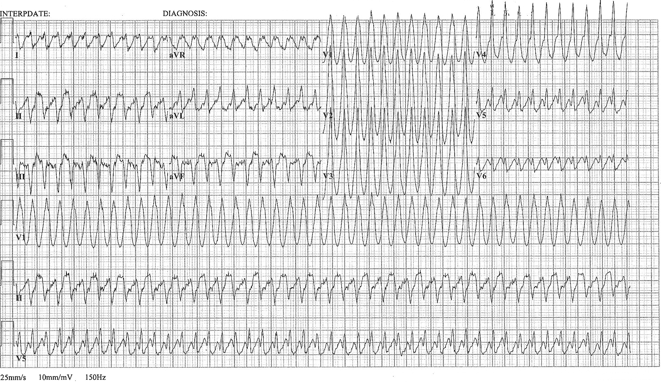
A 48 year old woman has acute chest pain and palpitations over the past several hours. She has felt similar palpitations in the past but never sought medical attention. She arrives to your ED alert and anxious. HR = 270, BP=130/100. ECG is below. What’s the diagnosis and treatment?
Show Answer

Most wide complex regular tachycardias are ventricular tachycardia (VT). However, supraventricular tachycardias can also cause wide complexes through aberrant conduction and accessory pathways.

The exact diagnosis of a tachydysrhythmia is often irrelevant in the ED management of unstable patients. Clinical stability is a more important determinant of treatment than the underlying rhythm. This patient was thought to have VT and was successfully cardioverted. 
 
Subsequent EP study confirmed Atrial Flutter with 1:1 conduction and Wolff-Parkinson-White (WPW) Syndrome.
 
Why is this important?
 
Accessory pathways can be concealed, and lack of delta waves do not rule out preexcitation syndromes such as WPW. 
 
Consider this diagnosis in patients with the triad of extremely rapid rate (>250 bpm), regular rhythm, and wide QRS complexes. 
 
This is particularly important if pharmacological treatment is chosen rather than cardioversion. Any treatment that slows AV nodal conduction may cause preferential conduction down the accessory pathway and precipitate cardiovascular collapse. 
 
Bottom-line: 
 
When treating extremely fast wide complex rhythms, avoid the ABCD Meds (Adenosine, Beta-Blockers, Ca2+ Channel Blockers, & Digoxin). Instead, consider using Procainamide or cardioversion!

Show References

Nelson JG, Zhu DW. Atrial Flutter with 1:1 Conduction in Undiagnosed Wolff-Parkinson-White Syndrome. The Journal of Emergency Medicine. January 2014. Pubmed Link

Want more emergency cardiology pearls?
Follow me on Twitter @alifarzadmd

Attachments

  • 1401260828_JEM.WCT.Jan14.pdf (1,636 Kb)


Title: Pacing Atrioventricular Block

Category: Cardiology

Posted: 1/19/2014 by Semhar Tewelde, MD

Pacing Atrioventricular Block

 - Atrioventricular (AV) block is classically treated with restoration of heart rate via right ventricular pacing, however high rates of right ventricular pacing is associated w/ left  ventricular systolic dysfunction  

- A recent multi-center randomized control trial (RCT) assessed the efficacy of right vs biventricular pacing in heart failure w/ AV block [BLOCK HF Trial]

- Primary outcomes of: morbidity, mortality, and adverse left ventricular remodeling were shown to be significantly lower in biventricular vs right ventricular pacing 

- In patients with a high rate of pacing and/or an  abnormally low left ventricular ejection fraction biventricular pacing may be more advantageous than conventional right ventricular pacing


Show References

Curtis A.B., Worley S.J., Adamson P.B.,et al: Biventricular pacing for atrioventricular block and systolic dysfunction. N Engl J Med 2013; 368: 1585-1593


Title: Origin of premature ventricular beats

Category: Cardiology

Keywords: PVC, Premature ventricular beats, Premature ventricular complexes (PubMed Search)

Posted: 1/12/2014 by Ali Farzad, MD (Updated: 3/10/2014)

Differentiation between right and left ventricular origin of premature beats can be useful clinically.

 
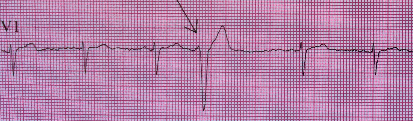
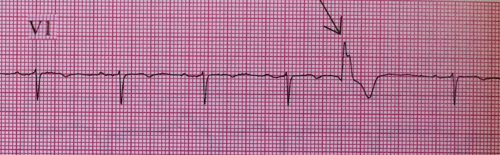
The origin of ectopic ventricular beats are recognized best in lead V1 (oriented to differentiate right vs. left sided cardiac activity).
 
  • PVCs arising from the right ventricle have a left bundle branch block morphology (dominant S wave in V1)
  • PVCs arising from the left ventricle have a right bundle branch block morphology (dominant R wave in V1)
Left Ventricular premature beats are more often associated with heart disease and may precipitate ventricular fibrillation, whereas right ventricular premature beats are commonly seen in individuals with normal hearts. 


Want more emergency cardiology pearls? Follow me @alifarzadmd

Show References

Wagner, Galen. Chapter 15 - Premature Beats. Marriott's Practical Electrocardiography, 12th Edition. 2013


Title: Assessment of Intermediate Coronary Lesions

Category: Cardiology

Posted: 1/5/2014 by Semhar Tewelde, MD

Assessment of Intermediate Coronary Lesions

- Coronary angiography alone to assess CAD is fraught with subjectivity

- Fractional flow reserve (FFR) has become the standard to assess/quantify obstructive CAD; it determines the myocardial flow in the presence of stenosis identifying the lesion responsible for ischemia

- FFR assesses focal stenosis, but does not consider diffuse atherosclerotic narrowing or microcirculatory dysfunction as contributors of ischemic heart disease

- An index of microcirculatory resistance (IMR) can be concomitantly measured with FFR during cardiac catheterization to specifically evaluate the microvasculature

- Coronary flow reserve (CFR) was the 1st proposed method for assessment of intermediate coronary lesion, but proved suboptimal because of its variability especially in patients with microvascular dysfunction (diabetes, prior MI, etc.)

- Utilization of FFR, IMR, and CFR together support the existence of differentiated patterns of ischemic heart disease & may help to determine future ischemic events 

Show References

Echavarria-Pinto M, Escaned J, Macias E, et al. Disturbed Coronary Hemodynamics in Vessels With Intermediate Stenosis Evaluated With Fractional Flow Reserve: A Combined Analysis of Epicardial and Microcirculatory Involvement in Ischemic Heart Disease. Circulation Volume 128(24), 17 December 2013, p 2557–2566


Title: How to measure ST elevation

Category: Cardiology

Keywords: ST-elevation, Cardiology, MI (PubMed Search)

Posted: 12/29/2013 by Ali Farzad, MD (Updated: 3/10/2014)

There is debate and confusion regarding where and how to measure ST elevation (STE). Do you measure the STE at the J-point? Or at 40 msec after the J-point? And how much STE is considered significant? The current guidelines have clarified this issue.

 - STE should be measured at the J-point.

- STEMI is defined by STE ≥ 1 mm in at least 2 contiguous leads, with the exception of leads V2-V3.

- STEMI is defined by STE ≥ 2 mm in leads V2-V3 in men.

- STEMI is defined by STE ≥ 1.5 mm in leads V2-V3 in women.

For more cardiology pearls from the 2013 literature , check out Amal Mattu's Articles You've Gotta Know!


Want more emergency cardiology pearls? Follow me @alifarzadmd

Show References

2013 ACCF/AHA Guideline for the Management of ST-Elevation Myocardial Infarction

O'Gara PT, Kushner FG, Ascheim DD, et al; American College of Cardiology Foundation/American Heart Association Task Force on Practice Guidelines Circulation. 2013;127:e362-e425 Pubmed Link


Title: Metoprolol Usage Cardioprotective

Category: Cardiology

Posted: 12/22/2013 by Semhar Tewelde, MD (Updated: 7/21/2026)

Metoprolol Usage Cardioprotective

  • Intravenous (IV) metoprolol is sparingly used in STEMI given concern about precipitation of cardiogenic shock (COMMIT/CCS-2 Trial)
  • A recent study (n=220) looked at usage of IV metoprolol versus controls in patients with STEMI and a killip class II or less prior to primary PCI
  • MRI was preformed 5-7 days after STEMI revealing reduced infarct size and increased left ventricular ejection fraction in the IV metoprolol group
  • IV beta-blockade appears cardioprotective in those with a low killip score and should be considered prior to primary PCI in certain subgroups  

Show References

Ibanez B, Sanchez-Brunete V, Pizarro G, et al. Effect of early metoprolol on infarct size in ST-segment-elevation myocardial infarction patients undergoing primary percutaneous coronary intervention: the Effect of Metoprolol in Cardioprotection During an Acute Myocardial Infarction (METOCARD-CNIC) trial. Circulation. 2013 Oct 1; 128(14):1495-503.


Title: The HEART score for ED patients with Chest Pain

Category: Cardiology

Keywords: ACS, Chest Pain, HEART score (PubMed Search)

Posted: 12/8/2013 by Ali Farzad, MD (Updated: 3/10/2014)

 
The diagnosis of non-STE ACS can be difficult to exclude in ED patients with chest pain. Consequently, over-diagnosis and unnecessary treatment are common. Risk stratification tools (ie. TIMI, GRACE) have been created to help risk stratify ACS patients and predict mortality. However, they are of limited utility in the ED and do not effectively differentiate low to intermediate risk patients in all-comers with chest pain.  
 
The HEART score was recently prospectively validated in an ED population and was able to quickly and reliably predict risk of major adverse cardiac events (MACE - AMI, PCI, CABG, & Death). 
  • 5 practical considerations (History, ECG, Age, Risk factors, & Troponin) are scored (0,1,or 2 points each) depending on the extent of the abnormality.
  • A HEART score (0-10) can be quickly determined without complex calculations
  • Low scores (0-3) exclude short term MACE with >98% certainty
  • High scores (7-10) have high (>50%) MACE rates
  • The HEART score performed significantly better than TIMI and GRACE scores 

Bottom-line: The HEART score can help to objectively risk stratify ED patients with chest pain into low, intermediate, and high risk groups. Using the HEART score can also facilitate more efficient and effective communication with colleagues.


Want more emergency cardiology pearls? Follow me @alifarzadmd


Show References

Backus BE, Six AJ, Kelder JC, et al. A prospective validation of the HEART score for chest pain patients at the emergency department. International Journal of Cardiology. 2013;168(3):2153–2158. 

Six AJ, Backus BE, Kelder JC. Chest pain in the emergency room: value of the HEART score. Neth Heart J. 2008;16(6):191–196.

Attachments

  • 1312081410_International_Journal_of_Cardiology_2013_Backus.pdf (371 Kb)
  • 1312081419_Neth_Heart_J_2008_Six.pdf (144 Kb)


Title: ECG Following Cardiac Transplant

Category: Cardiology

Keywords: Cardiac Transplant (PubMed Search)

Posted: 12/1/2013 by Semhar Tewelde, MD (Updated: 8/28/2014)

ECG Following Cardiac Transplant

  • Suturing of donor atria to the corresponding structures of a recipient’s residual atria produces two sets of P-waves:
    • A small native P-wave (often so small it may not been visualized)
    • Followed by a donor P-wave of normal size associated w/ a QRS complex
  • A complete or incomplete right bundle branch develops in >80% transplant recipients
  • ~7–25% of recipients also demonstrate a left anterior fascicular block (LAFB)
  • The transplanted heart contracts faster than the atrial remnant secondary to autonomic denervation frequently resulting in an increased resting heart rate 


Show References

Chou's Electrocardiography in Clinical Practice: Adult and Pediatric 6th Edition 

Attachments

  • 1312011353_TransplantECG.jpg (160 Kb)


Title: Too early to give hypothermia the cold shoulder

Category: Cardiology

Keywords: Therapeutic Hypothermia, ROSC, Cardiac Arrest, Resuscitation (PubMed Search)

Posted: 11/24/2013 by Ali Farzad, MD (Updated: 3/10/2014)

Hyperthermia after resuscitation from cardiac arrest is associated with poor outcomes and death. Induced mild hypothermia gained widespread use after two RCT's from 2002 (n=352) showed improved survival & neurological outcomes for select patients with OHCA. 
 
In a new RCT (n=939), patients with ROSC after arrest were assigned to targeted temperature management at either 33°C or 36°C. Survival (51%) and a good neurologic outcome (47 to 48%) did not differ significantly between groups. However, cooling to 36°C is not the same as not regulating temperature and allowing hyperthermia. 
 
In contrast to a decade ago, one half instead of one third of these patients can expect to survive hospitalization. Paying attention to temperature makes survival more likely than death when a patient is hospitalized after cardiac arrest. 
 

Want more emergency cardiology pearls? Follow me @alifarzadmd

Show References

Newly Published RCT & Editorial 
- Nielsen N, Wetterslev J, Cronberg T, et al. Targeted Temperature Management at 33°C versus 36°C after Cardiac Arrest. N Engl J Med. Nov 2013. 
- Rittenberger JC, Callaway CW. Temperature Management and Modern Post-Cardiac Arrest Care. N Engl J Med. Nov 2013. 
 
Original RCTs
- Hypothermia after Cardiac Arrest Study Group. Mild therapeutic hypothermia to improve the neurologic outcome after cardiac arrest. N Engl J Med. 2002. 
- Bernard SA, Gray TW, Buist MD, et al. Treatment of comatose survivors of out-of-hospital cardiac arrest with induced hypothermia. N Engl J Med. 2002. 
 
More on the deleterious effects of hyperthermia after cardiac arrest
- Gebhardt K, Guyette FX, Doshi AA, et al. Prevalence and effect of fever on outcome following resuscitation after cardiac arrest. Resuscitation. 2013;84(8):1062–1067.

Attachments

  • 1311241245_N_Engl_J_Med_2013_Nielsen.pdf (497 Kb)
  • 1311241245_N_Engl_J_Med_2013_Rittenberger.pdf (317 Kb)
  • 1311241246_N_Engl_J_Med_2002_Hypothermia_after_Cardiac_Arrest_Study_Group.pdf (172 Kb)
  • 1311241246_N_Engl_J_Med_2002_Bernard.pdf (102 Kb)
  • 1311241246_Resuscitation_2013_Gebhardt.pdf (551 Kb)


Title: Utility of Intra-Aortic Balloon Pump

Category: Cardiology

Keywords: Intra-Aortic Balloon Pump, Cardiogenic Shock (PubMed Search)

Posted: 11/17/2013 by Semhar Tewelde, MD (Updated: 11/17/2013)

Utility of Intra-Aortic Balloon Pump (IABP)

  • IABP therapy has not been proven to reduce mortality in all-comers with cardiogenic shock complicating acute myocardial infarction (IABP-SHOCK II)
  • A recent retrospective review of IABP therapy in patients with mechanical complications (ventricular septal rupture [VSR] or mitral regurgitation [MR]) following acute myocardial infarction has proven efficacious in this subset
    • IABP reduced mortality in patient with shock (61% vs 100%, p = 0.04)
    • IABP reduced preoperative mortality (11% vs 88%, p <0.001)
  • Post infarction VSR or MR with signs of cardiogenic shock should be considered for an IABP as a bridge to emergent surgical repair
  • Patients with mechanical complications without shock were not shown to benefit from an IABP and should undergo cardiac surgery after medical stabilization

Show References

Kettner J, Sramko M, Holek M, et al. Utility of Intra-Aortic Balloon Pump Support For Ventricular Septal Rupture and Acute Mitral Regurgitation Complicating Acute Myocardial Infarction. American Journal of Cardiology. 2013 Vol 112 Issue 11 Pages 1709-1713


Title: Diagnosis of STEMI in LBBB

Category: Cardiology

Keywords: AMI, LBBB, Sgarbossa criteria (PubMed Search)

Posted: 11/10/2013 by Ali Farzad, MD (Updated: 3/10/2014)

Diagnosis of STEMI in patients with LBBB can be challenging. Guidelines that previously recommended emergent reperfusion for these patients have been reconsidered to avoid inappropriate cath lab activation and fibrinolytic therapy.

The 2013 ACC/AHA STEMI guidelines no longer consider new or presumably new LBBB a STEMI equivalent. This dramatic change may prevent inappropriate therapy for some, but fail to help identify patients with LBBB who are having STEMI's. Delayed reperfusion in this population could be fatal and is estimated to affect 5,000-10,000 patients per year in the US alone.

The Sgarbossa ECG criteria are the most validated tool to aid in the diagnosis of STEMI in the presence of LBBB. A Sgarbossa score of ≥ 3 has high specificity (>98%) and positive predictive value for acute MI and angiography-confirmed coronary occlusion. The following algorithm has been recently proposed to identify the high-risk population in which reperfusion therapy would be denied by the 2013 STEMI guidelines.
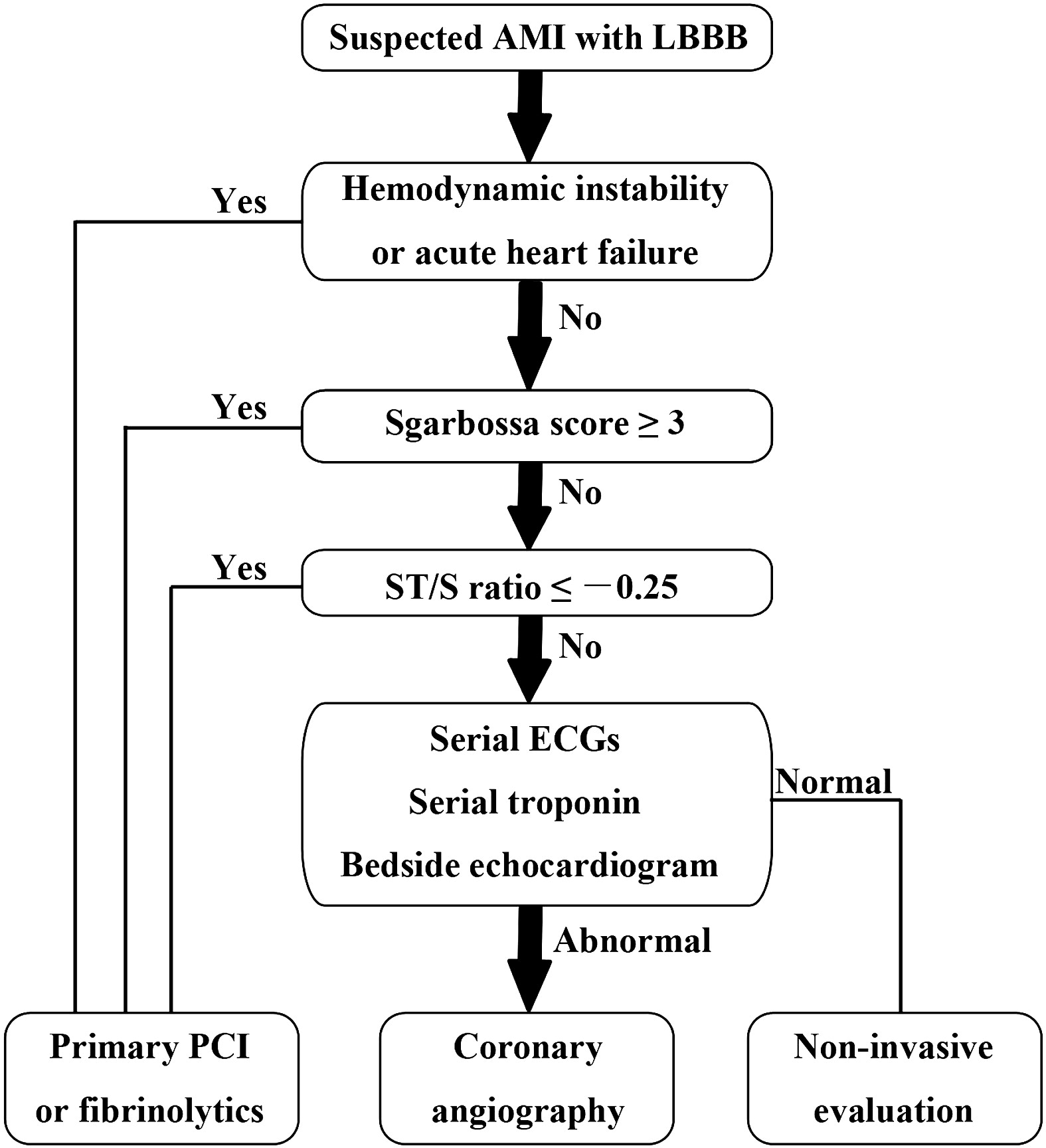
Watch this video to review Sgarbossa criteria and the modified Sgarbossa rule.


Want more emergency cardiology pearls? Follow me @alifarzadmd

Show References

The left bundle-branch block puzzle in the 2013 ST-elevation myocardial infarction guideline: From falsely declaring emergency to denying reperfusion in a high-risk population. Are the Sgarbossa Criteria ready for prime time? American Heart Journal. 2013;166(3):409–413.

Attachments

  • 1311092043_American_Heart_Journal_2013_MD.pdf (726 Kb)


Title: Secondary Prevention in Acute Myocardial Infarction (AMI)

Category: Cardiology

Posted: 11/3/2013 by Semhar Tewelde, MD (Updated: 11/16/2013)

Secondary Prevention in AMI

Just as aspirin is pivotal in the treatment of acute coronary syndrome, medications such as beta-blocker, statins, and angiotensin-converting enzyme inhibitors have been proven to be essential in secondary prevention of AMI.

Patients after AMI are typically discharged on appropriate secondary prevention medications; however the prescribed doses are often far below the proven efficacy based on clinical trials.

A review of 6,748 patients from 31 hospitals enrolled in 2 U.S. registries (2003 to 2008) illustrated that only 1 in 3 patients were prescribed these medications at goal doses.

Of patients not discharged on goal doses, up-titration during follow-up occurred infrequently ~25%.

Optimal medication dosing and appropriate titration is integral to prevention of further morbidity and mortality.

Show References

Arnold S, Spertus J, Masoudi F, et al. Beyond Medication Prescription as Performace Measure: Optimal Secondary Prevention Dosing After Acute Myocardial Infarction. JACC Nov 5, 2013 Vol 62:19;1791-1801


Title: Marked First Degree AV Block

Category: Cardiology

Keywords: AV Block (PubMed Search)

Posted: 10/27/2013 by Ali Farzad, MD (Updated: 3/10/2014)

First-degree atrioventricular (AV) block is defined as an abnormally prolonged PR-interval >200ms. Although traditionally considered to be a benign clinical entity, not all first degree AV blocks are treated the same.  

Markedly prolonged PR-intervals (PR >300ms) can cause symptoms and hemodynamic compromise due to inadequate timing of atrial and ventricular contractions. Consider the following ECG from a 32 YOF with intermittent episodes of syncope and dizziness…
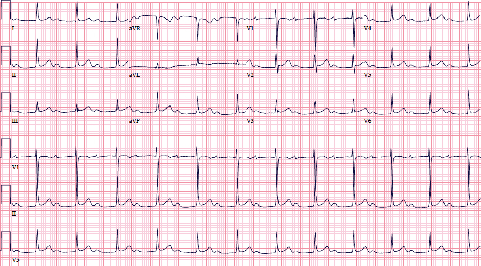
There is marked first degree AV block (PR=434 ms). When the PR-interval gets too long, AV dyssynchrony compromises ventricular filling and decreases cardiac output, similar to the so-called pacemaker syndrome.

Current ACC/AHA guidelines state that permanent pacemaker implantation is reasonable for marked first degree AV block with hemodynamic compromise or symptoms similar to those of pacemaker syndrome. (Class IIa, Level of Evidence B). The guidelines caution that pacemakers are not indicated in asymptomatic patients with isolated first degree AV block.

Want more emergency cardiology pearls? Follow me @alifarzadmd

Show References

Special thanks to Dr. Brandon Cole for submitting the ECG

References / Further Reading (Attached)

First-Degree AV Block-An Entirely Benign Finding or a Potentially Curable Cause of Cardiac Disease? Annals of Noninvasive Electrocardiology.2013;18(3):215–224. doi:10.1111/anec.12062.

ACC/AHA/HRS 2008 Guidelines for Device-Based Therapy of Cardiac Rhythm Abnormalities: A Report of the American College of Cardiology/American Heart Association Task Force on Practice Guidelines. Circulation. 2008;117(21):e350–e408. doi:10.1161/CIRCUALTIONAHA.108.189742.

Attachments

  • 1310272020_ACC:AHA_Pacemaker_Guidelines.pdf (1,524 Kb)
  • 1310272159_1st_Degree_AVB._Benign_or_Curable_Cardiac_Disease.pdf (247 Kb)


Title: Ebstein's Anomaly

Category: Cardiology

Posted: 10/20/2013 by Semhar Tewelde, MD

Ebstein's Anomaly

  • Congenital defect of the tricuspid valve (TV) and the right ventricle (RV)
  • TV septal and posterior leaflets are apically displaced resulting in "atrialization" of a portion of the right ventricle (ultimately a large right atrium and small right ventricle)
  • ~40-50% of individuals with Ebstein anomaly have evidence of Wolf-Parkinson-White, secondary to the atrialized right ventricle
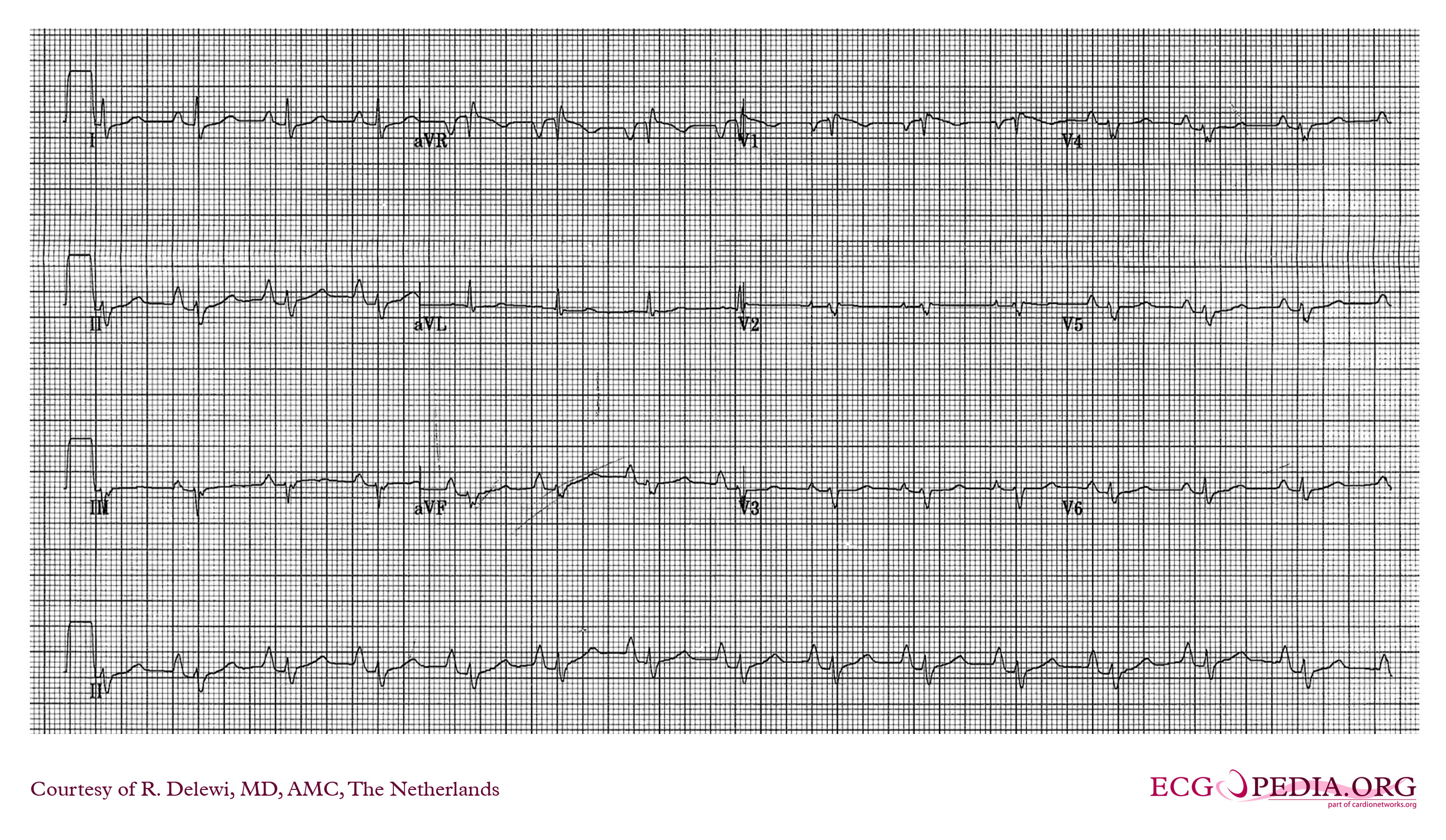
  • ECG abnormalities include:
    • Right atrial enlargement or tall and broad P waves (Himalayan P waves) 
    • Prolonged PR interval
    • Right bundle branch block 
    • Low amplitude QRS complexes in the right precordial leads
    • T wave inversions V1-V4 and/or Q waves V1-V4
Show References

Osiro S, Tiwari K, Mathenge N, et al. When Lithum Hurts: A Look at Ebstein Anomaly. Cardiology in Review. Oct 2013, Vol 21(5); pgs 257-263.


Title: What's the ECG abnormality?

Category: Cardiology

Keywords: Dyspnea, Chest Pain (PubMed Search)

Posted: 10/13/2013 by Ali Farzad, MD (Updated: 3/10/2014)

Question

A 48 year-old female presents to the ED with progressive dyspnea and chest discomfort over the past 3 months. HR = 105, BP = 100/60 mmHg, with mild JVD on exam. Her ECG is shown below. What ECG abnormalites are present? What does your differential diagnosis include? What is the best initial diagnostic test?

Show Answer

The ECG shows sinus tachycardia at rate of 107 with low QRS voltage diffusely.  Echocardiogram revealed significant pericardial effusion with signs of impending tamponade. Emergent pericardiocentesis was performed and her symptoms improved. 

Low QRS voltage (LQRSV)

  • Presence of QRS amplitudes which are < 0.5 mV (5 mm) in all of the limb leads (alternatively, voltages in leads I+II+III < 15 mm) or < 1.0 mV (10 mm) in all of the precordial leads (or, V1+V2+V3 < 30 mm)
  • Pericardial tamponade should be on top of your differential list as early diagnosis will change acute mangement and outcomes
  • Use bedside echo to quickly evaluate for significant effusions or tamponade
  • LQRSV + tachycardia = pericardial effusion until proven otherwise (electrical alternans is freqently absent)  

LQRSV Differential

Cardiac - impaired voltage generation, "low power"
  • Cardiomyopathies (i.e. ischemic, end stage dilated) 
  • Infiltrative (i.e. amyloidosis, scleroderma, myxedema, hemochromatosis)
  • Inflammatory diseases (i.e. myocarditis, constrictive pericarditis)
Extracardiac - impaired voltage transmission by fluid, fat, or air
  • Large pericardial effusion or tamponade, pleural effusions, anasarca
  • Obesity
  • COPD with hyperinflation (i.e emphyesema)
  • Pneumothorax and other forms of barotrauma (especially left-sided)

Want more emergency cardiology pearls? Follow me @alifarzadmd

Show References

Low TT et al. ECGs with small QRS voltages. Singapore Med J. 2012;53(5):299–303.
 
Madias JE. Low QRS voltage and its causes. Journal of Electrocardiology. 2008;41(6):498–500. 
 
Check out this previous ECG of the week video that demonstrates how diagnosing pericardial effusions saves lives!

Attachments

  • 1310132023_ECGs_with_small_QRS_voltages.pdf (578 Kb)
  • 1310132023_Low_QRS_voltage_and_its_causes.pdf (108 Kb)


Title: Acute Aortic Syndromes

Category: Cardiology

Keywords: Aortic Syndrome, Aortic Dissection, Intramural hematoma, Atheromatous ulcer (PubMed Search)

Posted: 10/6/2013 by Semhar Tewelde, MD

Acute Aortic Syndromes

Classically, aortic dissection is considered the primary culprit in patients with chest pain that radiates to the back (aortic pain) or chest pain combined with ischemia (cerebral, cardiac, peripheral), syncope, or cardiac arrest. However, it should not be your only concern: the rate of aortic rupture is much higher in penetrating atheromatous ulcer (42%) and intramural hematoma (35%) than in aortic dissection (types A 7.5% and type B 4.1%).

Chest pain with concomitant ischemic symptoms and acute decompensation should prompt consideration of several etiologies under the umbrella of aortic syndromes and not limited to dissection :

  1. Penetrating atheromatous ulcer - rupture of an atheromatous plaque through the internal elastic lamina, with subsequent localized medial disruption and potential dissection, pseudoaneurysm formation, or free rupture
  2. Intramural hematoma - rupture of the vasa vasorum or hemorrhage within an atherosclerotic plaque followed by aortic wall infarct
  3. Aortic dissection- an intimal tear with resultant propagation within the middle third of the medial layer of the aorta
  4. Aneurysm leak or rupture - progressive vessel dilation and increased wall tension
  5. Traumatic transection - rapid deceleration forces or direct trauma, commonly shearing distal to left subclavian artery at aortic isthmus where the aorta is fixed by ligamentum arteriosum

Show References

Sheikh A, Ali K, Mazhar S. Acute Aortic Syndrome. Circulation 2013;128:1122-1127

Coady MA, Rizzo JA, Elefteriades JA. Pathological Variants of thoracic aortic dissection. Penetrating atherosclerotic ulcers and intramural hematomas. Cardiol Clin. 1999;17:637-657


Previous  |  1 |  2 |  3 |  4 |  5 |  6 |  7 |  8 |  9 |  ... |  19 |  Next
University of Maryland School of Medicine logo
  • Departments
  • Programs
  • Research Centers
  • Institutes
  • Graduate Education
  • CIBR Cores
  • Offices of the Dean
    • University of Maryland, Baltimore
    • University of Maryland Medical Center
    • Medical Alumni Association of the University of Maryland
    • Health Sciences and Human Services Library
University of Maryland School of Medicine
655 W. Baltimore Street
Baltimore, MD 21201
Contact Us

© University of Maryland School of Medicine

    • Webmaster
    • Web Accessibility
    • Site Index
    • UMB Hotline
    • Employment
    • Map