
Search
Subscribe to daily UMEM Pearls (FREE!) via:
Question
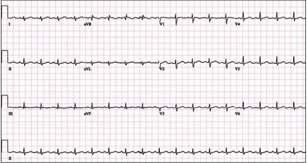
A 48 year-old female presents to the ED with progressive dyspnea and chest discomfort over the past 3 months. HR = 105, BP = 100/60 mmHg, with mild JVD on exam. Her ECG is shown below. What ECG abnormalites are present? What does your differential diagnosis include? What is the best initial diagnostic test?
Answer
The ECG shows sinus tachycardia at rate of 107 with low QRS voltage diffusely. Echocardiogram revealed significant pericardial effusion with signs of impending tamponade. Emergent pericardiocentesis was performed and her symptoms improved.
Low QRS voltage (LQRSV)
- Presence of QRS amplitudes which are < 0.5 mV (5 mm) in all of the limb leads (alternatively, voltages in leads I+II+III < 15 mm) or < 1.0 mV (10 mm) in all of the precordial leads (or, V1+V2+V3 < 30 mm)
- Pericardial tamponade should be on top of your differential list as early diagnosis will change acute mangement and outcomes
- Use bedside echo to quickly evaluate for significant effusions or tamponade
- LQRSV + tachycardia = pericardial effusion until proven otherwise (electrical alternans is freqently absent)
LQRSV Differential
Cardiac - impaired voltage generation, "low power"
- Cardiomyopathies (i.e. ischemic, end stage dilated)
- Infiltrative (i.e. amyloidosis, scleroderma, myxedema, hemochromatosis)
- Inflammatory diseases (i.e. myocarditis, constrictive pericarditis)
Extracardiac - impaired voltage transmission by fluid, fat, or air
- Large pericardial effusion or tamponade, pleural effusions, anasarca
- Obesity
- COPD with hyperinflation (i.e emphyesema)
- Pneumothorax and other forms of barotrauma (especially left-sided)
Want more emergency cardiology pearls? Follow me @alifarzadmd
References
Low TT et al. ECGs with small QRS voltages. Singapore Med J. 2012;53(5):299–303.
Madias JE. Low QRS voltage and its causes. Journal of Electrocardiology. 2008;41(6):498–500.
Check out this previous ECG of the week video that demonstrates how diagnosing pericardial effusions saves lives!
Attachments
- 1310132023_ECGs_with_small_QRS_voltages.pdf (578 Kb)
- 1310132023_Low_QRS_voltage_and_its_causes.pdf (108 Kb)