University of Maryland School of Medicine logo
Emergency Medicine
All Departments
UMSOM Assistant
UMEM Educational Pearls
  • Home
  • Departments
  • Emergency Medicine
  • CME
  • UMEM Educational Pearls
  • CME
  • Courses and Conferences
  • Free Open Access Medical Education
  • UMEM Educational Pearls

Search

281-300 of 321 results by Haney Mallemat

Previous  |  1 |  ... |  10 |  11 |  12 |  13 |  14 |  15 |  16 |  17 |  Next

Title: What's the Diagnosis? Images submitted by Dr. Joy Kay

Category: Visual Diagnosis

Posted: 8/8/2011 by Haney Mallemat, MD (Updated: 8/8/2011)

Question

13 year-old right-hand dominant male following assault with blunt object. Diagnosis?


Show Answer

Answer: Monteggia fracture


Monteggia Fracture

  • Ulnar fracture with dislocation of proximal radioulnar joint (often subtle); do not confuse with Galeazzi fracture (radial-shaft fracture with distal radioulnar dislocation)
  • Due to fall on outstretched hand with arm in hyper-pronation or with direct trauma to forearm as in defensive wounds (e.g., nightstick injury).
  • Ulnar fracture typically the proximal third (although any portion can be involved).
  • Relatively uncommon; 1-2% forearm fractures
  • Interosseous membrane (between radius and ulna) transmits forces to radioulnar joints and causes associated dislocations.
  • Suspected forearm fractures should always include Xray of the wrist, forearm, and elbow  
  • Bado classification system (Type I-IV) used; based on displacement of radial head.
  • Children may be treated with closed reduction and immobilization; adults usually require open reduction and internal fixation.
  • Radial head dislocations should be reduced within 6-8 hours as can lead to articular damage and/or nerve injury.
  • Radial, ulnar and/or median nerve neuropraxias (motor or sensory deficits) may complicate injury with resolution over several weeks.

Show References

Bruce H.E., Harvey J.P., Wilson J.C. Monteggia Fractures. J Bone Joint Surg Am. 1974;56:1563.

Reckling F.W. Unstable fracture-dislocation of the forearm (Monteggia and Galeazzi lesions). J Bone Joint Surg Am. 1982;64:857.

http://emedicine.medscape.com/article/1231438-overview
 

Follow me on Twitter (@criticalcarenow) or Google+ (+haney mallemat)


Title: Pregnancy Pearls in Trauma

Category: Critical Care

Keywords: trauma, resuscitaiton, pregnancy, IVC, supine hypoventilation, edema, intubation, RSI, desaturaiton (PubMed Search)

Posted: 8/2/2011 by Haney Mallemat, MD

Pregnancy causes many physiologic changes, which may be challenging during trauma resuscitations. A few pearls on the ABC’s:

Airway

  • Increased progesterone levels cause mucosal hyperemia and edema, increasing risk of bleeding and smaller (i.e., edematous) airway.
  • PEARL: Have smaller tubes ready and let the most experienced person intubate.

Breathing

  • The enlarging uterus pushes the diaphragms into the thorax, reducing the total lung capacity and the functional residual capacity.
  • PEARL: During intubation, patients in late pregnancy may have less oxygenation reserve and apnea time, desaturating faster during RSI.

Circulation

  • The late stage uterus can compress the IVC when supine, reducing venous return to the heart (i.e., the Supine-Hypotension syndrome) subsequently reducing cardiac output.
  • PEARL: Have a 30-degree wedge placed under patient's right hip, moving the uterus off IVC and improving venous return.
  • BONUS PEARL: During resuscitation, ask medical students to manually move the uterus midline, relieving the compressed IVC. They will appreciate that you got them clinically involved.

Show References

Chesnutt, A. Physiology of normal pregnancy. Crit Care Clinics 2004 Oct;20(4):609-15.

Follow me on Twitter (@criticalcarenow) or Google+ (+haney mallemat)


Title: What's the Daignosis?

Category: Visual Diagnosis

Posted: 7/25/2011 by Haney Mallemat, MD (Updated: 8/28/2014)

Question

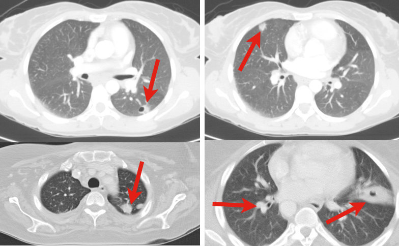
34 y.o. male with history of IVDA (intravenous drug abuse) complains of fever, chills and cough. Diagnosis?


Show Answer
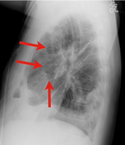
Answer: Lung Abscess (from septic pulmonary emboli)

Lung Abscess

  • Necrosis of lung parenchyma with pus and debris-filled cavities

  • Caused by direct injury (e.g., aspiration pneumonia) or secondary causes (e.g., tricuspid  endocarditis, bacteremia, etc.)

  • Suspect with:

    • Loss of airway reflexes (e.g., CVA, seizures, alcohol / narcotic abuse, etc)

    • Poor dentition

    • Immunosuppression

    • IVDA

  • Gram positives, negatives and anaerobic bacteria have all been implicated.

  • CXR may suggest diagnosis, but CT scan better identifies abscess, necrotic tissue, empyema, or other pathology (see image below).

  • After drawing blood cultures, broad-spectrum antibiotics should be started and narrowed once culture data is available; address underlying cause (e.g., valve replacement for endocarditis).

  • Prognosis is generally good with normal immune function and antibiotics, but mortality sharply increases with immunocompromise and treatment delay.

Show References

Mansharamani N, et al. Lung abscess in adults: clinical comparison of immunocompromised to non-immunocompromised patients. Respiratory Medicine. Mar 2002;96(3):178-85

Follow me on Twitter (@criticalcarenow) or Google+ (+haney mallemat)


Title: Heat Stroke? Time to Chill.

Category: Critical Care

Keywords: heat stroke, critical care, acute kidney injury, seizures, neurological (PubMed Search)

Posted: 7/19/2011 by Haney Mallemat, MD

Heat stroke is hyperthermia (>41.6 Celsius / 106 Fahrenheit) plus neurologic findings (e.g., altered mental status, seizures, coma, etc.); it also causes systemic inflammation response syndrome (i.e., cytokine release), coagulation disorders (e.g., thrombosis in end organs) and tissue abnormalities (e.g., acute kidney injury and rhabdomyolysis)

Two classifications exist:

  • Exertional heatstroke (young people engaged in strenuous physical activities in hot climates)
  • Non-exertional heatstroke occurring in sedentary people (elderly, debilitated, or chronically-ill patients) who are unprotected from the elements (e.g., trapped in apartments during heat waves)

Treatment includes:

  • Insertion of a continuous core thermometer
  • Supporting ABC’s
  • Cooling by at least to 0.2 degrees celsius per minute to 39 degrees (to avoid overshoot)
  • Benzodiazepines for sedation, shivering, and seizures
  • Antipyretics and phenytoin have not been shown beneficial
  • Support and protect end-organs with particular attention to kidneys; increased risk of kidney injury from rhabdomyolysis, ischemia and systemic inflammation.

Despite the most aggressive therapy, up to 30% survivors may have permanent neurologic or multi-organ system dysfunction months to years after recovery

Show References

Leon, L. Heat stroke: role of the systemic inflammatory response. Journal of Applied Physiology 2010 Dec;109(6):1980-8

http://emedicine.medscape.com/article/166320-overview

Follow me on Twitter: @criticalcarenow or Google +: @haney mallemat


Title: What's the Diagnosis? Written by Dr. Ari Kestler

Category: Visual Diagnosis

Posted: 7/11/2011 by Haney Mallemat, MD (Updated: 8/28/2014)

Question

48 year old male following 15 foot fall onto both feet. What is the diagnosis?
…and why is it called the “Lover’s Fracture”?
 

Show Answer
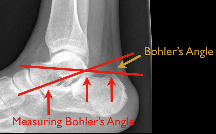
Answer: Calcaneus fracture; historically called the “Lover’s Fracture” for “lovers” jumping out of bedroom windows to evade suspicious spouses and landing directly on their feet.

Calcaneus fractures

  • Most commonly fractured tarsal bone
  • 2 types:
    • Extra-articular fracture from direct blow, twisting force and repetitive forces (causing stress fractures)
    • Intra-articular fracture from axial loading secondary to fall >6 feet, motor vehicle crash, etc; this is the classic “lover’s fracture”
  • 10% of axial loaded intra-articular fractures associated with:
    • Bilateral calcaneus fractures and/or,
    • Thoracic or lumbar compression fractures and/or,
    • Proximal femur or tibial plateau fractures
  • Ankle Xray is diagnostic for fractures and to measure Bohler’s angle (angle formed by intersection of lines connecting apex of anterior process with the apex of posterior facets and apex of posterior facet with the posterior tuberosity; see figure below)
    • Normally 20-40 degrees; <20 degrees increases suspicion for intra-articular fracture
  • Ankle CT in select cases; Xray may underestimate some injuries
  • Typically, extra-articular fractures treated with closed reduction and casting, while intra-articular fractures by open reduction and internal fixation (closed reduction in select cases)

Show References

Rosen's Emergency Medicine: Online Edition

Follow me on Twitter @criticalcarenow


Title: Amiodarone and Thyroid Disease

Category: Airway Management

Keywords: thyroid, hyperthyroid, hypothyroid, amiodarone (PubMed Search)

Posted: 7/5/2011 by Haney Mallemat, MD

Amiodarone is a class III anti-arrhythmic for tachyarrhythmias

Although most patients remain euthyroid on amiodarone, 4-18% develop thyroid disease months to years after exposure.

Amiodarone-induced thyroid disease occurs because amiodarone is structurally similar to triiodothyronine and thyroxine and each 200mg tablet contains 75 mg of iodine.

Two types of amiodarone-induced thyroid disease:

  • Amiodarone-induced hypothyroidism (AIH)
  • Amiodarone-induced thyrotoxicosis (AIT)

Amiodarone-induced hypothyroidism (AIH)

  • Presents with subtle to overt hypothyroidism 
  • Treat by discontinuing amiodarone; thyroid recovers within 3 months
  • If amiodarone cannot be discontinued, start levothyroxine

Amiodarone-induced thyrotoxicosis (AIT)

  • Sudden symptom onset months to years following exposure; mean 2-47 months post-exposure
  • Can be a life-threatening presentation (similar to thyroid storm) with severe cardiac manifestations and hemodynamic instability
  • Treatment (treat like thyroid storm, if severe)
    • Discontinue drug, if possible
    • Thionamides (inhibit enzyme producing thyroid hormones)
    • Methimazole or propylthiouracil
    • Beta-blockers
    • Steroids
    • Airway and hemodynamic support

Show References

Padmanabhan H. Amiodarone and Thyroid Dysfunction. South Med J. 2010 Sep; 103 (9): 922-30

Follow me on Twitter @criticalcarenow


Title: What's the diagnosis? Written by Dr. Ari Kestler

Category: Visual Diagnosis

Posted: 6/27/2011 by Haney Mallemat, MD

Question

49 y.o. female on Trimethoprim/sulfamethoxazole presents with rash and oral mucus membrane lesions. Diagnosis?

Show Answer

Answer: Steven-Johnson Syndrome

  • Malaise and fever prodrome, followed by erythematous or purpuric macules and plaques
    • <10% body-surface area (BSA) and mucosal membrane involvement (e.g., ocular, oral, genital)
    • Toxic Epidermal Necrolysis defined as >30% BSA involvement
  • Immune-complex hypersensitivity reaction from viral, bacterial, chemical, or drug exposure.
    • Common medications:
      • Antibiotics; Sulfonamides > PCN > Cephalosporins
      • NSAIDs
      • Anti-psychotics
  • Management:
    • Remove possible trigger(s)
    • Treat like burns; supportive care (hemodynamic support, electrolyte management, analgesia)
    • Strongly consider Burn ICU.

Show References

French LE. Toxic epidermal necrolysis and Stevens Johnson syndrome: our current understanding. Allergol Int. Mar 2006;55(1):9-16

Schöpf E. Toxic epidermal necrolysis and Stevens-Johnson syndrome. An epidemiologic study from West Germany. Arch Dermatol. 1991;127(6):839.

Roujeau JC. Severe adverse cutaneous reactions to drugs. N Engl J Med. 1994;331(19):1272.


Follow me on Twitter @criticalcarenow


Title: Cancer and Acute Kidney Injury (AKI)

Category: Critical Care

Keywords: AKI, critical care, ICU, cancer, renal failure, acute kidney injury (PubMed Search)

Posted: 6/21/2011 by Haney Mallemat, MD

Cancer patients admitted to ICUs with AKI or who develop AKI during their ICU stay have increased risk of morbidity and mortality. AKI in cancer patients is typically multi-factorial:

Causes indirectly related to malignancy

  • Septic, cardiogenic, or hypovolemic shock (most common)

  • Nephrotoxins:

    • Aminoglycosides

    • Contrast-induced nephropathy

    • Chemotherapy 

  • Hemolytic-Uremic Syndrome

Causes directly related to malignancy

  • Tumor-lysis syndrome

  • Disseminated Intravascular Coagulation

  • Obstruction of urinary tract by malignancy

  • Multiple Myeloma of the kidney

  • Hypercalcemia

Because AKI increases the already elevated morbidity and mortality in these patients, prevention (e.g., using low-osmolar IV contrast, avoiding nephrotoxins), early identification (e.g., strict attention to urine output and renal function), and aggressive treatment (e.g., early initiation of renal replacement therapy) is essential.

Show References

Benoit D. Acute kidney injury in critically ill patients with cancer. Critical Care Clinics 2010 Jan; 26(1): 151-79

Follow me on Twitter @Criticalcarenow


Title: What's the Diagnosis? Written by Dr. Katherine Baugher

Category: Visual Diagnosis

Posted: 6/13/2011 by Haney Mallemat, MD (Updated: 6/13/2011)

Question

13 y.o. with shoulder trauma (during basketball game). Arm held in adduction and exquisite scapular tenderness. Diagnosis?

Show Answer

Answer: Scapular Fracture


Scapular Fracture

  • Uncommon (1% of all fractures)

  • Extensive force required; high probability of associated injuries (pneumothorax, shoulder dislocation).

  • 5 types: body/spine, acromion, neck, glenoid, and coracoid

  • Arm held in adduction and pain with shoulder movement; may mimic rotator cuff tear.

  • Obtain X-rays (AP shoulder and lateral scapula) or CT (if displaced).

  • Conservative management for non-displaced fractures; Orthopedic reduction for displaced fractures.

  • Complications include post-traumatic arthritis or bursitis.

  • NSAIDs are first-line analgesics.

Show References

Rosen P, Barkin R. Emergency Medicine: Concepts and Clinical Practice. Mosby Year Book; 2010:573-574.

Zlowodzki M, Bhandari M, Zelle BA, Kregor PJ, Cole PA. Treatment of scapula fractures: systematic review of 520 fractures in 22 case series. J Orthop Trauma. Mar 2006;20(3):230-3.


Title: Controlling uremic bleeding

Category: Critical Care

Keywords: uremia, bleeding, ddavp, estrogens, epogen, cryoprecipitate (PubMed Search)

Posted: 6/7/2011 by Haney Mallemat, MD (Updated: 6/7/2011)

Bleeding associated with uremia is a spectrum, from mild cases (e.g., bruising or prolonged bleeding from venipuncture) to life-threatening (e.g., GI or intracranial bleed). The exact pathologic mechanisms are not understood, but are likely multi-factorial (e.g., dysfunctional von Willebrand’s Factor (vWF) and factor VIII, increased NO, etc.)

Besides dialysis, treatments for uremic bleeding include:

  1. DDAVP (fastest)
    1. 0.3-0.4 micrograms/kg IV or SC
    2. Increases vWF and factor VIII release
    3. Advantages: Begins < 1 hour
    4. Disadvantages: Tachyphylaxis; Stored factors deplete
  2. Cryoprecipitate
    1. Replaces fibrinogen, vWF, and factor VIII
    2. Advantages: Works 1-4 hours
    3. Disadvantages: transfusion reactions, infections, pulmonary edema, etc.
  3. Conjugated Estrogens
    1. Unclear mechanism; possibly increases ADP and thromboxane activity
    2. 0.6 mg/kg once daily x 5 days
    3. Advantages: Short and long-term effects
    4. Disadvantages: Hot flashes (males too!)
  4. Recombinant Erythropoietin (slowest)
    1. 40-150 U/kg three times weekly
    2. Multiple mechanisms
    3. Advantages: Helps anemia (common in renal failure) as well as bleeding complications.
    4. Disadvantages: Up to 7 days to observe effects

Show References

Hedges, SJ. Evidence-based treatment recommendations for uremic bleeding.NatClinPractNephrol.2007 Mar;3(3):138-53.


Follow me on Twitter: @criticalcarenow


Title: What's the diagnosis?

Category: Visual Diagnosis

Posted: 5/30/2011 by Haney Mallemat, MD

Question

13 y.o. female with ankle pain following fall down escalator. What's the diagnosis? (Hint: Look very closely)

Show Answer

Answer: Tri-plane ankle fracture

Tri-plane Fractures (Submitted and written by Dr. Michael Santiago)

  • Multi-planar ankle fracture in older children and adolescents during an 18 month window prior to distal tibial-physis closure.
  • 3 planes (see Xray below): coronal (A: tibal metaphysis), sagital (B: epiphysis), and transverse (C: growth plate)
  • Associated fibular spiral fracture (50% cases)
  • Commonly due to external rotational or "twisting" forces.
  • Xrays are helpfully, but CT scan may be indicated as fractures may be more displaced than radiographs suggest.
  • Non-operative treatment for non-displaced fractures:
    • Closed reduction with long-leg splint/cast
    • Reduction may slip once swelling has subsided
  • Indications for operative repair:
    • >2mm displacement of fracture segments
    • Intra-articular fracture


Title: Typhlitis

Category: Critical Care

Keywords: neutropenia, sepsis, abdominal pain, necrotizing enterocolitis (PubMed Search)

Posted: 5/24/2011 by Haney Mallemat, MD (Updated: 5/24/2011)

  • Necrotizing enterocolitis with predilection for cecum.
  • Occurs in the immunosuppressed, especially when neutropenic (<500 PMNs)
  • Typically a polymicrobial infection; gram positive cocci, gram negative rods, anaerobes, and/or fungal. 
  • Classically, right lower quadrant pain but can present with diffuse abdominal pain and peritoneal signs.
  • CT scan with IV and PO contrast is diagnostic (see below)
  • Treatment:
    • Culture and begin broad spectrum antibiotics (cover anaerobes) and antifungals (if suspected) 
    • Aggressive resuscitation
    • Surgical consult for GI perforation or clinical deterioration
  • High mortality (40-50%)

TIP: Suspect when abdominal pain presents 10-14 after chemotherapy (when PMNs are lowest).

Show References

Blijlevens NM, et al. Mucosal barrier injury: biology, pathology, clinical counterparts and consequences of intensive treatment for haematological malignancy: an overview. Bone Marrow Transplant 2000 Jun;25(12):1269-78

http://emedicine.medscape.com/article/375779-overview


Title: What's the Diagnosis?

Category: Visual Diagnosis

Posted: 5/23/2011 by Haney Mallemat, MD

Question

50 yo female s/p motor vehicle crash. Diagnosis?

Show Answer

Answer: Pneumomediastinum from pneumothorax


Pneumomediastinum


Mediastinal air has multiple causes:

  • Intrathoracic injury (e.g., Pneumothorax)
  • Upper respiratory tract (e.g., Injury during intubation) 
  • Digestive tract perforation (e.g., Boerhaave’s syndrome)
  • Intra-abdominal perforation (e.g., EGD complication)
  • Anaerobic infection at sites communicating with mediastinum
    • Ludwig’s angina
    • Pneumoperitoneum
    • Bacterial mediastinitis

Listen for Hamman's sign ("crunching" with cardiac auscultation) and feel for crepitus (distinct feeling on palpation).

Treat the precipitating cause.

Usually resolves without mediastinal decompression; air travels along tissue planes decompressing increased mediastinal pressure.


Title: Treating Clostriudium difficile in the critically-ill

Category: Critical Care

Keywords: Clostridium difficile, diarrhea, critical, ICU, sepsis, abdominal pain, vanocmycin,metronidazole, fidaxmicin (PubMed Search)

Posted: 5/10/2011 by Haney Mallemat, MD

Although oral metronidazole is indicated for mild to moderate Clostridium difficile associated diarrhea, oral vancomycin should be considered first-line therapy in critically-ill patients with moderate to severe disease. Vancomycin dosing should begin at 125mg PO q6 and increased to 250mg q6 if poor enteral absorption exists. Consider adding metronidazole IV if either reduced enteral absorption or severe disease exists. 

Recently, fidaxomicin has been shown to be non-inferior to oral vancomycin in the treatment of mild to moderate C. difficile. While promising, the study population was not critically-ill and extrapolation should be avoided.

Show References

Riddle, D. Clostridium difficile infection in the intensive care unit. Infect Dis Clin North Am. 2009 Sep;23(3):727-43.


Title: What is the Diagnosis?

Category: Visual Diagnosis

Posted: 5/9/2011 by Haney Mallemat, MD

Question

70 yo female from nursing home with fever. RUQ ultrasound is shown below. Diagnosis?

Show Answer

Acalculous Cholecystitis

Acalculous cholecystitis is due to gallbladder (GB) wall ischemia and GB stasis (reduced GB contraction from reduced PO intake)

which all leads to “sludging” (Figure 1; A), thickened GB walls (B), and pericholecystic fluid (C).


Risk factors include critical illness (especially sepsis), acute cholecystitis, total parenteral nutrition, diabetes, and GB dysmotility.


Treatment includes antibiotics covering enteric / biliary pathogens and cholecystectomy (if a surgical candidate) or percutaneous

cholecystostomy if unsuitable for the OR. 

Complications include perforation, GB gangrene, and extrabiliary abscess.


Title: Are Two Drugs Better Than One?

Category: Critical Care

Keywords: sepsis, shock, antimicrobials, combination, antibiotics (PubMed Search)

Posted: 4/26/2011 by Haney Mallemat, MD

A mortality benefit from combination antimicrobial therapy has not been clearly demonstrated in sepsis. However, when only the most severely-ill patients (i.e., septic shock) are considered in subgroup analysis, there appears to be a mortality benefit to using two antimicrobials against a suspected organism.

Combination antimicrobial therapy may reduce mortality through three mechanisms.

  1. Increased probability that the causative organism will respond to at least one drug. 
  2. Preventing emergence of antimicrobial resistance.
  3. Two antimicrobials may act synergistically.

Always obtain appropriate cultures before initiating therapy. Although identification and susceptibility of the organism may take some time, eventually narrowing antimicrobial therapy to monotherapy in the ICU is still recommended. 

Show References

Abad, C. Antimicrobial Therapy of Sepsis and Septic Shock: When are Two Drugs Better Than One? Crit Care Clinic 27 (2011) e1-e27.


Title: What's the Diagnosis?

Category: Visual Diagnosis

Posted: 4/25/2011 by Haney Mallemat, MD (Updated: 7/21/2026)

Question

Patient presents with the following X ray after yawning. Diagnosis?

Show Answer

Diagnosis: Mandibular Dislocation

The link below demonstrates an interesting alternative to the traditional reduction method; prospective randomized trails are pending.

http://www.youtube.com/watch?v=Kp8AzHIC0hM

Show References

Thanks to Dr. George Kochman for the image and Dr. Christopher Doty (EM Program Director at SUNY Downstate / Kings County Hospital Center) for the video.


Title: Vancomycin Alternatives

Category: Critical Care

Keywords: Vancomycin, Daptomycin, Linezolid, MRSA, gram positive, infections, sepsis, pneumonia (PubMed Search)

Posted: 4/12/2011 by Haney Mallemat, MD

Vancomycin is often started empirically for gram-positive and MRSA coverage. Although effective and generally well-tolerated, emerging resistance and side-effect profiles limit its use in some patients. Two alternatives are Linezolid and Daptomycin.


Linezolid

  • 600 mg IV every 12 hours
  • No renal dosing
  • Better lung penetration in pneumonia (compared to Vancomycin)
  • Side effects: Serotonin Syndrome (w/ concurrent MAOIs), hypersensitivity reaction, and myelosuppresssion


Daptomycin

  • 4 mg/kg IV once daily (skin/subcutaneous tissues infection), 6 mg/kg IV once daily (bacteremia or endocarditis), or 6-8mg/kg IV once daily (bacteremia with intravascular line)
  • Renally dosed by altering administration frequency; no change in dose.
  • NEVER use for pneumonia; pulmonary surfactant binds and inactivates drug.
  • Side effects: Reversible rhabdomyolysis (requires weekly CPK levels)

Show References

Alder, J. The Use of Daptomycin for Staphylococcus Aureus Infection in the Critical Care Medicine. Crit Care Clin 24(2008); 349-363.


Title: What's the diagnosis? Written by John Greenwood, MD

Category: Visual Diagnosis

Posted: 4/11/2011 by Haney Mallemat, MD (Updated: 4/11/2011)

Question

60 y/o male transferred from local rehab facility c/o abdominal pain.


Show Answer

Ogilvie's syndrome / Acute colonic pseudo-obstruction (ACPO)

Syndrome of decreased GI motility believed to be an autonomic imbalance (specifically, parasympathetic suppression) predominantly in the cecum and right colon. ACPO is also commonly due to narcotic overuse / abuse. 

Differential diagnosis:

  • Mechanical large bowel obstruction
  • Toxic megacolon / colitis
  • Ileus 

Abdominal XR:

  • Gaseous distention without fluid levels (UNLIKE obstruction)
  • Small bowel dilation is typically absent (UNLIKE ileus)
  • Preserved haustral markings and smooth inner contour (UNLIKE colitis)

Treatment:

  • Treat underlying cause (e.g., discontinue narcotics) 
  • Neostigmine
  • Therapeutic Colonoscopy
  • Surgical decompression (if severe)

Show References

Fazel A, Verne GN.  New solutions to an old problem: Acute colonic pseudo-obstruction. JClin Gastroenterol 2005; Vol 39(1): 17-20.

Ozkurt H, Yilmaz F, et al.  Acute colonic pseudo-obstruction (Ogilvie's syndrome): radiologic diagnosis and medical treatment with neostigmine.  Report of 4 cases.  Am J of Emer Med 2009; Vol 27: 757.e1 - 757e4.


Title: Non-invasive Ventilation (NIV): What s the Evidence?

Category: Critical Care

Keywords: bilevel ventilation, bipap, cpap, respiratory failure, respiratory distress, copd, acute pulmonary edema (PubMed Search)

Posted: 3/29/2011 by Haney Mallemat, MD

Emergency Medicine physicians are gaining experience with non-invasive ventilation (i.e., Bi-level ventilation and continuous positive-pressure ventilation) in managing respiratory distress and failure. Although NIV is commonly used across a variety of pathologies, the best data exists for use with COPD exacerbation and cardiogenic pulmonary edema (CHF, not an acute MI) 


Although other indications for NIV have been studied, the data is less robust (eg., smaller study size, weak control groups, etc.). If there are no contraindications, however, many experts still support a trial of NIV in the following populations:

  • Asthma
  • Severe community acquired pneumonia
  • Acute lung injury / Acute Respiratory Distress Syndrome
  • Chest trauma (lung contusion, rib fractures, flail chest,etc)
  • Immunosuppression with acute respiratory failure
  • Neuromuscular respiratory failure (eg., Myesthenia Gravis)
  • Cystic Fibrosis
  • Pneumocystis Jiroveci Pneumonia
  • “Do not intubate” status


Failure to clinically improve during a NIV trial should prompt invasive mechanical ventilation.

Show References

Keenan, S. et al. Clinical practice guidelines for the use of noninvasive positive-pressure ventilation and noninvasive continuous positive airway pressure in the acute care setting. CMAJ. 2011 Feb 22;183(3):E195-214. Epub 2011 Feb 14.


Previous  |  1 |  ... |  10 |  11 |  12 |  13 |  14 |  15 |  16 |  17 |  Next
University of Maryland School of Medicine logo
  • Departments
  • Programs
  • Research Centers
  • Institutes
  • Graduate Education
  • CIBR Cores
  • Offices of the Dean
    • University of Maryland, Baltimore
    • University of Maryland Medical Center
    • Medical Alumni Association of the University of Maryland
    • Health Sciences and Human Services Library
University of Maryland School of Medicine
655 W. Baltimore Street
Baltimore, MD 21201
Contact Us

© University of Maryland School of Medicine

    • Webmaster
    • Web Accessibility
    • Site Index
    • UMB Hotline
    • Employment
    • Map