University of Maryland School of Medicine logo
Emergency Medicine
All Departments
UMSOM Assistant
UMEM Educational Pearls
  • Home
  • Departments
  • Emergency Medicine
  • CME
  • UMEM Educational Pearls
  • CME
  • Courses and Conferences
  • Free Open Access Medical Education
  • UMEM Educational Pearls

Search

41-60 of 81 results by WanTsu Wendy Chang

Previous  |  1 |  2 |  3 |  4 |  5 |  Next

Title: Occipital Nerve Block for Migraine?

Category: Neurology

Keywords: occipital nerve block, migraine, headache (PubMed Search)

Posted: 2/15/2018 by WanTsu Wendy Chang, MD (Updated: 2/15/2018)

  • Greater occipital nerve (GON) block with local anesthetics is an alternate treatment option for headaches.
  • Zhang et al. conducted a systematic review and meta-analysis of 7 randomized controlled trials assessing the efficacy of GON block for migraine.
  • Pooled outcome suggests that GON block: 
    • Reduces pain intensity (mean difference -1.24 [-1.98, -0.49], p=0.001)
    • Decreases analgesia medication consumption (mean difference -1.10 [-2.07, -0.14], p=0.02)
    • Has no significant impact on headache duration (mean difference -6.96 [-14.09, 0.18], p=0.06)

Show References

Zhang H, Yang X, Lin Y, Chen L, Ye H. The efficacy of greater occipital nerve block for the treatment of migraine: a systematic review and meta-analysis. Clin Neurol Neurosurg. 2018;165:129-133.

Follow me on Twitter @EM_NCC


Title: Reversible Cerebral Vasoconstriction Syndrome (RCVS)

Category: Neurology

Keywords: RCVS, thunderclap headache, migraine, SAH (PubMed Search)

Posted: 1/10/2018 by WanTsu Wendy Chang, MD

  • Reversible cerebral vasoconstriction syndrome (RCVS) is the second most common cause of thunderclap headache after aneurysmal subarachnoid hemorrhage (SAH) and the most common cause of recurrent thunderclap headaches.
  • Up to 40% of patients with RCVS have a history of migraine.
  • It is associated with selective serotonin reuptake inhibitors (SSRIs), triptans, cocaine, marijuana, tacrolimus, oral contraceptives, as well as the peripartum period.
  • Symptoms are often triggered by emotional stress, sexual activity, showering, straining, and physical exertion.
  • Although the vasoconstriction is reversible, it can cause intracranial hemorrhage, seizures, stroke, and coma.
  • Diagnosis is by history, cerebral angiography and exclusion of aneurysmal SAH.

Bottom Line: Consider RCVS in the differential of thunderclap headache and in patients who present with worse than usual migraine headache.

Show References

Arrigan MT, Heran MKS, Shewchuk JR. Reversible cerebral vasoconstriction syndrome: an important and common cause of thunderclap and recurrent headaches. Clin Radiol. 2017 Dec 21 [Epub ahead of print]

Follow me on Twitter @EM_NCC


Title: A New DAWN for Stroke Intervention?

Category: Neurology

Keywords: DAWN, thrombectomy, mismatch, wake-up, stroke, penumbra (PubMed Search)

Posted: 12/13/2017 by WanTsu Wendy Chang, MD

  • The DAWN trial was a multicenter, randomized, open-label study comparing endovascular thrombectomy plus standard medical care with standard medical care alone for patients with:
    • Acute stroke symptoms
    • Last known well 6 to 24 hours earlier
    • Evidence of intracranial ICA or proximal MCA occlusion
    • Mismatch between clinical deficit and infarct volume on CTA or MRA
  • The study found that patients receiving thrombectomy plus standard medical care had improved functional independence at 90 days as defined by modified Rankin Scale (mRS) of 0, 1, or 2 (49% vs 13%).
  • The trial was stopped early based on prespecified interim analysis intended with the adaptive trial design.
  • While the two treatment groups were similar, with median NIHSS score of 17, they had small infarct volumes and short time from symptom observation (4.8 vs 5.6 hours) compared to time of patient's last known well (12.2 vs 13.3 hours). 
  • 88% of the patients had unwitnessed stroke onset (including wake-up strokes), thus it is possible that these patients had actual ischemia times closer to 6 hours, thereby reproducing similar results as prior thrombectomy trials.

Bottom Line: The use of neuroimaging to identify an ischemic penumbra that may benefit from thrombectomy may be considered even for patients with time of last known well beyond 6 hours.

Show Additional Information

  • Endovascular thrombectomy is recommended for patients with large vessel occlusion within 6 hours of stroke symptom onset.
  • Eligibility has been based on the time that patient was last known to be well.
  • Prior studies suggest that patients who have evidence of ischemic brain tissue that has not yet infarcted could benefit from reperfusion even when performed more than 6 hours after patient was last known to be well.
  • The DAWN (DWI or CTP Assessment with Clinical Mismatch in the Triage of Wake-Up and Late Presenting Strokes Undergoing Neurointervention with Trevo) trial enrolled 206 patients from 26 sites over 29 months.  
  • It is unclear how many patients were screened for enrollment.
  • This study was sponsored by Stryker Neurovascular and only allowed the use of their Trevo endovascular stent retriever device for the thrombectomy group.

Show References

Nogueira RG, Jadhav AP, Haussen DC, et al. Thrombectomy 6 to 24 hours after stroke with a mismatch between deficit and infarct. N Engl J Med. 2017 Nov 11. [Epub ahead of print]

Follow me on Twitter @EM_NCC


Title: Isolated Aphasia - Is It a Stroke?

Category: Neurology

Keywords: aphasia, stroke, middle cerebral artery, MCA, mimic, NIHSS (PubMed Search)

Posted: 11/8/2017 by WanTsu Wendy Chang, MD

  • A retrospective single center study reviewed 788 patients who presented to the ED with concern of stroke and found 21 (3%) patients had only aphasia symptoms by the NIHSS.
  • None of these patients had evidence of infarct on neuroimaging.
  • 3 of these patients were diagnosed with possible transient ischemic attack (TIA) though also had other possible diagnoses.
  • Toxic/metabolic disturbances (39%), followed by seizure (11%), syncope (11%), and chronic medical problems (11%) were the most commonly diagnosed stroke mimics.

Take Home Point: This small but interesting study looked at the incidence of isolated aphasia presenting for concern of stroke. They found that none of their patients had evidence of an infarct, suggesting that strokes affecting language without motor or sensory deficits are uncommon.

Show Additional Information

  • Aphasia can be caused by ischemia of the left middle cerebral artery (MCA) territory.
  • However, a stroke of the left MCA territory is usually accompanied by some component of contralateral motor and sensory deficits, gaze deviation, or visual field cut.
  • Aphasia can also be caused by stroke mimics such as infection, toxic/metabolic abnormalities, dementia, migraine, or seizure.

Show References

Casella G, Llinas RH, Marsh EB. Isolated aphasia in the emergency department: The likelihood of ischemia is low. Clin Neurol Neurosurg 2017:163:24-26.

Follow me on Twitter @EM_NCC


Title: Traumatic Brain Injury in Older Adults - The Silver Tsunami?

Category: Neurology

Keywords: traumatic brain injury, TBI, fall, subdural hematoma, SDH, elderly (PubMed Search)

Posted: 10/11/2017 by WanTsu Wendy Chang, MD

Traumatic brain injury (TBI) is associated with close to half of major trauma admissions in adults over age 65 in the U.K.

Falls accounted for 85% of all TBIs, while 45% of patients had subdural hematomas (SDH).

More than 3/4 of patients were treated conservatively, though outcomes were not significantly better than those who underwent neurosurgical intervention.

Higher age is associated with higher mortality and greater disability.

Bottom Line: Trauma in older adults is increasing and fall prevention is important in reducing significant injuries.

Show References

Hawley C, Sakr M, Scapinello S, et al. Traumatic brain injuries in older adults - 6 years of data for one UK trauma centre: retrospective analysis of prospectively collected data. Emerg Med J 2017;0:1-8.


Follow me on Twitter @EM_NCC


Title: IV vs. Non-IV Benzodiazepines for Cessation of Seizures

Category: Neurology

Keywords: seizure, status epilepticus, benzodiazepine, RAMPART, pediatric (PubMed Search)

Posted: 9/13/2017 by WanTsu Wendy Chang, MD (Updated: 9/14/2017)

IV vs. Non-IV Benzodiazepines for Cessation of Seizures

  • A meta-analysis by Alshehri et al. included 11 studies with a total of 1633 patients, comparing IV vs. non-IV benzodiazepines from any route (buccal, intranasal, intramuscular) for seizure cessation in status epilepticus.
  • They found that non-IV benzodiazepine is more effective than IV benzodiazepine in patients presenting without IV access.
  • The largest and highest quality study included in the meta-analysis was the RAMPART study, which was also the only study to include adults.
  • When considering pediatric studies only, there is no difference between IV vs. non-IV benzodiazepine in seizure cessation for status epilepticus.

Show Additional Information

  • Benzodiazepine is first-line therapy for treatment of status epilepticus.
  • The RAMPART study in 2012 by Silbergleit et al. demonstrated that prehospital treatment of status epilepticus with IM midazolam was more effective than IV lorazepam, even though the time from drug administration to seizure cessation was shorter for IV lorazepam.
  • It is important to note that there is a 30% treatment failure rate for a single appropriately dosed benzodiazepine in treatment of status epilepticus.

Show References

  • Alshehri A, Abulaban A, Bokhari R, et al. Intravenous vs. nonintravenous benzodiazepines for the cessation of seizures: a systematic review and meta-analysis of randomized controlled trials. Acad Emerg Med 2017;24(7):875-83.
  • Silbergleit R, Durkalski V, Lowenstein D, et al. Intramuscular versus intravenous therapy for prehospital status epilepticus. N Engl J Med 2012;366(7):591-600.

Follow me on Twitter @ EM_NCC


Title: Rapid detection of bacterial meningitis using point-of-care glucometer

Category: Neurology

Keywords: meningitis, CSF, glucose, glucometer (PubMed Search)

Posted: 8/24/2017 by WanTsu Wendy Chang, MD

 
Rapid detection of bacterial meningitis using point-of-care glucometer

  • CSF:blood glucose ratio is a useful characteristic in differentiating bacterial meningitis from viral meningitis. 
  • Normal CSF glucose is at least 2/3 of serum glucose level.
  • In bacterial meningitis, CSF:blood glucose ratio is usually <0.4
  • Rousseau et al. conducted a study comparing CSF:blood glucose ratio obtained using a bedside glucometer with the laboratory.
  • They found the optimal cutoff of CSF:blood glucose ratio using a bedside glucometer is 0.46 compared to 0.44 using the laboratory.
  • This proof-of-concept study suggests that a point-of-care glucometer can be used for rapid diagnosis of abnormal CSF:blood glucose ratio in the evaluation of meningitis.

Show References

Rousseau G, Asmolov R, Grammatico-Guillon L, et al. Rapid detection of bacterial meningitis using a point-of-care glucometer. Eur J Emerg Med 2017 Aug 10. [Epub ahead of print]
 
Follow me on Twitter @EM_NCC


Title: What is the cause of this patient's decreased vision?

Category: Neurology

Keywords: Terson syndrome, vitreous hemorrhage, intraocular hemorrhage, subarachnoid hemorrhage (PubMed Search)

Posted: 7/12/2017 by WanTsu Wendy Chang, MD

Question

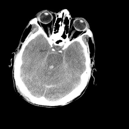
50 YOF with acute onset of worst headache of life associated with nausea and vomiting.  Patient is somnolent, will rouse to noxious stimuli and complains of a headache as well as decreased vision.
Show Answer

  • There is a vitreous hemorrhage of the left globe associated with subarachnoid hemorrhage (SAH), intraventricular hemorrhage, and diffuse cerebral edema on this non-contrast head CT.
  • This is also known as Terson syndrome, reported to occur in 15-30% of patients with SAH.
  • The mechanism by which this intraocular hemorrhage occurred is thought to be from a sudden increase in intracranial pressure causing obstruction of the central retinal vein.

Show References

Image courtesy of Dr. Nasir Siddiqui, Radiopaedia.org. From the case rID: 36469


Follow me on Twitter @EM_NCC


Title: What is the role of EEG for first-time seizures in the ED?

Category: Neurology

Keywords: seizure, electroencephalogram, EEG, epilepsy, antiepileptic (PubMed Search)

Posted: 6/14/2017 by WanTsu Wendy Chang, MD


What is the role of EEG for first-time seizures in the ED?

  • Wyman and colleagues performed a prospective trial on the use of 30-minute routine electroencephalogram (EEG) in the ED after a first-time seizure or recurrent seizure without performance of a previous EEG to guide decision making in the initiation of antiepileptic medication.
  • A diagnosis of epilepsy based on EEG findings was made for 21% of patients (n=15/71).
  • Antiepileptic medication was initiated in 24% of patients (n=17/71), including 2 patients with abnormal but not epileptic EEG findings.

Take Home Point:  A 30-minute routine EEG in the ED in adults with an uncomplicated first-time seizure revealed a substantial number of epilepsy diagnosis and can change ED management with immediate initiation of antiepileptic medication.

Show Additional Information

Background:

  • Seizures account for 1.2% of all ED visits with 24% representing first-time seizures.
  • The 2014 ACEP Clinical Policy on the evaluation and management of patients presenting to the ED with seizures recommend that antiepileptic medication not be initiated for uncomplicated first-time seizure.
  • Literature suggests that epileptiform activity on EEG predicts seizure recurrence while earlier EEG performance has a higher rate of finding epilepsy than delayed EEG performance.

Show References

  • Wyman AJ, Mayes BN, Hernandez-Nino J, Rozario N, Beverly SK, Asimos AW. The first-time seizure emergency department electroencephalogram study. Ann Emerg Med 2017;69(2):184-191.
  • Huff JS, Melnick ER, Tomaszewski CA, et al. Clinical policy: critical issues in the evaluation and management of adult patients presenting to the emergency department with seizures. Ann Emerg Med 2014;63(4):437-447.
  • Krumholz A, Wiebe S, Gronseth G, et al. Practice parameter: evaluating an apparent unprovoked first seizure in adults (an evidence-based review): report of the Quality Standards Subcommittee of the American Academy of Neurology and the American Epilepsy Society. Neurology 2007;69(21):1996-2007.


Follow me on Twitter @EM_NCC


Title: Vasogenic Cerebral Edema

Category: Neurology

Keywords: vasogenic cerebral edema, white matter, blood-brain-barrier, steroids (PubMed Search)

Posted: 4/26/2017 by WanTsu Wendy Chang, MD

 
Vasogenic Cerebral Edema
  • Vasogenic cerebral edema is most commonly seen with brain tumors and cerebral abscesses.
  • It mainly involves the white matter.
  • Gray-white differentiation is maintained, so the edema has a finger-like pattern on CT (see Figure).
  • It is caused by disruption of the blood-brain-barrier, thus responds to treatment with steroids.


Show References

Case image courtesy of Dr David Cuete, Radiopaedia.org, rID: 23178

Follow me on Twitter @EM_NCC!

Attachments

  • 1704262312_20170426_Figure.jpg (60 Kb)


Title: Simplified GCS vs. Full GCS? Which One To Use?

Category: Neurology

Keywords: Glasgow Coma Scale, GCS, motor GCS, mGCS, Simplified Motor Scale, SMS (PubMed Search)

Posted: 4/12/2017 by WanTsu Wendy Chang, MD

 
Simplified GCS vs. Full GCS?  Which One To Use?

  • The Glasgow Coma Scale (GCS) is an instrument widely used to assess level of consciousness by EMS.
  • The motor GCS (mGCS) and Simplified Motor Scale (SMS) have been proposed to simplify EMS triage.
  • A number of retrospective studies have compared these scales.
  • Chou et al. performed a systematic review and meta-analysis of 18 studies with a total number of 1.7 million patients to compare the predictive utility of these scales for identification of patients with severe traumatic injury.
  • The total GCS was slightly better than the mGCS or SMS on predicting mortality, neurosurgical intervention, severe traumatic brain injury, and emergent intubation.

Bottom Line:  The motor GCS and Simplified Motor Scale (SMS) have similar discrimination when compared with the total GCS, and may be easier to use.

Show References

Chou R, Totten AM, Carney N, et al. Predictive Utility of the Total Glasgow Coma Scale Versus the Motor Component of the Glasgow Coma Scale for Identification of Patients with Serious Traumatic Injuries. Ann Emerg Med. 2017 Jan 11. [Epub ahead of print].


Follow me on Twitter @EM_NCC


Title: IV Fluids for Headache?

Category: Neurology

Keywords: headache, migraine, intravenous fluids, IVF (PubMed Search)

Posted: 3/8/2017 by WanTsu Wendy Chang, MD

 
IV Fluids for Headache?
  • Headache is the 4th most common ED visit in the US.
  • Clinical experience suggests that IV fluids (IVF) are commonly used as adjunctive treatment for headaches, however, the efficacy is unknown.
  • A retrospective study using the National Hospital Ambulatory Medical Care Survey (NHAMCS) found that ED length of stay was significantly greater in patients who received IVF than in those who did not (202 min vs. 131 min, p<0.001) even after adjusting for initial pain score, sex, age, and mode of arrival. 
  • A post-hoc analysis of data collected from 4 ED-based migraine trials found that IVF was not associated with improvement of pain score or sustained headache freedom.
  • There is no current evidence to suggest a direct analgesic effect of IVF in the treatment of headaches.


Show References

  • Jones CW, et al. Epidemiology of intravenous fluid use for headache treatment: Findings from the National Hospital Ambulatory Medical Care Survey. Am J Emerg Med. 2017. [Epub ahead of print]
  • Balbin JEB, et al. Intravenous fluids for migraine: a post hoc analysis of clinical trial data. Am J Emerg Med. 2016;34:713-6.

Follow me on Twitter @EM_NCC


Title: Back to the Basics: Aphasia

Category: Neurology

Keywords: aphasia, fluency, comprehension, repetition, Broca's aphasia, Wernicke's aphasia, conduction aphasia (PubMed Search)

Posted: 2/8/2017 by WanTsu Wendy Chang, MD

 
Back to the Basics: Aphasia
  • Aphasia is an impairment of language
  • 3 important assessments in an aphasic patient are fluency, comprehension, and repetition (see attached figure)
  • Patients with fluent speech are able to generate speech spontaneously, though the content of their speech may have errors
  • Patients with non-fluent speech have difficulty initiating speech
  • Patients who have fluent speech but are unable to repeat have a problem with comprehension or a disconnect between the sensory and motor components of language
    • In Wernicke’s aphasia, patients cannot comprehend what they read and hear 
    • In conduction aphasia, patients can comprehend what they read and hear


Show References

Types of aphasia [Online image]. Retrieved February 8, 2017 from https://www.aphasia.org/

Follow me on Twitter @EM_NCC

Attachments

  • 1702082322_20170208_Figure.jpg (84 Kb)


Title: Driving after concussion: Is it safe to drive after symptoms resolve?

Category: Neurology

Keywords: concussion, driving performance, cognitive impairment (PubMed Search)

Posted: 1/11/2017 by WanTsu Wendy Chang, MD

 
Driving after concussion: Is it safe to drive after symptoms resolve?
  • Limited data is available to guide when individuals should return to driving after a concussion.
  • Cognitive impairments in reaction time, executive function, and attention can persist even after symptoms of a concussion resolve.
  • Schmidt et al. compared driving performance between individuals within 48 hours following symptom resolution after a concussion with matched controls using simulated driving.
  • They found that concussed individuals had poorer driving performance despite being asymptomatic.
  • This study is limited by a small sample size (n=28), however, it raises interesting questions regarding whether driving should be restricted following concussions and how should readiness to return to driving be determined.


Show References

Schmidt JD, Hoffman NL, Ranchet M, et al. Driving after concussion: Is it safe to drive after symptoms resolve? J Neurotrauma. 2016 Dec 13. [Epub ahead of print]

Follow me on Twitter @EM_NCC


Title: ED Pharmacist on Time to Thrombolysis

Category: Neurology

Keywords: pharmacist, thrombolysis, door-to-needle time, acute ischemic stroke (PubMed Search)

Posted: 12/14/2016 by WanTsu Wendy Chang, MD

Impact of an ED pharmacist on time to thrombolysis in acute ischemic stroke

  • Prior studies showed that incorporation of ED pharmacists within ED clinical teams lead to more rapid treatment of trauma, stroke, and STEMI.
  • A recent retrospective study conducted by Montgomery et al. showed that having an ED pharmacist on the stroke alert team increased the number of patients meeting goal door-to-needle time of 60 minutes.

Show Additional Information

  • This retrospective study compared the number of patients meeting goal door-to-needle (DTN) time of 60 minutes with and without an ED pharmacist participating on the stroke alert team.
  • A higher proportion of patients with an ED pharmacist met goal DTN time of 60 minutes (71% vs. 39%, p=0.002, 95% CI 0.10-0.50).
  • Patients with an ED pharmacist had an average 20-minute decrease in door-to-needle time (p=0.004, 95% CI 6.6-33.4).

Show References

Montgomery K, Hall AB, Keriazes G. Impact of an emergency medicine pharmacist on time to thrombolysis in acute ischemic stroke. Am J Emerg Med 2016;34:1997-9.

Follow me on Twitter @EM_NCC


Title: Subarachnoid Hemorrhage -- Or Is It?

Category: Neurology

Keywords: subarachnoid hemorrhage, mimic, pseudosubarachnoid hemorrhage, cerebral edema (PubMed Search)

Posted: 11/9/2016 by WanTsu Wendy Chang, MD

Question

Patient found pulseless after submersion in water for 20 minutes.  After ROSC, patient’s GCS was 3 and pupils are dilated and nonreactive.

Show Answer

  • There is increased attenuation of the basal cisterns and subarachnoid space as well as diffuse cerebral edema.
  • At first glance, it appears to be a subarachnoid hemorrhage.
  • However, the Hounsfield unit of the density is lower than blood.
  • This is a pseudosubarachnoid hemorrhage sign.  
  • This can be seen in anoxic injury with cerebral edema, pyogenic meningitis, venous sinus thrombosis, bilateral large subdural hemorrhages.

Show References

Kim JM, Eom TH. The pseudosubarachnoid hemorrhage: clinical implications of subarachnoid hemorrhage misdiagnosis. Pediatr Emerg Care. 2016 May 12. [Epub ahead of print]

Follow me on Twitter @EM_NCC


Title: Updated Guidelines for Traumatic Brain Injury

Category: Neurology

Keywords: Brain Trauma Foundation, BTF, guideline, traumatic brain injury, TBI (PubMed Search)

Posted: 10/12/2016 by WanTsu Wendy Chang, MD

Updated Guidelines for Traumatic Brain Injury

The Brain Trauma Foundation (BTF) Guidelines for the Management of Severe Traumatic Brian Injury (TBI) was recently updated and published in September 2016.

Updated recommendations include:

  • Prophylactic hypothermia is not recommended (Level IIB).
  • Phenytoin is recommended for seizure prophylaxis (Level IIA).
    • There is insufficient evidence to recommend levetiracetam over phenytoin.
  • Maintain SBP 100 mmHg for patients 50-69 years old or 110 mmHg for patients 15-49 or >70 years old (Level III).
  • Treat intracranial pressure (ICP) > 22 mmHg (Level III)
  • Target cerebral perfusion pressure (CPP) between 60-70 mmHg (Level IIB).

For the executive summary and complete guidelines, go to https://braintrauma.org/guidelines/guidelines-for-the-management-of-severe-tbi-4th-ed#/


Title: My Patient Won't Open His/Her Eyes!

Category: Neurology

Keywords: eyelid apraxia, eye opening apraxia (PubMed Search)

Posted: 9/14/2016 by WanTsu Wendy Chang, MD

 
My patient won't open his/her eyes!
 
  • Beware of the patient who can't open his/her eyes but is otherwise awake!
  • This coma mimic is the result of eyelid apraxia, which is the inability to voluntarily open eyes despite intact frontalis muscle contraction and absent oculomotor dysfunction.
  • This can be seen in injuries of the nondominant hemisphere (e.g. R MCA stroke), medial frontal lobe, bilateral thalami (e.g. bilateral thalami stroke), and brainstem (e.g. progressive supranuclear palsy).
  • When asking these patients to open their eyes, they may use their forehead muscles to try and raise their eyelids.


Title: What's the cause of this patient's hemiplegia?

Category: Neurology

Keywords: Uncal herniation, ipsilateral hemiplegia, Kernohan's notch, Kernohan's sign (PubMed Search)

Posted: 8/10/2016 by WanTsu Wendy Chang, MD

Question

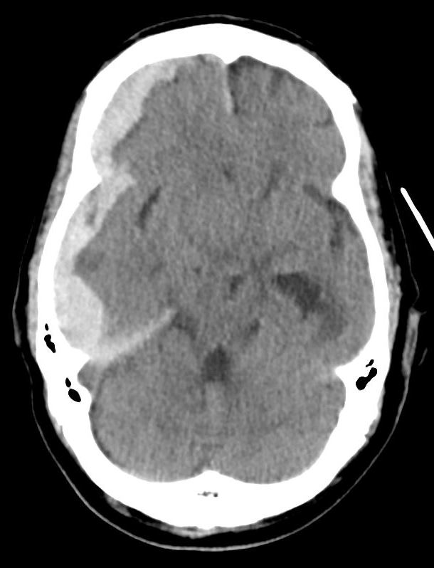
Patient presents after a fall confused, not moving his right side, but moving his left side spontaneously.  What's the diagnosis?
 

Show Answer

  • The patient has a right subdural hematoma (SDH) with midline shift and right uncal herniation. 
  • The classic presentation of a cerebral lesion with uncal herniation is ipsilateral mydriasis, contralateral hemiparesis, and abnormal extensor posturing.
  • In this patient’s case, there is compression of the contralateral cerebral peduncle of the midbrain by the edge of the tentorium cerebelli producing an ipsilateral hemiplegia (see attachment for Figure 2).
  • This false localizing sign is named Kernohan’s notch syndrome, Kernohan’s notch phenomenon, or Kernohan’s sign.

Show References

Diagram modified from "Localised Neurological Disease and Its Management A Intracranial". clinicalgate.com/localised-neurological-disease-and-its-management-a-intracranial/. Accessed 10 Aug 2016.


Follow me on Twitter @EM_NCC

Attachments

  • 1608101928_20160810_Figure_2.jpg (38 Kb)


Title: Screening Tool for Large Vessel Occlusion Strokes?

Category: Neurology

Keywords: Large vessel occlusion stroke, endovascular intervention, Field Assessment Stroke Triage for Emergency Destination, FAST-ED, NIHSS, Rapid Arterial Occlusion Evaluation, RACE, Cincinnati Prehospital Stroke Severity scale, CPSS (PubMed Search)

Posted: 7/13/2016 by WanTsu Wendy Chang, MD

 
Screening Tool for Large Vessel Occlusion Strokes (LVOS)?
 
  • Endovascular intervention for acute ischemic stroke from ICA or proximal MCA occlusion is a Level IA recommendation1.
  • Identification of patients who may benefit from endovascular intervention begins in the prehospital setting.
  • Several prehospital stroke scales exist, but have not been validated using arterial imaging to determine the presence of LVOS.
  • The Field Assessment Stroke Triage for Emergency Destination (FAST-ED) scale (see Table 1) was designed based on items of the NIH Stroke Scale (NIHSS) with higher predictive value for LVOS.
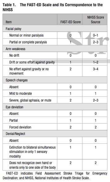
  • The FAST-ED scale has comparable accuracy to predict LVOS to the NIHSS, and higher accuracy compared to the Rapid Arterial Occlusion Evaluation (RACE) and the Cincinnati Prehospital Stroke Severity (CPSS) scale
  • The FAST-ED scale also provides 3 distinct groups for the likelihood of LVOS:
    • Score 0 or 1: <15%
    • Score 2 or 3: 30%
    • Score >= 4: >60%

Bottom Line: Additional assessment of gaze deviation, aphasia and neglect, as included in the FAST-ED scale, increases the accuracy of predicting LVOS.  

Show References

  1. Powers WJ, Derdeyn CP, Biller J, et al. 2015 American Heart Association/American Stroke Association Focused Update of the 2013 Guidelines for the Early Management of Patients with Acute Ischemic Stroke Regarding Endovascular Treatment: A Guideline for Healthcare Professionals From the American Heart Association/American Stroke Association. Stroke. 2015;46(10):3020-35.
  2. Lima FO, Silva GS, Furie KL, et al. Field Assessment Stroke Triage for Emergency Destination: A Simple and Accurate Prehospital Scale to Detect Large Vessel Occlusion Strokes. Stroke. 2016 Jun 30. [Epub ahead of print]


Follow me on Twitter @EM_NCC


Previous  |  1 |  2 |  3 |  4 |  5 |  Next
University of Maryland School of Medicine logo
  • Departments
  • Programs
  • Research Centers
  • Institutes
  • Graduate Education
  • CIBR Cores
  • Offices of the Dean
    • University of Maryland, Baltimore
    • University of Maryland Medical Center
    • Medical Alumni Association of the University of Maryland
    • Health Sciences and Human Services Library
University of Maryland School of Medicine
655 W. Baltimore Street
Baltimore, MD 21201
Contact Us

© University of Maryland School of Medicine

    • Webmaster
    • Web Accessibility
    • Site Index
    • UMB Hotline
    • Employment
    • Map