University of Maryland School of Medicine logo
Emergency Medicine
All Departments
UMSOM Assistant
UMEM Educational Pearls
  • Home
  • Departments
  • Emergency Medicine
  • CME
  • UMEM Educational Pearls
  • CME
  • Courses and Conferences
  • Free Open Access Medical Education
  • UMEM Educational Pearls

Search

61-80 of 382 results with category "Neurology"

Previous  |  1 |  2 |  3 |  4 |  5 |  6 |  7 |  8 |  9 |  ... |  20 |  Next

Title: Neuroimaging in Syncope - Is It Necessary?

Category: Neurology

Keywords: Syncope, neurological, neuroimaging, CT, MRI (PubMed Search)

Posted: 6/13/2018 by WanTsu Wendy Chang, MD (Updated: 7/21/2026)

  • The use of neuroimaging in syncope-related ED visits increased from 21% in 2001 to 45% in 2010.
  • A recent single-center retrospective study of 1114 patients who presented to the ED with syncope found that 62.3% patients underwent CT, while 10.2% underwent MRI.
  • A subset of patients (10.4%) sustained mild head trauma (GCS 14-15) due to syncope and all received neuroimaging.
  • Neuroimaging studies were not found to be beneficial in patients without features of:
    • Confusion
    • Amnesia
    • Focal neurological deficit
    • Dizziness
    • Severe headache
    • Nausea and vomiting
    • Signs of serious head injury
    • Intracranial malignancies
    • Use of anticoagulant drugs

Bottom Line: Consider obtaining neuroimaging in patients presenting with syncope only if clinical features suggest probable neurological syncope.

Show References


  • Idil H, Kilic TY. Diagnostic yield of neuroimaging in syncope patients without high-risk symptoms indicating neurological syncope. Am J Emerg Med. 2018 May 16 [Epub ahead of print]
  • Probst MA, Kanzaria HK, Gbedemah M, Richardson LD, Sun BC. National trends in resource utilization associated with ED visits for syncope. Am J Emerg Med. 2015;33(8):998-1001.

Follow me on Twitter @EM_NCC


Title: Lhermitte's Sign

Category: Neurology

Keywords: myelopathy, myelitis, physical exam (PubMed Search)

Posted: 5/23/2018 by Danya Khoujah, MBBS (Updated: 7/21/2026)

Lhermitte’s phenomenon is as a sign of cervical spinal cord demyelination. It is considered positive if flexion of the neck causes a tingling sensation moving down the limbs or trunk, and may be reported as a symptom or elicited as a sign. This is due to stretching of the dorsal column sensory fibers, the commonest cause of which is multiple sclerosis. Other causes include other myelopathies, such as B12 deficiency, radiation and toxic (due to chemotherapy) or idiopathic myelitis. Its sensitivity is low at 16%, but its specificity for myelopathy is high at 97%.

Show References

Kempster PA, Rollinson RD. The Lhermitte phenomenon: variant forms and their significance. J Clin Neurosci 2008;15(4):379–81.

Khare S, Seth D. Lhermitte's Sign: The current status. Ann Indian Acad Neurol. 2015 Apr-Jun; 18(2): 154-156.


Title: Predicting ICH Expansion

Category: Neurology

Keywords: Intracerebral hemorrhage, ICH, hematoma expansion, prediction score, BAT score (PubMed Search)

Posted: 5/9/2018 by WanTsu Wendy Chang, MD (Updated: 7/21/2026)

  • CT angiography (CTA) spot sign is a strong predictor of intracerebral hemorrhage (ICH) expansion.
  • However, since CTA is not part of the routine diagnostic workup of acute ICH, other predictors using noncontrast head CT have been reported in the past.
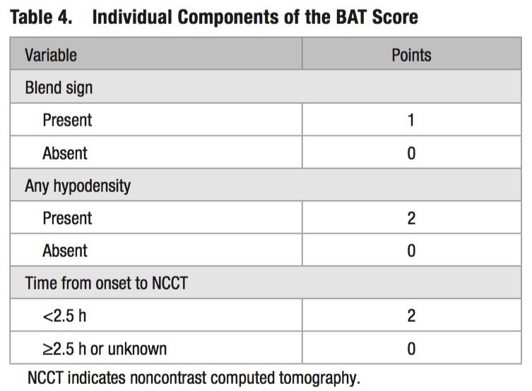
  • A 5-point BAT score can be used to identify patients at high risk of hematoma expansion:
  •  
  • Patients with a score ≥ 3 have a higher risk of hematoma expansion. 

Show Additional Information

  • 14 Variables including patient age, sex, history of HTN, initial SBP and DBP, ICH volume at baseline, baseline INR, time from hemorrhage onset, and noncontrast head CT findings (hemorrhage location, hypodensities, blend sign, irregular shape, heterogeneous density, and fluid level) were analyzed by bivariate analysis and stepwise logistical regression using bootstrap samples.
  • AUC for the BAT score was 0.77 in the development sample, 0.65 and 0.70 in two validation cohorts.
  • Patients with coagulopathy were excluded in the development and validation of the BAT score to maximize the applicability of the score, though coagulopathy is a known predictor of hematoma expansion. 

Show References

Morotti A, Dowlatshahi D, Boulouis G, et al. Predicting intracerebral hemorrhage expansion with noncontrast computed tomography: The BAT score. Stroke 2018;49(5):1163-9. 

Follow me on Twitter @EM_NCC


Title: Atypical Stroke Symptoms

Category: Neurology

Keywords: stroke, altered mental status, gender, sex, confusion (PubMed Search)

Posted: 3/28/2018 by Danya Khoujah, MBBS (Updated: 7/21/2026)

Patients may present atypically with ischemic strokes, reporting symptoms such as face or hemibody pain, lightheadedness, mental status change, headache and non-neurological symptoms.

Up to 25% of patients will have these symptoms.

Women are more likely than men to present with these atypical (or “nontraditional”) symptoms, especially altered mental status.

Show References

Labiche LA, Chan W, Saldin KR, Morgenstern LB. Sex and acute stroke presentation. Ann Emerg Med. 2002;40(5):453-460.

Lisabeth LD, Brown DL, Hughes R, et al. Acute stroke symptoms: comparing women and men. Stroke. 2009;40(6):2031-2036.


Title: Prehospital Stroke Scales for Large Vessel Occlusion

Category: Neurology

Keywords: stroke, prehospital, large vessel occlusion, NIHSS, RACE, LAMS, VAN (PubMed Search)

Posted: 3/14/2018 by WanTsu Wendy Chang, MD (Updated: 7/21/2026)

  • A recent systematic review evaluated the diagnostic accuracy of 19 prehospital stroke scales.
  • Arm motor strength is the most frequently evaluated item by the scales (15/19), followed by gaze (13/19) and language (13/19).
  • Only 4 scales (RACE, LAMS, VAN, sNIHSS-EMS) were performed by paramedics in their original studies.
  • The NIHSS, LAMS, and VAN appear to have better results in predicting large vessel occlusion.
  • The presence of hemineglect, a sign of cortical involvement, improved the accuracy of the scale.

Show Additional Information

  • There is growing evidence for mechanical thrombectomy in acute ischemic stroke patients with large vessel occlusion.
  • Identification of potential eligible patients begin in the prehospital setting in order for preferential transport of these patients to comprehensive stroke centers.
  • Selected prehospital stroke scales:
    • RACE = Rapid Arterial Occlusion Evaluation Scale
    • LAMS = Los Angeles Motor Scale
    • VAN = Stroke Vision, Aphasia, Neglect Assessment
    • NIHSS = National Institute of Health Stroke Scale

Show References

 
  • Vidale S, Agostini E. Prehospital stroke scales and large vessel occlusion: A systematic review. Acta Neurol Scand. 2018 Feb 11. [Epub ahead of print]

Follow me on Twitter @EM_NCC


Title: Headache in the Bodybuilder

Category: Neurology

Keywords: headache, steroids, bleed (PubMed Search)

Posted: 2/28/2018 by Danya Khoujah, MBBS

Benign headaches are common in bodybuilders. However, several less benign headaches are worth noting:

  • Low cerebrospinal fluid (CSF) pressure headache: caused by a small dural tear mostly at the thoracic level. Similar to postdural headache. Treated by recumbency, and blood patches if recalcitrant.
  • Subarachnoid hemorrhage (SAH)
  • Spontaneous intracranial hemorrhage
  • Ischemic stroke
  • Dural sinus thrombosis

All except the first two are exclusively reported in patients on anabolic steroids, growth hormone, and/or “energy” supplements. Make sure to ask your patient about these risk factors.


Show References

Busche K. Neurologic Disorders Associated with Weight lifting and Bodybuilding. Neurology Clinics. 26 (2008) 309–324


Title: Occipital Nerve Block for Migraine?

Category: Neurology

Keywords: occipital nerve block, migraine, headache (PubMed Search)

Posted: 2/15/2018 by WanTsu Wendy Chang, MD (Updated: 2/15/2018)

  • Greater occipital nerve (GON) block with local anesthetics is an alternate treatment option for headaches.
  • Zhang et al. conducted a systematic review and meta-analysis of 7 randomized controlled trials assessing the efficacy of GON block for migraine.
  • Pooled outcome suggests that GON block: 
    • Reduces pain intensity (mean difference -1.24 [-1.98, -0.49], p=0.001)
    • Decreases analgesia medication consumption (mean difference -1.10 [-2.07, -0.14], p=0.02)
    • Has no significant impact on headache duration (mean difference -6.96 [-14.09, 0.18], p=0.06)

Show References

Zhang H, Yang X, Lin Y, Chen L, Ye H. The efficacy of greater occipital nerve block for the treatment of migraine: a systematic review and meta-analysis. Clin Neurol Neurosurg. 2018;165:129-133.

Follow me on Twitter @EM_NCC


Title: Brain Tumor Imaging 101

Category: Neurology

Keywords: edema, hemorrhage, tumor, CT, MRI, contrast (PubMed Search)

Posted: 1/24/2018 by Danya Khoujah, MBBS (Updated: 7/21/2026)

Although MRI is more sensitive for identifying tumors of the CNS, CT is usually the first line imaging modality in the ED. Some pearls:

  • Hyperattenuation = bright = dense (blood)
  • Hypoattenuation = dark = radiolucent (fluid, air, lipid, scar)
  • Masses that are darker + increased volume or mass effect = edema (image 1)
  • Masses that are darker + decreased volume = scar tissue or atrophy (image 2)
  • Masses that are bright + edema = hemorrhage (image 3)
  • Adding IV contrast improves detection of tumors: abnormal enhancement from disruption of blood brain barrier, necrosis or increased vascularity. (Image 4)
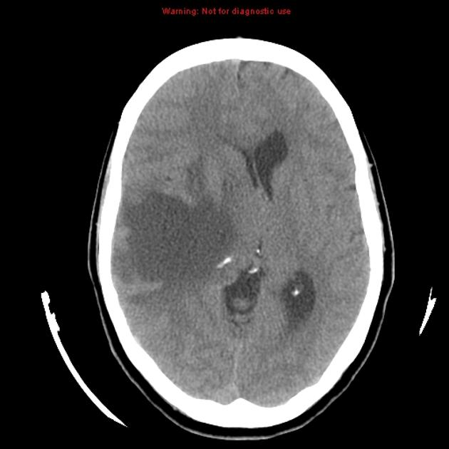
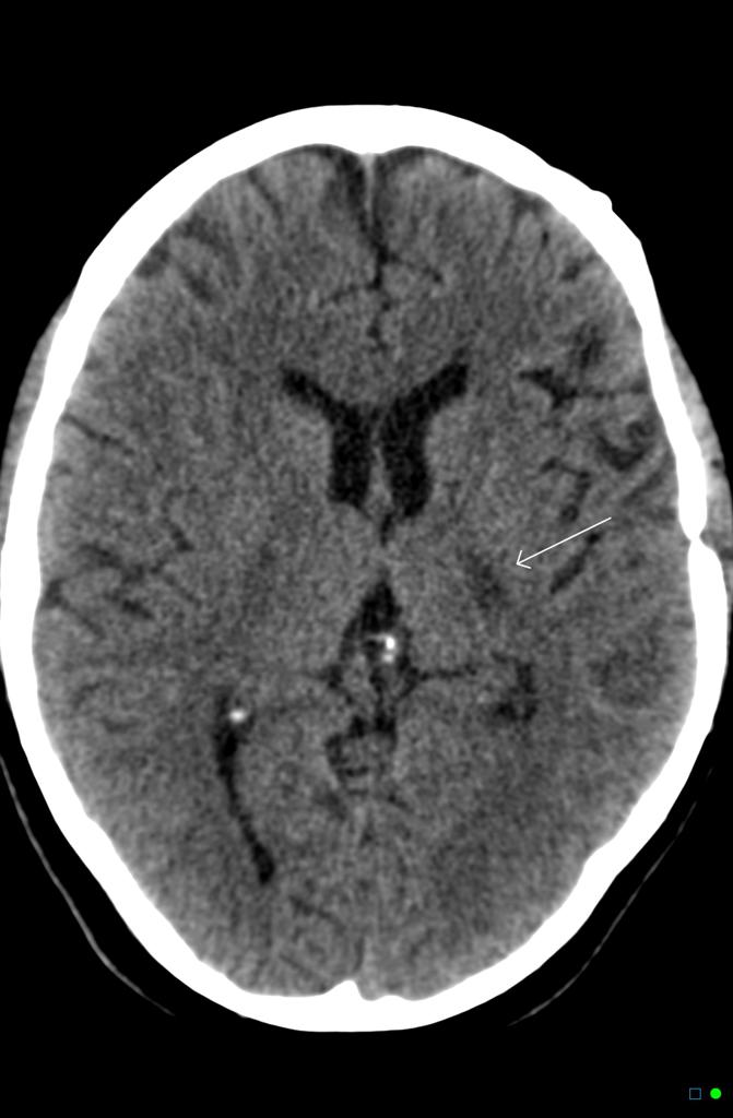
Image 1 Courtesy of Radiopedia.orgImage 2: courtesy of Dr Chris O'Donnell, Radiopaedia.orgImage 3: courtesy of Dr David Cuete, Radiopaedia.orgImage 4: Courtesy of David Kernick, and Stuart Williams Br J Gen Pract2011;61:409-411

Show References

Klein JP, Dietrich J. Neuroradiologic Pearls for Neuro-oncology. Continuum 2017;23(6):1619-1634.


Title: Reversible Cerebral Vasoconstriction Syndrome (RCVS)

Category: Neurology

Keywords: RCVS, thunderclap headache, migraine, SAH (PubMed Search)

Posted: 1/10/2018 by WanTsu Wendy Chang, MD

  • Reversible cerebral vasoconstriction syndrome (RCVS) is the second most common cause of thunderclap headache after aneurysmal subarachnoid hemorrhage (SAH) and the most common cause of recurrent thunderclap headaches.
  • Up to 40% of patients with RCVS have a history of migraine.
  • It is associated with selective serotonin reuptake inhibitors (SSRIs), triptans, cocaine, marijuana, tacrolimus, oral contraceptives, as well as the peripartum period.
  • Symptoms are often triggered by emotional stress, sexual activity, showering, straining, and physical exertion.
  • Although the vasoconstriction is reversible, it can cause intracranial hemorrhage, seizures, stroke, and coma.
  • Diagnosis is by history, cerebral angiography and exclusion of aneurysmal SAH.

Bottom Line: Consider RCVS in the differential of thunderclap headache and in patients who present with worse than usual migraine headache.

Show References

Arrigan MT, Heran MKS, Shewchuk JR. Reversible cerebral vasoconstriction syndrome: an important and common cause of thunderclap and recurrent headaches. Clin Radiol. 2017 Dec 21 [Epub ahead of print]

Follow me on Twitter @EM_NCC


Title: Cerebral Venous Thrombosis (CVT)

Category: Neurology

Keywords: headache, seizure, stroke, neurological deficit, thrombogenic (PubMed Search)

Posted: 12/27/2017 by Danya Khoujah, MBBS (Updated: 7/21/2026)

Cerebral venous thrombosis is a rare (but dangerous) cause of headaches and strokes in patients below the age of 50. It includes thrombosis of the cerebral veins and major dural sinuses. 
A d-dimer can NOT be used to rule it out, as it would be falsely negative in up to 40% of patients. A dry head CT is completely normal in 30% of patients, with nonspecific changes present in another 30%.

Take home: If you are considering the diagnosis, obtain a CT venography (95% sensitive) and don’t rely on a negative dimer or dry head CT.


Show References

Long B, Koyfman A, Runyon MS. Cerebral Venous Thrombosis: A Challenging Neurologic Diagnosis. Emerg Med Clin N Am 35 (2017) 869–878


Title: A New DAWN for Stroke Intervention?

Category: Neurology

Keywords: DAWN, thrombectomy, mismatch, wake-up, stroke, penumbra (PubMed Search)

Posted: 12/13/2017 by WanTsu Wendy Chang, MD

  • The DAWN trial was a multicenter, randomized, open-label study comparing endovascular thrombectomy plus standard medical care with standard medical care alone for patients with:
    • Acute stroke symptoms
    • Last known well 6 to 24 hours earlier
    • Evidence of intracranial ICA or proximal MCA occlusion
    • Mismatch between clinical deficit and infarct volume on CTA or MRA
  • The study found that patients receiving thrombectomy plus standard medical care had improved functional independence at 90 days as defined by modified Rankin Scale (mRS) of 0, 1, or 2 (49% vs 13%).
  • The trial was stopped early based on prespecified interim analysis intended with the adaptive trial design.
  • While the two treatment groups were similar, with median NIHSS score of 17, they had small infarct volumes and short time from symptom observation (4.8 vs 5.6 hours) compared to time of patient's last known well (12.2 vs 13.3 hours). 
  • 88% of the patients had unwitnessed stroke onset (including wake-up strokes), thus it is possible that these patients had actual ischemia times closer to 6 hours, thereby reproducing similar results as prior thrombectomy trials.

Bottom Line: The use of neuroimaging to identify an ischemic penumbra that may benefit from thrombectomy may be considered even for patients with time of last known well beyond 6 hours.

Show Additional Information

  • Endovascular thrombectomy is recommended for patients with large vessel occlusion within 6 hours of stroke symptom onset.
  • Eligibility has been based on the time that patient was last known to be well.
  • Prior studies suggest that patients who have evidence of ischemic brain tissue that has not yet infarcted could benefit from reperfusion even when performed more than 6 hours after patient was last known to be well.
  • The DAWN (DWI or CTP Assessment with Clinical Mismatch in the Triage of Wake-Up and Late Presenting Strokes Undergoing Neurointervention with Trevo) trial enrolled 206 patients from 26 sites over 29 months.  
  • It is unclear how many patients were screened for enrollment.
  • This study was sponsored by Stryker Neurovascular and only allowed the use of their Trevo endovascular stent retriever device for the thrombectomy group.

Show References

Nogueira RG, Jadhav AP, Haussen DC, et al. Thrombectomy 6 to 24 hours after stroke with a mismatch between deficit and infarct. N Engl J Med. 2017 Nov 11. [Epub ahead of print]

Follow me on Twitter @EM_NCC


Title: Guillain-Barre's less evil twin - CDIP!

Category: Neurology

Keywords: GBS, weakness, intubation, CSF, LP (PubMed Search)

Posted: 11/22/2017 by Danya Khoujah, MBBS

CDIP, or chronic inflammatory demyelinating polyradiculoneuropathy, is an immune-mediated polyneuropathy which presents similarly to Guillain-Barré Syndrome (GBS). However, it is not as dangerous as GBS. Patients present with symmetric proximal and distal weakness with reduced or absent deep tendon reflexes, just like GBS. The difference is that in typical CDIP, patients have prominent sensory signs, no autonomic dysfunction, no facial weakness, no preceding infectious illness, and most importantly no respiratory failure. It also continues to progress past 4 weeks.

CSF is not diagnostic, and may show albuminocytologic dissociation. The diagnostic test is nerve conduction studies. 

Show References

Allen JA. Chronic Demyelinating Polyneuropathies. Continuum 2017;23(5):1310–1331


Title: Isolated Aphasia - Is It a Stroke?

Category: Neurology

Keywords: aphasia, stroke, middle cerebral artery, MCA, mimic, NIHSS (PubMed Search)

Posted: 11/8/2017 by WanTsu Wendy Chang, MD

  • A retrospective single center study reviewed 788 patients who presented to the ED with concern of stroke and found 21 (3%) patients had only aphasia symptoms by the NIHSS.
  • None of these patients had evidence of infarct on neuroimaging.
  • 3 of these patients were diagnosed with possible transient ischemic attack (TIA) though also had other possible diagnoses.
  • Toxic/metabolic disturbances (39%), followed by seizure (11%), syncope (11%), and chronic medical problems (11%) were the most commonly diagnosed stroke mimics.

Take Home Point: This small but interesting study looked at the incidence of isolated aphasia presenting for concern of stroke. They found that none of their patients had evidence of an infarct, suggesting that strokes affecting language without motor or sensory deficits are uncommon.

Show Additional Information

  • Aphasia can be caused by ischemia of the left middle cerebral artery (MCA) territory.
  • However, a stroke of the left MCA territory is usually accompanied by some component of contralateral motor and sensory deficits, gaze deviation, or visual field cut.
  • Aphasia can also be caused by stroke mimics such as infection, toxic/metabolic abnormalities, dementia, migraine, or seizure.

Show References

Casella G, Llinas RH, Marsh EB. Isolated aphasia in the emergency department: The likelihood of ischemia is low. Clin Neurol Neurosurg 2017:163:24-26.

Follow me on Twitter @EM_NCC


Title: Guillain- Barr Syndrome

Category: Neurology

Keywords: weakness, infection, paralysis, intubation, influenza, vaccine (PubMed Search)

Posted: 10/25/2017 by Danya Khoujah, MBBS

It's respiratory infection and flu vaccine season! Time to brush up on Guillain-Barré Syndrome..

- It is the most common cause of acute or subacute flaccid weakness worldwide

- 70% of cases are preceded by an infection in the past 10-14 days, but most are minimized or forgotten by the patient. 40% of these infections are by Campylobacter jejuni.

- 30% develop respiratory failure requiring intubation and ventilation

- Half of the patients will develop their maximum weakness by 2 weeks, most will develop it by 4 weeks.

Show References

Donofrio PD. Guillain-Barré Syndrome. Continuum 2017;23(5):1295–1309.


Title: Traumatic Brain Injury in Older Adults - The Silver Tsunami?

Category: Neurology

Keywords: traumatic brain injury, TBI, fall, subdural hematoma, SDH, elderly (PubMed Search)

Posted: 10/11/2017 by WanTsu Wendy Chang, MD

Traumatic brain injury (TBI) is associated with close to half of major trauma admissions in adults over age 65 in the U.K.

Falls accounted for 85% of all TBIs, while 45% of patients had subdural hematomas (SDH).

More than 3/4 of patients were treated conservatively, though outcomes were not significantly better than those who underwent neurosurgical intervention.

Higher age is associated with higher mortality and greater disability.

Bottom Line: Trauma in older adults is increasing and fall prevention is important in reducing significant injuries.

Show References

Hawley C, Sakr M, Scapinello S, et al. Traumatic brain injuries in older adults - 6 years of data for one UK trauma centre: retrospective analysis of prospectively collected data. Emerg Med J 2017;0:1-8.


Follow me on Twitter @EM_NCC


Title: tPA Contraindications

Category: Neurology

Keywords: stroke, tPA, thrombolytics, ICH, hemorrhage, adverse events (PubMed Search)

Posted: 9/28/2017 by Danya Khoujah, MBBS

Classically, the list of contraindications for tPA in stroke has been extensive and excludes a significant percentage of patients. This scientific statement from AHA clarifies the evidence behind these contraindications, and in short, expands the population of patients that should be considered for tPA.
The following is NOT considered a contraindication for tPA: 
- Age over 80 
- Severe stroke (NIHSS >25)
- Improving symptoms, if patient remains moderately impaired and potentially disabled
- A small (<10 mm) unruptured and unsecured intracranial aneurysm (NOT other vascular malformations)
- Extra-axial intracranial neoplasms (e.g. meningiomas, pituitary adenomas)
- Blood glucose of >400mg/dL that is subsequently normalized
- Seizure at onset of stroke if residual impairment is secondary to stroke not a postictal phenomenon 

Show References

Demaerschalk BM, Kleindorfer DO, Adeoye OM, et al; American Heart Association Stroke Council and Council on Epidemiology and Prevention. Scientific Rationale for the Inclusion and Exclusion Criteria for Intravenous Alteplase in Acute Ischemic Stroke: A Statement for Healthcare Professionals From the American Heart Association/American Stroke Association. Stroke 47(2):581–641, 2016. 


Title: IV vs. Non-IV Benzodiazepines for Cessation of Seizures

Category: Neurology

Keywords: seizure, status epilepticus, benzodiazepine, RAMPART, pediatric (PubMed Search)

Posted: 9/13/2017 by WanTsu Wendy Chang, MD (Updated: 9/14/2017)

IV vs. Non-IV Benzodiazepines for Cessation of Seizures

  • A meta-analysis by Alshehri et al. included 11 studies with a total of 1633 patients, comparing IV vs. non-IV benzodiazepines from any route (buccal, intranasal, intramuscular) for seizure cessation in status epilepticus.
  • They found that non-IV benzodiazepine is more effective than IV benzodiazepine in patients presenting without IV access.
  • The largest and highest quality study included in the meta-analysis was the RAMPART study, which was also the only study to include adults.
  • When considering pediatric studies only, there is no difference between IV vs. non-IV benzodiazepine in seizure cessation for status epilepticus.

Show Additional Information

  • Benzodiazepine is first-line therapy for treatment of status epilepticus.
  • The RAMPART study in 2012 by Silbergleit et al. demonstrated that prehospital treatment of status epilepticus with IM midazolam was more effective than IV lorazepam, even though the time from drug administration to seizure cessation was shorter for IV lorazepam.
  • It is important to note that there is a 30% treatment failure rate for a single appropriately dosed benzodiazepine in treatment of status epilepticus.

Show References

  • Alshehri A, Abulaban A, Bokhari R, et al. Intravenous vs. nonintravenous benzodiazepines for the cessation of seizures: a systematic review and meta-analysis of randomized controlled trials. Acad Emerg Med 2017;24(7):875-83.
  • Silbergleit R, Durkalski V, Lowenstein D, et al. Intramuscular versus intravenous therapy for prehospital status epilepticus. N Engl J Med 2012;366(7):591-600.

Follow me on Twitter @ EM_NCC


Title: Rapid detection of bacterial meningitis using point-of-care glucometer

Category: Neurology

Keywords: meningitis, CSF, glucose, glucometer (PubMed Search)

Posted: 8/24/2017 by WanTsu Wendy Chang, MD

 
Rapid detection of bacterial meningitis using point-of-care glucometer

  • CSF:blood glucose ratio is a useful characteristic in differentiating bacterial meningitis from viral meningitis. 
  • Normal CSF glucose is at least 2/3 of serum glucose level.
  • In bacterial meningitis, CSF:blood glucose ratio is usually <0.4
  • Rousseau et al. conducted a study comparing CSF:blood glucose ratio obtained using a bedside glucometer with the laboratory.
  • They found the optimal cutoff of CSF:blood glucose ratio using a bedside glucometer is 0.46 compared to 0.44 using the laboratory.
  • This proof-of-concept study suggests that a point-of-care glucometer can be used for rapid diagnosis of abnormal CSF:blood glucose ratio in the evaluation of meningitis.

Show References

Rousseau G, Asmolov R, Grammatico-Guillon L, et al. Rapid detection of bacterial meningitis using a point-of-care glucometer. Eur J Emerg Med 2017 Aug 10. [Epub ahead of print]
 
Follow me on Twitter @EM_NCC


Title: Pituitary Apoplexy

Category: Neurology

Keywords: pituitary apoplexy, subarachnoid hemorrhage, meningitis, headache, CT, MRI (PubMed Search)

Posted: 8/9/2017 by Danya Khoujah, MBBS (Updated: 7/21/2026)

Pituitary apoplexy is a sudden hemorrhage or infarction of the pituitary.
  • It most commonly occurs in patients with preexisting pituitary adenomas, but 3 out of 4 patients with pituitary adenomas are unaware of their diagnosis.
  • Patients may acutely present with thunderclap headache, with or without visual field deficits or cranial nerve dysfunction. They may also have meningeal symptoms due to extravasation of blood into the subarachnoid space.
  • Endocrine dysfunction is common but not readily diagnosed in the ED.
  • Symptoms may be triggered by some hormonal treatments (e.g. GnRH agonists for prostate CA), head trauma, angiographic procedures, or anticoagulation therapy.
  • CT is diagnostic in only one-third of cases, but can reveal the intrasellar mass in 80% of cases, and therefore should be the initial test. Blood may be missed in subacute cases.
  • MRI is the test of choice, with a sensitivity of over 90%.  

Bottomline: Keep pituitary apoplexy in your differential when considering SAH or meningitis, especially in the presence of risk factors, and have a low threshold to order an MRI. 

Show References

Ishii, M. Endocrine Emergencies With Neurologic Manifestations. Continuum 2017;23(3):778–801. 


Title: What is the cause of this patient's decreased vision?

Category: Neurology

Keywords: Terson syndrome, vitreous hemorrhage, intraocular hemorrhage, subarachnoid hemorrhage (PubMed Search)

Posted: 7/12/2017 by WanTsu Wendy Chang, MD

Question

50 YOF with acute onset of worst headache of life associated with nausea and vomiting.  Patient is somnolent, will rouse to noxious stimuli and complains of a headache as well as decreased vision.

Show Answer

  • There is a vitreous hemorrhage of the left globe associated with subarachnoid hemorrhage (SAH), intraventricular hemorrhage, and diffuse cerebral edema on this non-contrast head CT.
  • This is also known as Terson syndrome, reported to occur in 15-30% of patients with SAH.
  • The mechanism by which this intraocular hemorrhage occurred is thought to be from a sudden increase in intracranial pressure causing obstruction of the central retinal vein.

Show References

Image courtesy of Dr. Nasir Siddiqui, Radiopaedia.org. From the case rID: 36469


Follow me on Twitter @EM_NCC


Previous  |  1 |  2 |  3 |  4 |  5 |  6 |  7 |  8 |  9 |  ... |  20 |  Next
University of Maryland School of Medicine logo
  • Departments
  • Programs
  • Research Centers
  • Institutes
  • Graduate Education
  • CIBR Cores
  • Offices of the Dean
    • University of Maryland, Baltimore
    • University of Maryland Medical Center
    • Medical Alumni Association of the University of Maryland
    • Health Sciences and Human Services Library
University of Maryland School of Medicine
655 W. Baltimore Street
Baltimore, MD 21201
Contact Us

© University of Maryland School of Medicine

    • Webmaster
    • Web Accessibility
    • Site Index
    • UMB Hotline
    • Employment
    • Map