
Search
Subscribe to daily UMEM Pearls (FREE!) via:
121-140 of 479 results with category "Orthopedics"
Diagnostic performance of Ultrasonography for detection of pediatric elbow fracture
Elbow fractures account for approximately 15% of pediatric fractures
Fat pads are traditionally taught as a marker of fracture
In a cadaveric study:
Elbow effusions of 1-3 mL could be identified with ultrasound
Elbow effusions of 5-10 mL could be identified with plain film
Pediatric plain films are sometimes challenging to obtain and interpret compared to adults
-More likely to be uncooperative in obtaining required views
-Non-ossified epiphyses
Ultrasound may be used to detect
-Cortical disruption and irregularity
-Growth plate widening
-Hematoma interposed between fracture fragments
-Elevated posterior fat pad
Absence of elbow fracture was indicated by
-Lack of cortical disruption
-Absence of posterior fat pad sign
Meta-analysis of 10 articles totaling 519 patients using ultrasonography to detect elbow fractures
Sensitivity 96%
Specificity 89%
False negative rate 3.7%
For comparison, plain radiographs
Interpreted by peds EM physicians (87.5% sensitive and 100% specific)
Interpreted by radiology (96% sensitive, 100% specific)
Consider using ultrasound as a noninvasive, radiation-free modality for accurate diagnosis of pediatric elbow fractures.
Show References
Pronator Teres Syndrome
A compressive neuropathy of the median nerve in the region of the elbow
The median nerve passes through the cubital fossa and passes between the superficial and deep heads of the pronator teres muscle.
Rare compared to other compressive neuropathies such as carpal tunnel syndrome.
More common in women and in fifth decade of life
May be seen with weight lifters, arm wrestlers, rowers, tennis, archery, professional cyclists, dentists, fiddlers, pianists, harpists
Also associated with well-developed forearm muscles
History:
Forearm pain – unlike carpal tunnel
Paresthesias in median distribution
No night symptoms – unlike carpal tunnel
Physical exam:
Sensory loss in medial nerve distribution.
Involves the thenar eminence!
Unlike carpal tunnel syndrome which doesn’t involve sensory loss in thenar eminence.
Pain may be made worse with resisted forearm pronation
Compression/Tinel’s sign over pronator mass reproduces symptoms
Treatment:
Splinting which limits pronation and NSAIDs
Steroid injection
Surgical nerve decompression is non operative treatment fails after greater than 6 months (rare)
Treatment for carpal tunnel syndrome (CTS)
The management of CTS depends of the severity of the disease
If symptoms or on the mild to moderate range, a trial of conservative treatment is encouraged.
Possible therapeutic approaches can include splinting in wrist neutral position. Some even extend to keep the CMP joints extended. Extreme flexion and extension can increase pressure within the carpal tunnel. Usually for nighttime use only. May be used during day based on work and activity demands.
Has been shown to improve electrophysiologic findings after 12 weeks of use in moderate CTS.
Formal hand physical therapy (by an experienced therapist) may also be of some benefit including carpal bone mobilization, ultrasound and nerve glide exercises.
There is small evidence for the benefit of prednisone (20mg/d) as it has been shown to be more effective than placebo with improvements lasting an average of 8 weeks.
There is no benefit to NSAIDs or diuretics.
There is poor evidence for therapeutic ultrasound and acupuncture.
While more invasive than the above modalities, steroid injections may decrease inflammation and pressure in the carpal tunnel. Patients randomized to steroid injection may do better than those randomized to nighttime splinting.
Early referral in those with positive electrodiagnostic findings is encouraged as they do best with earlier surgical release and have better recovery.
If however the patient has severe, progressive or persistent symptoms or there is known evidence of nerve injury on diagnostic testing, referral for surgical decompression is warranted.
Sickle cell trait (SCT) is common and often overlooked clinically
-7.3% African Americans
-0.7% Hispanics
-0.3% Caucasians
SCT is a leading cause of exertional death in athletes who play football
The exact mechanism is unknown but likely involves a combination of high intensity exercise, dehydration, heat strain and inadequate opportunity for cardiovascular recovery leading to microvascular erythrocyte sickling.
This leads to hypoxia, cell death, hyperkalemia, and death from arrhythmia.
Presentation often involves rhabdomyolysis and exertional collapse.
In August of 2010 the NCAA enacted legislation requiring documentation of SCT status of all Division 1 athletes (2012 for Division 2 and 2014 for Division 3)
They also mandated education, counseling and issued guidelines for proper conditioning
Sudden death in athletes with SCT was first observed in military recruits in 1970.
Death in African American military recruits was 28 times more likely in those with SCT than in those without.
A 2012 study of football athletes found the risk of exertional death to be 37 times higher in athletes with SCT than in those without.
Despite game/competition situations being more intense, deaths occur almost exclusively during practice and conditioning drills.
Following the 2010 legislation, there has been a 89% decrease in death from SCT in NCAA D1 football.
Workout plans need to account for heat/humidity, the athletes level of conditioning and allow for adequate rest, recovery, hydration. SCT screening is only part of the solution.
Show References
Post concussion musculoskeletal injuries
Sport related concussion (SRC) impairs numerous functions of the CNS.
Traditional research has focused on risk of repeat concussion following clearance and return to sport
Several studies have shown a consistent elevated risk of lower extremity injuries from 90 days up to one year following SRC.
These include lateral ankle sprains and ACL injuries. Risk ranges, 1.3-3.4x.
This risk may be greater in those with multiple concussions.
This elevated rate has been seen in populations ranging from high school, college to professional athletes and has also been seen in the general population.
Persistent neurological deficits in cognitive and postural control, stability and gait deviations have been postulated as potential mechanisms.
These may be potential modifiable risk factors before return to play/activity. This may be a role best served by sport physical therapists to assist with sport specific rehabilitation post concussion.
MRI for Concussion Testing in the ED
The increased sensitivity of MRI may have a role in detecting more subtle intracranial injuries.
135 patients with mild TBI were prospectively evaluated for acute head injury in emergency departments of 3 LEVEL I trauma.
27% of these patients with a normal initial head CT had an abnormal brain MRI including contusions and microhemorrhages. A greater number of these subtle findings was associated with neuropsychological defects on both short-term memory function and with poorer 3 month cognitive outcomes. Inherent difficulties of access, actionable results and reimbursement issues prevent application of MRI for concussion evaluation in the ED.
Note: Mild TBI defined as GCS 13-15 is not the same as sport or activity related concussion which I consider to be GCS 14-15.
Take home: There is currently no role for MRI in the acute evaluation of concussion in the ED.
Show References
Comparison of Oral Ibuprofen at Three Single-dose Regimens for Treating Acute Pain in the Emergency Department: A Randomized Controlled Trial
Ibuprofen is one of the most commonly used medications in the ED for the acute treatment of pain. Analgesic ceiling doses are not well supported. Some adverse effects of NSAIDs are dose dependent (GI and cardiovascular).
A recent study looked to compare the analgesic effect of oral ibuprofen at 3 different doses
Population: Adult ED patients (aged 18 and older) with acute pain.
Methods: Randomized double-blind trial.
Goal: To examine the efficacy of ibuprofen at 400, 600 and 800mg.
Only 225 patients enrolled (75 per group). Outcome was difference in pain scores at 60 minutes.
Results: Difference in mean pain scores at 60 minutes between 400 and 600mg (0.14), 400 and 800mg (0.14) and 600 and 800mg (0.00).
Conclusion: Reduction in pain scores was similar between all 3 dosing groups. Consider lower dosing of ibuprofen in ED patients presenting with acute pain.
This analgesic ceiling dose is lower than recommended by the FDA and most EM textbooks.
Consider using the 400mg ibuprofen dose for ED patients with acute pain
Show References
Question
A recent study looked at
Back pain is the most common pain complaint worldwide
-Accounted for about 9% of all ED visits.
Summary: ED use of tramadol for back pain doubled from 2007 to 2016 despite an overall decrease in opioid use (in that period)
Tramadol -- either administered in the ED or prescribed -- was used in 8.4% of back pain visits in 2016, up from 4.1% in 2007 (P=0.001).
In 2007, overall opioid use was 53.5%; in 2016, it was 46.5% (P=0.001). The largest drop was in hydrocodone use.
A recent study in JAMA looked at the risk of death in 90,000 people one year after filling a Rx for tramadol vs. one of several other analgesics such as naproxen, diclofenac or codeine.
All patients were aged 50 years or older and has osteoarthritis.
Initial Rx for tramadol was associated with a higher rate of mortality than with NSAIDs (but not compared to codeine).
Show Answer
Show References
Studying the demographics of all both sports and recreation related injuries is important for the development of effective preventive strategies.
Methods: National electronic injury surveillance system all injury program from 2005 to 2013 (367,300 sports and recreation related ED visits).
18 common sports and recreational activities in the United States
Results: A fracture occurred in 20.6% and a joint dislocation in 3.6% in ED visits for a sport related visit
Most of the fractures occurred in football (22.5%) and occurred in autumn and summer. Most fractures occurred in arm/hand (finger most common). Most fractures occurred in school or sporting venues.
The OR for fracture was greatest for inline skating (6.03), males (1.21) and those between 10 and 14 years of age and those older than 84 years (4.77).
Dislocations were highest in basketball (25.7%) and occurred in the autumn and on weekends. Most dislocations occurred in school or sporting venues.
The OR for dislocation was greatest in gymnastics (4.08), males (1.50) and those aged 20 to 24 years (9.04)
The most common fracture involved the finger and the most common dislocation involved the shoulder, followed by finger and knee.
Show References
Taking an accurate history to diagnose Cauda Equina Syndrome (CES)
Classic teaching is to inquire specifically about bowel and bladder function, sexual dysfunction, and/or loss of sensation in the groin.
Rather than asking about urinary incontinence, clinicians should ask specifically about difficulty passing urine, new leakage and retention.
Discussing issues related to sexual dysfunction are difficult for both clinicians and patients.
Rather than asking if there are any issues with sexual function, a more direct and informative way would be to ask if the patient has a “change in ability to achieve an erection or ejaculate” or “loss of sensation in genitals during sexual intercourse.”
Saddle anesthesia has the highest predictive value in diagnosing MRI-proven CES. Loss of sensation may be incomplete and patchy. Ask about change in sensation with wiping after a bowel movement.
Show References
Timeliness of Concussion Referral
Do patients with a self-limited diagnosis of “concussion” require specialty follow up?
If so, is there a benefit to earlier evaluation?
Recently published research from the University of Pittsburgh Sports Medicine Concussion Program suggests so.
Subjects: 162 concussed athletes between the ages of 12 and 22
Findings: Athletes treated in the first week after injury recovered faster than those who did not receive care until 8 to 21 days post injury.
Note: Once in care the length of time spent recovering was the same for both groups. This suggests that the amount of time prior to the initiation of care may explain the longer recovery time of the 2nd group.
Earlier recovery can help minimize effects on mood, quality of life and lost time in school/work.
Take home: Consiuder early follow up referral to a qualified provider for all concussed patients seen in the ED
Show References
Slipped Capital Femoral Epiphysis (SCFE)
- Progressive, posterior medial displacement (slipping) of the proximal femoral epiphysis
- Complicated by AVN and premature physis closure
http://www.raymondliumd.com/images/SCFE%20illustrated%20and%20cropped.jpg

Early Diagnosis:
- Allows best chance for intervention and good functional outcome
- Subtle and difficult with X-ray
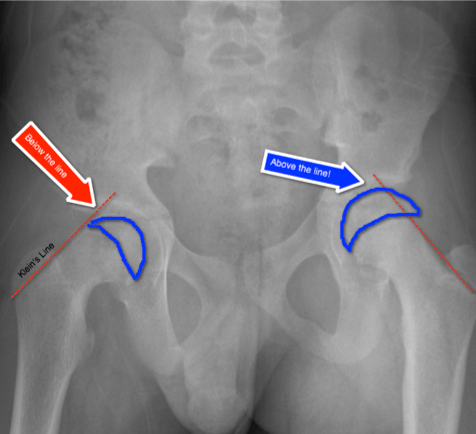
- Classic teaching is Klein’s line
Klein’s Line on AP view
- A line drawn from the superior aspect of the femoral neck will not intersect the femoral head epiphysis
- Modified line
- >2mm difference in width lateral to line between each side
https://pedemmorsels.com/wp-content/uploads/2018/01/Slipped-Capital-Femoral-Epiphysis-3.png
Another virtual line may assist in diagnosis
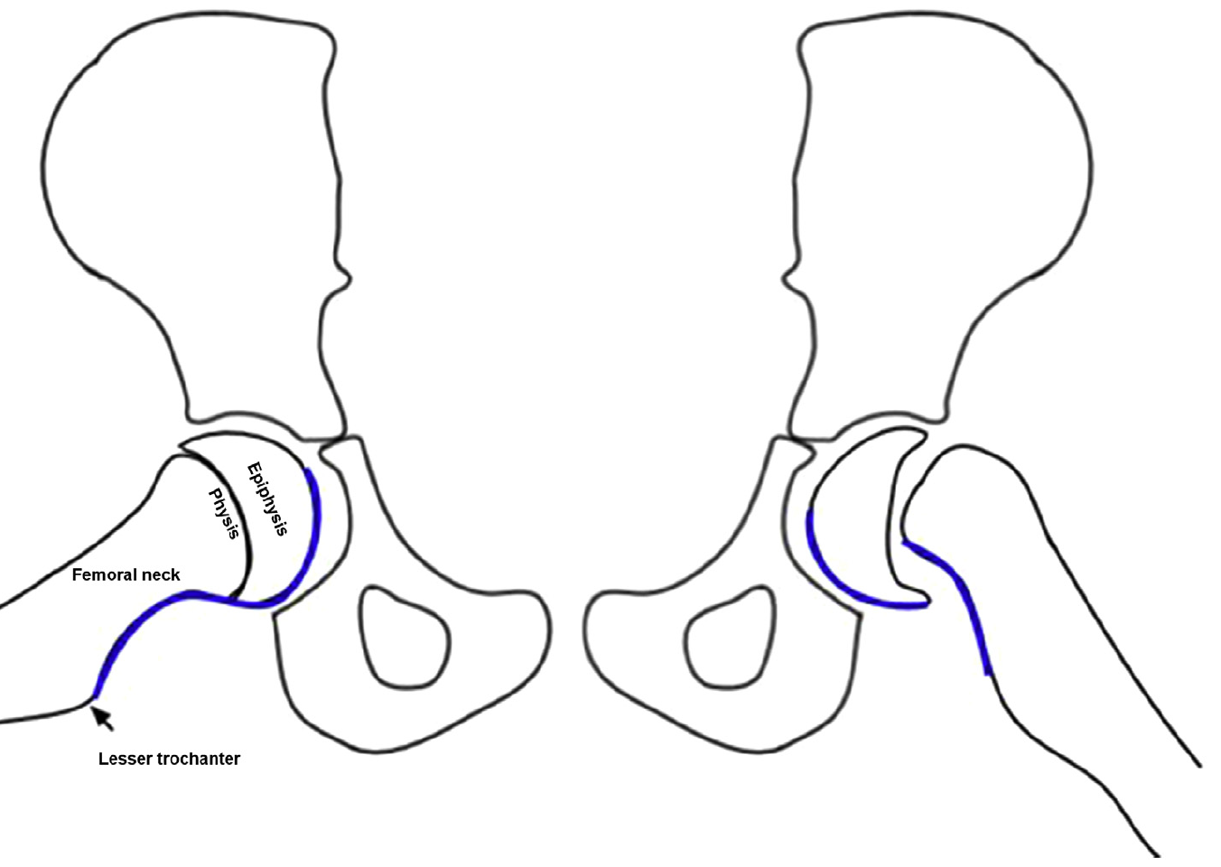
S-sign
- The S-sign is a curvilinear line drawn on the inferior margin of the proximal femoral head neck junction along the proximal femoral physis.
- Discontinuity or an abrupt sharp turn are abnormal
Klein's line and S-sign
- A group of 20 orthopedic surgeons, radiologists, and pediatricians viewed 35 radiographs of SCFE using Klein's line on the AP view and the S-sign on frog-leg lateral view to make the diagnosis.
- Overall diagnostic accuracy was better with the S-sign than Klein's line, 92% vs 79%.
- Sensitivity of the S-sign was 89%, specificity 95%.
- Sensitivity of Klein's line was 68%, specificity 89%.
- Combined S-sign + Klein's line sensitivity was 96%, specificity 85%.
Consider adding both of these virtual lines/signs to your review of the pediatric hip plain film
Show References
The role of skeletal muscle relaxants in the management of lower back pain in the ED
Patients with lower back pain (LBP) presenting to the ED are often treated with NSAIDs plus skeletal muscle relaxants.
A recent study in Annals of Emergency Medicine compared functional outcomes and pain in ED patients with acute non radicular LBP with 4 different treatment regimens.
- Ibuprofen plus placebo
- Ibuprofen plus baclofen
- Ibuprofen plus metaxalone
- Ibuprofen plus tizanidine
Conclusion: Adding a muscle relaxant to ibuprofen did not improve pain or improve function at 1 week following an ED visit for LBP.
Note: Prior studies have found no benefit to adding opioids or diazepam to NSAIDs for ED patients with acute non radicular LBP
Show References
- Decreased hepatic function
- Decreased renal function
- Multiple comorbidities and polypharmacy that can affect pharmokinetics of pain medications.
Therefore, pain medications must be dosed carefully, which runs the risk of underdosing. Pain medications can also contribute to delerium, and decreased functional status.
Recommendations:
- Start with non-opioid medications in most cases. Consider combination acetaminophen and ibuprofen/naproxen.
- Consider regional nerve blocks where applicable due to the decreased risk of systemic side effects and excellent analgesic properties.
- If using opioids, start low and reassess and use the lowest dose possible. Remember half-lifes are often prolonged so patient may not need the standard dosing interview.
Lateral hip pain is a common presentation of hip pain.
Typically seen in runners and women over the age of 40 who start unaccustomed exercise.
Pain from OA of the hip which is typically medial (groin pain)
Lateral hip pain has traditionally been diagnosed at trochanteric bursitis.
Research suggests that lateral hip pain may be multifactorial and better termed Greater trochanteric pain syndrome.
Pain from the gluteal medius and/or minimus due to non-inflammatory tendonopathy is likely causative. This may cause a secondary bursitis.
Pain is insidious, gradual worsens and is variable based on activity type.
Also, can be seen after a fall resulting in tearing.
Pain is described as a deep ache or bruise. It can stay localized or radiate down lateral thigh towards knee.
Patients report night/early morning pain and when rolling over onto the outer hip on affected side.
Fatigue from prolonged sitting, walking and single leg loading activities such as walking up stairs.
Provoking activities and postures cause compressive forces on the involved tendons.
These generally occur when the hip is adducted across midline such as with
Side sleeping,
Place pillow between legs to align pelvis and keep knee and hip in line
Crossed leg sitting
Sit w/ knees at hip distance and feet on floor
Selfie poses - Standing w a hitched hip (pushing hip to the side).
Attempt to correct biomechanical issues before progressing directly to bursal steroid injection
May only be a temporary fix if underlying issue not addressed.
A helpful clinical guide
https://bjgp.org/content/bjgp/67/663/479/F1.large.jpg?download=true

A recent epidemiology study in Pediatrics looked at concussions in 20 high school sports during the 2013–2014 to 2017–2018 school years.
For every athlete, one practice or competition was counted as one exposure.
Overall, 9542 concussions were reported for an overall rate of 4.17 per 10 000 athletic exposures (AEs).
Football continues to have the highest incidence with a concussion rate of 10.40 per 10 000 AEs.
As in previous studies, rates in competition (33.19 to 39.07 per 10 000 AEs) are increasing and higher than rates in practice which are lower and decreasing over the study period (5.47 to 4.44 per 10 000 AEs).
This may reflect better reporting or increasing injury rate
In all 20 sports, recurrent concussion rates decreased from 0.47 to 0.28 per 10 000 AEs.
Confirming prior studies, among sex-comparable sports, concussion rates were higher in girls than in boys (3.35 vs 1.51 per 10 000 AEs).
Also, among sex-comparable sports, girls had larger proportions of concussions that were recurrent than boys (9.3% vs 6.4%).
This study may reflect effective implementation of strategies to reduce concussion incidence such as mandatory removal from play and more stringent requirements associated with return to play.
Show References
Synthetic turf playing surfaces have been growing in popularity over the last decade and seem to have become a new standard.
Due to the need for durable fields that can accommodate multiple teams/activities, in addition to the high cost of maintaining grass and the need to conserve water, many parks and schools have switched from grass to turf. Turf is advertised as maintenance free but ….this is not the case.
Locally, at M&T Bank Stadium, groundskeepers drive a LitterKat turf sweeper across the field for 4 hours 2-3 times a week to ensure that the synthetic rubber is cleaned and distributed evenly. The field is also repainted every 4 games because the paint may become hard. The cost of this level of maintenance is beyond what many parks and local high schools can afford.
A recent study examined high school concussion data at almost 2000 high schools with over 14,000 recorded concussions. Researchers concluded that more concussions occurred in games than practices. Interestingly, they also found that playing surface was significantly associated with concussion. Almost 90% of all injuries occurred on turf-based surfaces. Turf outweighed all other mechanisms of injury, including helmet-to-helmet hits and grass playing surface. Between 10 and 15.5% of concussions occur from helmet to ground contact. In the NFL, this mechanism accounts for about 1 in 7 concussions.
Attempting to limit total exposure time in practice and games on turf surfaces may be beneficial until more study is needed.
Intersection Syndrome
De Quervain’s is a common tenosynovitis is involving the the 1st dorsal compartment of the wrist/forearm.
Intersection syndrome is a tenosynovitis that occurs at the intersection of the 1st and 2nd dorsal compartments.
Pathology located at crossing point of the 1st compartment structures (APL and EBP) with the radial wrist extensors (ECRB and ECRL)
Occurs most commonly from repetitive wrist extension and is common in rowers, weight lifters, and in those playing racquet sports.
Occurs about 4 to 6cm proximal to the radiocarpal joint VERSUS De Quervain’s which occurs near the level of the radial styloid.
Pain worse with resisted wrist and thumb extension
Radiographs not required
Splint and start NSAIDs
Recalcitrant cases can be referred for corticosteroid injection
https://stemcelldoc.files.wordpress.com/2012/09/intersection-syndrome-referral-pain-pattern1.jpg

Imaging of Lisfranc Injuries
Tarsometatarsal fracture-dislocation
Anatomy
3 Columns of the midfoot, divided by the tarsometatarsal joints
- Medial
- First TMT joint
- Middle
- 2nd and 3rd TMT joints
- Lateral
- 4th and 5th TMT joints
The Lisfranc ligament
- Extends from the 2nd MT to the medial cuneiform
- Critical to structure and stabilization of the 2nd MT and the midfoot arch
Imaging
Plain films: AP/lateral/oblique
Consider weight bearing view with contralateral comparison if high suspicion
CT: Can be useful to confirm abnormal plain films
MRI: not done in ED but can be used to diagnose pure ligament injuries
Below is a review of the lines of the foot which will ensure not missing this diagnosis. May be helpful to review with sample imaging.
Plain films findings: https://prod-images.static.radiopaedia.org/images/49189279/86408d5bae08ab80ae9ef377337ab7_big_gallery.jpeg

On AP view:
- Discontinuity of a line drawn from the medial part of 2nd MT to the medial side of the 2nd cuneiform
- Widening of the interval between the 1st and 2nd ray
- Bony fragment in 1st MT space (fleck sign) – Lisfranc ligament avulsion
On Lateral view:
- Dorsal displacement of the proximal 1st or 2nd MT (may be subtle)
On the Oblique view:
- Discontinuity of a line drawn from the medial border of the 3rd cuneiform with the medial border of the 3rd MT
- Discontinuity of a line drawn from the medial side of the 4th MT with the medial side of the cuboid
Remember that the lateral margin of the 5th MT can project lateral to the cuboid (up to 3 mm)
Lines drawn on 2 view foot for review
https://radiopaedia.org/cases/lisfranc-ligament-normal-alignment
Show References
Tarsometatarsal fracture-dislocation
The Lisfranc ligament is critical for stabilization of the midfoot arch and the 2nd MT
Injuries can range from mild (sprains) to severe (gross dislocation)
Injury may be purely ligamentous injuries or a fracture-dislocations
Difficult diagnosis to make
https://www.aafp.org/afp/1998/0701/afp19980701p118-f4.jpg

Mechanisms: MVAs, fall from height or athletic injuries
Common athletic mechanism: Axial load to a hyperplantar flexed forefoot

Injury severity is often underestimated
Severe pain and inability to weight bear
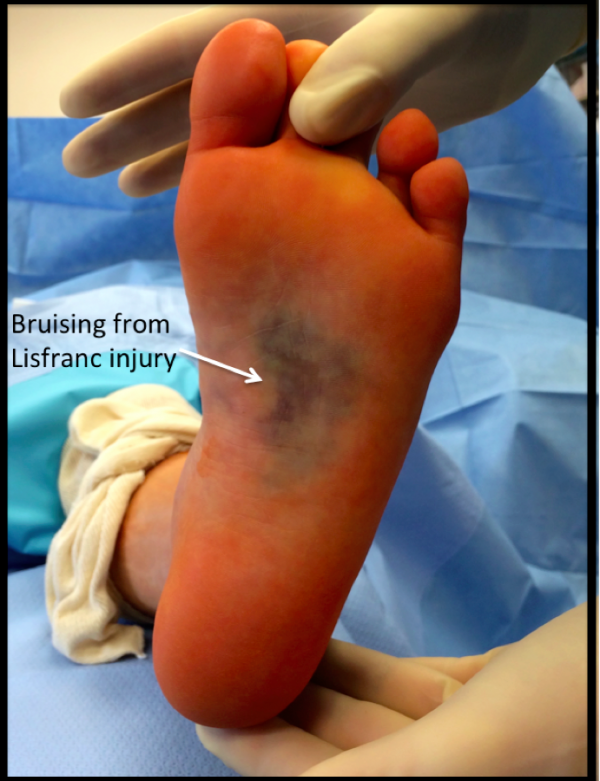
Plantar bruising and bruising throughout midfoot
No specific tests as exam is limited due to pain
Midfoot stress tests
-Often positive but unlikely to be allowed by patient due to pain
https://www.youtube.com/watch?v=v8SGVwz2RHs
Midfoot instability test
Grasp metatarsal heads and apply dorsal force to forefoot.
Other hand palpates the TMT joints and feels for dorsal subluxation