
Search
Subscribe to daily UMEM Pearls (FREE!) via:
201-220 of 479 results with category "Orthopedics"
22yo college track athlete presents with 3 weeks of gradual onset groin and thigh pain, worse with running, better with rest.
Stress fractures are a common cause of groin pain in athletes, particularly in long distance runners
Fractures occur in the pubic rami and femoral neck
Ask about a sudden change in training regimens
PE: check for tenderness to deep palpation over the pubic ramus. Ask athlete to stand and support full weight on affected leg or perform one legged hop on affected side. Pain out of proportion to physical examination findings.
Imaging: XR usually negative. Bone scans can be positive as early as 4 to 8 days after symptom onset. MRI used to diagnose and rule out other causes of groin pain.
Treatment: Rest for 4 to 6 weeks. Consider making patient non weight bearing if walking causes pain especially with femoral neck fractures on the superior side. Inferior side neck fractures may benefit from prophylactic fixation.
Show References
The Essex-Lopresti injury pattern is the lesser known of the triad of forearm injuries (Monteggia & Galeazzi).
It follows the “rule of the ring” aka the life saver candy rule: You can’t break a life saver in just one place.
These injury patterns are frequently missed because our eyes are drawn to the fracture and miss the associated dislocation.
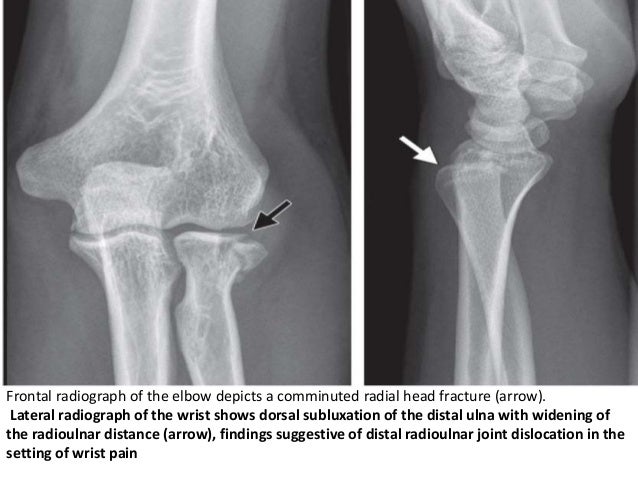
The Essex-Lopresti fracture pattern involves a fracture of the radial head with concomitant dislocation of the distal radio-ulnar joint (DRUG)
-With associated interosseous membrane disruption
Think of it as the Maisonneuve fracture of the forearm.
Mechanism: fall from height/high energy forearm trauma.
PE: Suspect if patient has significant tenderness at the DRUG with a radial head fx.
Patients have worse outcomes if injury is missed on initial presentation due to radial migration and instability.
Take home point: Remember the rule of the ring. Remember to exam the elbow with wrist injuries and the wrist with all elbow injuries
Treatment of Low Back Pain
A recent recommendation from the American College of Physicians (Internal Medicine) now recommends nonpharmacologic therapies as the first line treatment of acute or subacute lower back pain lasting 12 weeks or less. This might bring more people to our Emergency Departments so it is important that we know their current recommendations.
Some nonpharmacologic therapies recommended are:
- Moderate Evidence: Superficial heat
- Low quality evidence: Massage, Spinal manipulation, or accupuncture
For acute back pain they recommend:
- NSAIDs or muscle relaxants
- Acetominophen is NOT recommended. No evidence it is beneficial
For chronic back pain:
- Start with NSAIDs—>tramadol—>duloxetine.
- Opioids are only recommended for treatment failures.
Show References
During a busy ED shift, your 40yo charge nurse asked you to look at his hand. He is known avid mountain biker. He has pain in his right 4th and 5th digits. . He feels a lack of coordination and a feeling of “clumsiness” of the hand. Where is his possible nerve compression and what do you expect to find on exam?
Ulnar nerve entrapment is sometimes called “handlebar palsy.”
Compression location is Guyon’s canal.
The ulnar nerve supplies the intrinsic muscles of the hand AND the extrinsic muscles for flexion of the 4th and 5th digits. This is what aids in a “power grip” and why he may have diminished grip strength on exam.
Also innervates the ADDuctor pollicis and 1st dorsal interosseous muscles (pinch)
Note the ulnar nerve also passes through the radial tunnel at the elbow. Entrapment here is called Radial tunnel syndrome or Cubital tunnel syndrome and causes forearm pain and paresthesias in the 4th and 5th digits with grossly normal motor and sensory function.
You are covering a sporting event or working an ED shift when a young adolescent athlete without significant PMH presents with SOB and wheezing associated with exercise.
You immediately think exercise-induced asthma, prescribe a short-acting bronchodilator and pat yourself on the back.
While you may be right, there is increasing recognition of an alternative diagnosis
Exercise-induced laryngeal obstruction (EILO)
During high intensity exercise, the larynx can partially close, thereby causing a reduction in normal airflow. This results in the reported symptoms of SOB and wheezing.
This diagnosis has previously been called exercise induced vocal cord dysfunction. As the narrowing most frequently occurs ABOVE the level of the vocal cord, EILO is a more correct term.
While exercise induced bronchoconstriction has a prevalence of 5-20%, EILO is less common with a prevalence of 5-6%.
Patients are typically adolescents, with exercise associated wheezing and SOB, frequently during competitive or very strenuous events. Wheezing is inspiratory and high-pitched. Symptoms are unlikely to be present at time of medical contact unless you are at the event as resolution occurs within 5 minutes though associated cough or throat discomfort can persist after exercise cessation. EIB symptoms typically last up to 30 minutes following exercise.
Inhaler therapy is unlikely to help though some athletes report subjective partial relief. This may be explained as approximately 10% of individuals have both EIB and EILO.
In athletes with respiratory symptoms referred to asthma clinic, EILO was found in 35%.
Consider EILO in athletes with unexplained respiratory symptoms especially in those with ongoing symptoms despite appropriate therapy for EIB.
Show References
Take Home Point:
- According to a recent article in the NEJM there does not seem to be any difference in the rate of symptomatic venous thromboembolism (VTE) in patients given low molecular weight heparin that underwent arthorscopy or had lower leg casting at 3 months.
- Overall, the rates of VTE were really low ( casting: 1.4% vs. 1.8%; arthroscopy: 0.7% vs. 0.4%), so there is probably not need for prophalaxis in these patients.
Show Additional Information
Show References
Concussions are associated with an elevated risk of musculoskeletal injury
Significant associations were found between concussion and
Lateral ankle sprain (P = 0.012)
Knee injury (P = 0.002)
Lower extremity muscle injury (P = 0.031)
Keep in mind that 50 – 80% of concussions may go undiagnosed or unreported.
A discussion about risks of early return after concussion should include mention of risks beyond repeat head injury/2nd impact syndrome
Study limits: Retrospective design limits ability to establish causation/reporting bias
Show References
Incidence and Cost of Ankle Sprains US Emergency Departments
In a sample of 225,114 ED patients with ankle sprains:
Lateral ankle sprains represent the vast majority of all ankle sprains (91%).
Lateral ankle sprains incur greater ED charges than medial sprains ($1008 vs. $914).
Lateral ankle sprains were more likely to have associated pain in the limb, sprain of the foot and abrasions of the hip/leg than medial sprains.
Medial sprains were more likely to include imaging.
Hospitalizations were more likely with high ankle sprains than lateral sprains.
There is a higher incidence of ankle sprains in younger patients (≤25 years) and in female patients (57%).
Show References
Question
https://images.radiopaedia.org/images/3173801/1ee24da1a6fe907a27d2bf20481174.jpg

Young toddler presents with left lower leg pain. What is the diagnosis??
Show Answer

Many of us use exercise as a coping strategy when emotionally stressed or to blow off steam when angry. This may place your heart at risk.
A recent observational study in Circulation surveyed 12,000 first MI patients about potential triggers. The associations didn't depend on age, smoking status, hypertension, or baseline physical activity.
Anger or emotional upset in the hour before onset elevated odds of MI 2.44 fold
A similar 2.31 fold elevation was observed form heavy exertion
However, the combination of the two raised the odds to 3.05 fold (P<0.001 for interaction)
Show References
Davos Shoulder Reduction Technique
Take Home Points
- Uses the patients own weight to reduce their anterior shoulder dislocation.
- No sedation is required
- Provider exerts no effort and only sits on the patients foot.
Interested, well find out more by watching this video by Larry Mellick https://www.youtube.com/watch?v=u2MsnjVNoPM or clicking the link below.
Show Additional Information
Recurrence depends on age and activity level
27% if >30yo and 72% if <23yo
Surgical Recommendations:
Large bony Bankart lesion, glenoid or humeral head defect >25%, recurrent instability, event near the end of season
Non surgical return to play:
If event occurs at beginning/early in season
Rehabilitation for 2 to 3 weeks (most return to play in this time frame)
Immobilization for 3 to 7 days in simple sling, gentle range of motion, cryotherapy
Physical therapy to strengthen dynamic stabilizers
Shoulder stabilization brace for non overhead throwing and contact sports
Show References
Retroperitoneal hemorrhage
The pathophysiology is unknown. Some hypothesize that occult vasculopathy and arteriosclerosis of the small vessels in the retroperitoneum may render them friable and therefore prone to rupture. This can be seen in minor trauma in sports and forceful vomiting or coughing. Spontaneous bleeding starts at the microvascular level, and large vessels become disrupted or stretched as the hematoma enlarges.
Retroperitoneal hemorrhage occurs in a variety of clinical circumstances, including spontaneous hemorrhage into a pre-existing benign adrenal cyst or bleeding from a left inferior phrenic artery, tumors of the adrenal gland and kidney, rupture of any blood vessel (most commonly infrarenal aorta); percutaneous interventions (such as cardiac catheterization), trauma, and polycythemia vera,
It is most commonly seen in association with patients with bleeding abnormalities, in HD patients and with anticoagulation therapy,. Risk is much greater with unfractionated heparin therapy than with warfarin. In most of the heparin patients studied, their coagulation parameters were in the therapeutic range.
Patients may present to the non acute area of the ED with back, lower abdominal or groin discomfort, Over time, this may progress to hemodynamic instability, and a fall in hemoglobin, Early identification is crucial to improving patient morbidity and mortality. Early symptoms depend on the location of the bleeding.
Hematoma near or within the iliopsoas muscle usually presents as femoral neuropathy (groin pain or leg weakness).
Femoral neuropathy caused by retroperitoneal hematoma can present with sudden onset severe pain in the affected groin and hip, with radiation to the anterior thigh and the lumbar region. This can easily be missed as the presentation is similar to a pulled msucle or strained hip/back. Iliopsoas muscle spasm often results in the characteristic flexion and external rotation of the hip, and any attempt to extend the hip will result in severe pain. Over time, pain and parasthesia in the antero-medial thigh and leg is seen.
Show References
Patellofemoral Syndrome Treatment options
Patients do best with a combined intervention (ie, exercise therapy, education, manual therapy and taping) plan or patellofemoral bracing may improve outcomes for people with patellofemoral syndrome and the subtype of patellofemoral osteoarthritis.
For for the ED, we can start NSAIDs, and then have them follow up with Physical Therapy, A sports trainer if in organized sports, or with a sports medicine physician/PCP. Physical therapy is targeted at strengthening the quadricep muscle particularly vastus medialis, which improves the patella’s tracking with knee flexion.
Young athletes, especially around the age of puberty, are at higher risk for pelvic avulsion fractures
Often seen in sports that require sprinting, rapid changes in movement or jumping
Caused by sudden, forceful contraction of the muscles of the abdominal, the hip and thigh or the hamstring
Avulsion fractures can occur at many areas of the pelvis.
A mnemonic is: Alabama’s stoned rappers got ill hunting armadillos
· Iliac crest: Abdominal muscles
· Anterior superior iliac crest: Sartorius
· Anterior inferior iliac crest: Rectus femoris
· Greater trochanter: Gluteal muscles
· Lesser trochanter: Iliopsoas **(rare in adults)
· Ischial tuberosity: Hamstrings
· Pubic symphysis: Adductor group
http://roentgenrayreader.blogspot.com/2010/07/pelvic-avulsion-fractures.html
** Isolated nontraumatic avulsion fractures of the lesser trochanter in adults is a pathognomonic sign of metastatic disease
This site has some good images of common injury patterns
http://radiopaedia.org/articles/apophyseal-avulsion-fractures-of-the-pelvis-and-hip
Show References
According to the 4th International Patellofemoral Pain Research Retreat recently published in British Journal of Sports Medicine, the core criterion required to define Patelofemoral Pain (PFP) syndrome is pain around or behind the patella, which is aggravated by at least one activity that loads the patellofemoral joint during weight bearing on a flexed knee (eg, squatting, stair ambulation, jogging/running, hopping/jumping).
Additional criteria (not essential):
- Crepitus or grinding sensation emanating from the patellofemoral joint during knee flexion movement
- Tenderness on patellar facet palpation
- Small effusion
- Pain on sitting, rising on sitting, or straightening the knee following sitting
PFP is common in young adolescents, with a prevalence of 7–28%, and incidence of 9.2%.
Stay tuned for recommendations on treatment and diagnosis.
Show References
47yo M chef presents to your ED with 2 days of worsening left hand pain after sustaining a puncture wound to hand at work. The hand is red and swollen and he complains of pain. Interestingly, his index and middle digits are in an ABducted position at rest.
Collar Button Abscess
Web space infection of the palmer AND dorsal hand
The Palmer aponeurosis prevents volar extension (but promotes dorsal encroachment)
The pus spreads between the MC bones and erupts dorsally....creating a DORSAL abscess.
Loss of palmer concavity is seen.
ABduction of the adjacent digits, resulting in a "V" configuration with the apex pointing to the site of infection. This would not happen from simple pus in the dorsal space!
Can be missed if only focused on the dorsal hand. The palm will show the original injury (splinter, cut, foreign body)
Treatment is urgent surgical drainage.
http://www.eplasty.com/article_images/eplasty16ic06_fig1.gif
Show References
30yo male weight lifter who 10 days ago had a painful left shoulder injury after bench press. The next morning his left anterior chest wall and left upper arm were bruised and swollen. He went to see his PCP who diagnosed him with a muscle strain. 8 days later the bruising and swelling have resolved but he still cant move his shoulder and comes to the ED.
http://321gomd.com/wp-content/uploads/2015/01/pec-major-tears.jpg
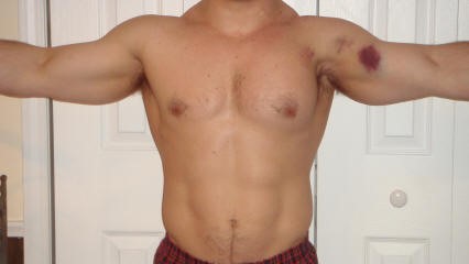
The pec major attaches to the humerus and originates from the sternum and clavicle
Injury is usually due to tendon rupture off the humerus but can also occur at the muscle tendon junction or within the muscle belly itself.
Injury is becoming increasingly common due to the popularity in power lifting sports.
Mechanism: excessive tension on a maximally eccentrically contracted muscle.
Patients will complain of pain and weakness of the shoulder.
PE: Swelling and bruising to anterior medial arm. Palpable defect and deformity or anterior axially fold (may be hidden by swelling).
Weakness and pain with adduction and internal rotation and forward flexion
Chronic presentations can be challenging to diagnose. Consider ultrasound
Non operative treatment may be indicated for partial tears (sling, ice, NSAIDs)
Operative repair of tendon avulsions is very successful. Patients age, occupation/activity level and location of injury and condition of tear are considered.
Sesamoid Injuries
Unlike other bones in the human body that are connected to each other at joints, sesamoid bones are only connected via tendons (or are imbedded in muscle).
The largest sesamoid bone is the patella.
2 small sesamoid bones lie on the plantar foot near the great toe
Sesamoid bones can fracture and the surrounding tendons can become inflamed (sesamoiditis)
Traumatic injury is usually due to hyperextension and axial loading
Sx: Pain located under the great toe on the ball of the foot (Gradual with sesamoiditis and acutely with a fracture).
There may be associated swelling and bruising. Pain with palpation, flexion and extension.
The medial/tibial sesamoid is larger, has great weight bearing status and is more commonly injured that its lateral counterpart.
In many people (10 - 25%) the medial sesamoid of the foot has two parts (bipartite). This finding is bilateral in 25% of people.
This may confuse some providers as it may appear to be a fracture
Look for a smooth contour to the bones and clinically correlate (bruising, soft tissue swelling, etc.) if it is an incidental finding.
Other radiographic clues include
1) The fractured sesamoid is usually slightly larger than the lateral sesamoid while the bipartite sesamoid has a much larger medial sesamoid than lateral sesamoid
2) The fractured sesamoid shows a sharp, radiolucent, uncorticated line between the two fragments while the bipartite sesamoid has two corticated components
3) The fractured sesamoid fragments often fit together like pieces of a puzzle while the bipartite sesamoid has two components that do not fit together snugly
4) Other means to differentiate the two involve MRI and bone scanning
Treatment involves a stiff-soled shoe or applying a cushioning pad or J-shaped pad around the area to relieve pressure.
It may take months for the pain to subside.
http://www.apfmj-archive.com/afm5_3/afm50.htm#F1
Show References
Plain films are commonly used to screen children for pelvic fractures or dislocations following blunt torso trauma
The sensitivity of this common screening practice is unknown
A recent study looked at this question.
Of 451 patients with pelvic fractures or dislocations, 382 had AP radiographs. Injury was correctly identified in 297 patients (sensitivity 78%).
The sensitivity was greater in the sicker subgroups :92% for those requiring operative intervention and 82% for those with hypotension
Plain AP pelvic radiographs should have a limited role in the sole evaluation of children with blunt torso trauma.
They should be incorporated in the assessment of hemodynamically unstable children and those in whom the clinician is not planning on otherwise obtaining an abdominal/pelvis CT.