
Search
Subscribe to daily UMEM Pearls (FREE!) via:
341-360 of 889 results with category "Critical Care"
Noninvasive Ventilation in De-Novo Respiratory Failure
- Noninvasive ventilation (NIV) is a primary therapy for patients with acute hypercapnic respiratory failure, especially those with an acute COPD exacerbation.
- Notwithstanding its benefits in COPD and acute cardiogenic pulmonary edema, NIV should be used cautiously in patients with "de-novo" respiratory failure.
- Many patients with de-novo respiratory failure will meet criteria for ARDS and have a high rate of intubation (30% - 60%).
- The use of NIV with delayed intubation in this patient population has been associated with increased mortality.
Show References
Hyperoxia has been repeatedly demonstrated to be detrimental in a variety of patients, including those with myocardial infarction, cardiac arrest, stroke, traumatic brain injury, and requiring mechanical ventilation,1-4 and the data that hyperoxia is harmful continues to mount:
- Systematic review and meta-analysis of 16,000 patients admitted to hospital with sepsis, trauma, MI, stroke, emergency surgery, cardiac arrest: liberal oxygenation strategy (supplemental O2 for average SpO2 96%, range 94-100%) associated with increased in-hospital and 30-day mortality compared to conservative strategy.5
- ED patients requiring mechanical ventilation admitted to ICU: hyperoxia defined as PaO@ >120mmHg. Patients with hyperoxia in the ED had higher mortality than not only normoxic but hypoxic patients (30% v 19% v 13% respectively), and longer vent days and ICU/hospital LOS.6
- ICU patients, majority respiratory failure, 60% requiring mechanical ventilation; hyperoxia defined as PaO2 >100mmHg. Just ONE episode of hyperoxia an independent risk factor for ICU mortality (OR 3.80, 95% CI 1.08-16.01, p=0.047).7
Bottom Line: Avoid hyperoxia in your ED patients, both relatively stable and critically ill. Remove or turn down supplemental O2 added by well-meaning pre-hospital providers and nurses, and wean down ventilator settings (often FiO2). A target SpO2 of >92% (>88% in COPD patients) or PaO2 >55-60 is reasonable in the majority of patients.8
Show References
A few (out of 10) tips for the care of sick patients with liver failure:
- Use of albumin is indicated to improve outcomes in spontaneous bacterial peritonitis (SBP), large-volume paracentesis, and hepatorenal syndrome (HRS).
- Norepinephrine remains the vasopressor of choice for nonhemorrhagic shock. Use vasopressin or terlipressin (outside the U.S.) in AKI due to HRS to maintain a target MAP and for splanchnic vasoconstriction.
- INR does not correctly reflect coagulation performance. Platelet count and fibrinogen are the best predictors of bleeding, and thromboelastography (via TEG/ROTEM) can reduce blood products administered for hemorrhage without affecting mortality.
- If a nasogastric tube is indicated (administration of lactulose, decompression of SBO, etcetera), presence of [non-recently banded] esophageal varices is not a contraindication.
Show References
Identifying Critically Ill Cancer Patients in the ED
- Immunosuppressed patients with malignancy are at high risk of complications and rapid decompensation.
- Select pearls in identifying ED patients with cancer that are at high risk of critical illness include:
- Patients with profound neutropenia (< 100/mm3) are at high risk for fungal infections (i.e., aspergillosis)
- Hypoxemia that requires oxygen is a predictor of later ICU admission.
- Patients with bilateral infiltrates on CXR are at high risk of decompensation. Consider ICU admission.
- Patients with promyelocytic leukemias are at high risk of DIC. Patients with this complication should be admitted to the ICU.
Show References
The most recent AHA guidelines for goal blood pressure after return of spontaneous circulation (ROSC) post-cardiac arrest recommend a definite mean arterial pressure (MAP) goal of > 65 mmHg.1 There is no definitive data to recommend a higher specific goal, but there is some evidence to indicate that maintaining higher MAPs may be associated with better neurologic outcomes.2
A recently published prospective, observational, multicenter cohort study looked at neurologic outcomes corresponding to different MAPs maintained in the initial 6 hours post-cardiac arrest.3
Findings:
1. Compared to lower blood pressures (MAPs 70-90 mmHg), the cohort with MAPs > 90 mmHg had:
- a higher rate of good neurologic function at hospital discharge (42 vs.15%, p < 0.001)
- a higher rate of survival to 72 hours (86 vs. 74%, p=0.01) and hospital discharge (57 vs 28%, p < 0.001)
2. The association between MAP > 90 mmHg and good neurologic outcome was stronger among patients with a previous diagnosis of hypertension, and persisted regardless of initial rhythm, use of vasopressors, or whether the cardiac arrest occured in or out of hospital.
3. There was a dose-response increase in probability of good neurologic outcome among all MAP ranges above 90 mmHg, with MAP >110 mmHg having the strongest association with good neurologic outcome at hospital discharge.
Note: The results of a separate trial, the Neuroprotect post-CA trial, comparing MAPs 85-100 mmHg to the currently recommended MAP goal of >65 mmHg, are pending.4
Bottom Line: As per current AHA guidelines, actively avoid hypotension, and consider use of vasopressor if needed to maintain MAPs > 90 mmHg in your comatose patients post-cardiac arrest, especially those with a preexisting diagnosis of hypertension.
Show References
We know that high flow nasal cannula is an option in the management of acute hypoxic respiratory failure without hypercapnea. A newer iteration of high flow, "high velocity nasal insufflation" (HVNI), may be up-and-coming.
According to its makers (Vapotherm), it is reported to work mainly by using smaller bore nasal cannulae that deliver the same flows at higher velocities, thereby more rapidly and repeatedly clearing dead space, facilitating gas exchange and potentially offering ventilatory support.
In an industry-sponsored non-inferiority study published earlier this year:
- 204 adult patients in 5 EDs
- Any acute respiratory failure deemed by the treating physician to require non-invasive positive pressure ventilation (NPPV)
- Patients randomized to either NPPV (bilevel positive airway pressure) or HVNI
- Rate of HVNI treatment failure (26%) and intubation @ 72 hours (7%) fell within predefined noninferiority margins
- Rates of PCO2 clearance were similar between HVNI and NPPV groups
- The study was not powered to detect differences between different etiologies for respiratory failure
- Authors concluded that HVNI is noninferior to NPPV for all-comer respiratory failure.
Bottom Line:
The availability of a nasal cannula that helps with CO2 clearance would be great, and an option for patients who can't tolerate the face-mask of NPPV would be even better.
HVNI requires more investigation with better studies and external validation before it can really be considered noninferior to NPPV, but it certainly is interesting.
Show References
Sedating Mechanically Ventilated Patients
- Providing appropriate analgesia and sedation to mechanically ventilated patients is of paramount importance.
- In a recent systematic review and meta-analysis, Stephens et al. assessed the impact of deep sedation within the first 48 hours of initiation of mechanical ventilation.
- In 9 studies that included over 4,500 patients, deep sedation within the first 48 hours of initiation of mechanical ventilation was associated with increased mortality, increased ICU LOS, and increased frequency of delirium.
- Take Home Points
- When possible, target lighter levels of sedation in mechanically ventiilated patients.
- Though no universally accepted definition of light sedation exists, most studies use a RASS of -2 to +1
Show References
The recently published BICAR-ICU study looked at the use of bicarb in critically ill patients with severe metabolic acidemia...
- Multicenter, open-label, RCT, 26 French ICUs
- Adult patients with pH < 7.2 not secondary to hypercapnia, serum bicarb < 20 not due to bicarb wasting process
- SOFA score > 4 or lactate > 2
- No bicarb versus 4.2% sodium bicarb infusion titrated to pH >7.3
- Primary outcome: Composite measure of 28-mortality and presence of any organ failure at 7 days post-randomization
- Secondary outcomes: Need for/length of life support measures (renal-replacement, vasopressors, mechanical ventilation), SOFA score after enrollment, electrolyte effects, occurrence of ICU-acquired infections, and ICU length of stay
- Major findings:
- No difference in primary outcome overall
- No difference in pressor-free days, days off RRT, dialysis dependence at ICU discharge, ICU LOS
- Bicarb group had less need for RRT during ICU stay (35 vs 52%, p=0.0009)
- In patients with AKI and AKIN score 2-3*, the bicarbonate group had a decrease in both 28-day mortality (46 vs 63%, p=0.0166) and presence of any organ failure at day 7 (66 vs 82%, p=0.0142)
- Limitations:
- Unblinded
- A quarter of the control group actually received bicarb
- No data regarding vent settings, ABGs to r/o ventilation effects on pH
- 4.2% is not a standard concentration of bicarb used in the U.S.
Bottom Line:
Consider administration of sodium bicarbonate for your critically ill ED patients with severe metabolic acidosis and AKI, especially if acidosis &/or renal function is not improved with usual initial measures (such as IVF, etc).
*Acute Kidney Injury Network Staging Criteria

Show References
Does Lactated Ringer's Raise Serum Lactate?
- Intravenous fluid administration is a cornerstone of resuscitation and the treatment of many critically ill ED patients.
- Recent publications have suggested that balanced crystalloid solutions may be better than 0.9% normal saline (NS) for select conditions.
- Lactated Ringer's (LR) is a common balanced crystalloid solution often used for fluid resuscitation in critically ill patients.
- AS LR contains approximately 28 mmol/L of sodium lactate, the question of whether LR elevates serum lacate is frequently asked.
- In a recent small, randomized, double-blind, controlled trial, investigators randomized healthy volunteers to receive 30 ml/kg of either 0.9% NS or LR. The authors report no statistical difference in the mean serum lactate when comparing LR to 0.9% NS.
Show References
Critical Post-Arrest Interventions
- Critical interventions to optimize neurologic outcome in the post-cardiac arrest patient include optimizing hemodynamics, preventing lung injury, maintaining normal O2 and CO2 tensions, targeted temperature management, and treating the underlying cause of the arrest.
- Current guidelines recommend the following:
- Target MAP > 70 mm Hg with IVFs, vasopressors, and inotropes.
- Use a low tidal volume strategy of 6 to 8 ml/kg predicted body weight.
- Decrease FiO2 to maintain SpO2 94% to 97%.
- Adjust RR to maintain PaCO2 35 to 45 mm Hg
- Initiate TTM with the goal temperature between 32 to 36o C
Show References
The highly-awaited PARAMEDIC2 trial results are in:
- Multicenter, double-blinded, randomized controlled trial of prehospital OHCA care
- 1mg IV epinephrine vs saline placebo, every 3-5 minutes
- 8014 OHCA patients over the age of 16 (excluded pregnant patients, anaphylactic and asthmatic cardiac arrests)
- Primary outcome: 30 day survival
- Secondary outcomes:
- Survival to hospital admission
- ICU and hospital LOS
- Survival to hospital discharge and at 3 months
- Neurologic outcomes at hospital discharge and at 3 months, "favorable" if mRS≤3
- Results:
- Higher 30 day survival in Epi group (3.2 vs 2.4%, unadj OR 1.39; 95% CI 1.06 to 1.82; P=0.02)
- No difference in ICU or hospital LOS
- No difference in favorable neurologic outcomes at discharge or 3 month
- Worse neurologic outcomes in the epinephrine survivors (mRS 4 or 5 in 31% of epi group vs. 17.8% of placebo)
Interestingly, the authors also queried the public as to what mattered to them most:

Bottom Line:
- As has been demonstrated in previous studies, use of bolus-dose epinephrine results in increased rates of ROSC.
- This survival comes with the trade-off of worsened neurologic function, a condition not in a majority of patients' personal wishes.
- Epinephrine "1mg every 3-5 minutes'" should no longer be the dogma of OHCA resuscitation.
Show Additional Information
Show References
Respiratory alkalosis is the most common acid-base disturbance in acute severe asthma.
Lactic acidosis is also extremely common, developing in up to 40%. This may be related to:
- tissue hypoxia
- increased respiratory muscle usage related to work of breathing
- beta agonist therapy
The first report of beta agonist administration associated with hyperlactatemia was in 1981 in patients treated for preterm labor with terbutaline. Since then, numerous case reports and studies have linked IV and inhaled beta agonist administration with the development/worsening of lactic acidosis in severe asthmatics in the ICU and in the ED.
The exact mechanism is unclear, but is thought to be related to adrenergic stimulation leading to increased conversion of pyruvate to lactate.
In a study published in Chest in 2014, investigators evaluated plasma albuterol levels and serum lactate levels, as well as FEV1.
They found plasma albuterol levels correlated with lactate concentration and maintained significant association after adjusting for asthma severity (suggesting the association was independent of work of breathing/respiratory muscle usage).
Furthermore, several reports have suggested that dyspnea may improve in patients with elevated lactate and acidosis after beta agonists are withheld.
Take Home Points:
- Beta agonist therapy may contribute to lactic acidosis.
- Lactic acidosis may contribute to respiratory distress.
- In patients on prolonged, high-dose beta agonist therapy, consider checking a serum lactate periodically. If elevated, consider whether worsening lactic acidosis is contributing to respiratory distress and contemplate transitioning to less frequent treatments.
-Patients with severe asthma exacerbation and elevated serum lactate must have thorough evaluation for true tissue hypoxia/hypoperfusion. **Beta agonist associated hyperlactatemia should be a diagnosis of exclusion.**
Show References
Improving Analgesia in Mechanically Ventilated ED Patients
- An analgosedation approach for mechanically ventilated patients has been shown to decrease the duration of mechanical ventilation and ICU LOS.
- The latest guidelines from the Society of Critical Care Medicine recommend an opioid as the initial agent, followed by a non-benzodiazepine sedative.
- Benzodiazepines have been shown to increase ICU delirium, increase the duration of mechanical ventilation, and increase ICU LOS.
- In a recent cohort study, ED physicians increased the use of opioid analgesics and markedly decreased the use of benzodiazepines in mechanically ventilated ED patients through an educational campaign and implementation of an electronic orderset.
- Take Home Point: An electronic health record orderset for mechanically ventilated ED patients can be helpful to guide clinicians and utilize an analgosedation approach.
Show References
When a do-not-intubate (DNI) hospice patient arrives in the ED with respiratory distress, consideration of non-invasive positive pressure ventilation (NIPPV) could invoke either a “What other option do I have?” or “Why torture the patient and prolong the dying process?” sentiment.
But what’s the data?
A recently-published meta-analysis1 found that in DNI patients receiving NIPPV, there was a 56% survival rate to hospital discharge and 32% survival to 1-year.
- Higher survival was seen in patients with COPD and pulmonary edema as the cause of their respiratory failure, as opposed to pneumonia or malignancy.
- In surviving patients, there was no decrease in quality of life at 3 months; quality of life was not assessed in the time before death in nonsurvivors.
- In comfort-measures only (CMO) patients, patients receiving NIPPV had a mildly lower dyspnea score with less opiates required/administered.
Independent studies have demonstrated:
- Better survival with NIPPV for DNI COPD and CHF patients2,3,4 who are awake and have a good cough.4
- No decrease in health-related quality of life or post-ICU psychological burden (symptoms of PTSD, anxiety, or depression) in DNI survivors receiving NIPPV.3
- 63% survival to hospital discharge & 49% survival to 90 days in DNI patients receiving NIVV, with no decrease in health-related quality of life in survivors. Survival was lower for CMO patients (14% and 0% at discharge and 90 days, respectively).5
Bottom Line:
- NIPPV can benefit DNI patients -- most identifiably those with COPD or cardiogenic pulmonary edema as the etiology for their respiratory distress.
- Mild benefits to dyspnea have been seen in CMO patients, without survival benefit. A trial of NIPPV therapy may be reasonable (especially in COPD or CHF) after frank discussion with the patient and his/her loved ones, with quick cessation if comfort is not achieved and/or more discomfort is caused.
Show References
Legionella is an important cause of community-acquired pneumonia. It ranks among the three most common causes of severe CAP leading to ICU admission and carries a high mortality rate – up to 33%. Resulting from inhalation of aerosols containing Legionella species and subsequent lung infection, it is often associated with contaminated air conditioning systems, and other hot and cold water systems.
Recommended antibiotic regimens include a fluoroquinolone, either in monotherapy or combined with a macrolide (typically Levaquin +/- or Azithromycin).
A retrospective, observational study published in the Journal of Antimicrobial Chemotherapy in 2017 looked at 211 patients admitted to the ICU with confirmed severe legionella pneumonia treated with a fluoroquinolone vs a macrolide and monotherapy vs combination therapy. Combination therapy included fluoroquinolone + macrolide, fluoroquinolone + rifampicin, or macrolide + rifampicin.
Of these 211 cases, 146 (69%) developed ARDS and 54 (26%) died in the ICU. Mortality was lower in the fluoroquinolone-based group (21%) than in the non-fluoroquinolone based group (39%), and in the combination therapy group (20%) than in the monotherapy group (34%). In a multivariable analysis, fluoroquinolone-based therapy, but not combination therapy was associated with a reduced risk of mortality (HR 0.41).
Take Home Points:
-Remember, our usual blanket coverage with vanc + zosyn in the ED does not cover atypicals!
-Consider Levaquin instead of Azithro if there is clinical concern for Legionella PNA
-hyponatremia, abnormal LFTs may be clues in the appropriate context
Show References
Volume Responsiveness, Carotid Ultrasound, and the PLR
- Passive Leg Raise (PLR) is accomplished by starting with the patient at a 45’ semi recumbent position, lowering the body to horizontal, passively raising the patients legs to 45’ for 30-90 seconds, then returning the patient to the semi-recumbent position.
- To assess volume responsiveness using PLR, you must assess cardiac output (CO) and not simply look at the changes in blood pressure or heart rate.
- Previous papers have shown EtCO2 to be a reasonable surrogate of CO with PLR when ventilation is unchanged.
- Another option for measuring CO is carotid ultrasound. One study demonstrated good correlation between carotid ultrasound and invasive measurements on ICU patients. It is calculated using the equation Diameter * VTi, where VTI is the velocity time integral.
- Take Home Point - Be sure to measure CO with a PLR to help determine volume responsiveness- EtCO2 or carotid ultrasound can be considered as surrogates of CO.
Show References
Although not specifically a part of current recommendations due to lack of data, the AHA has previously recommended shifting upward on the sternum during CPR in the pulseless pregnant patient in order to account for upward displacement of the heart by a gravid uterus. Should the same be done for our obese patients?
Lee et al. performed a retrospective study that reviewed chest CTs to determine the location on the sternum that corresponded to the optimal point of maximal left ventricular diameter (OPLV), in both obese and non-obese patients.
They found that the OPLV was higher (more cranial) on the sternum for obese patients than for patients with normal weight, although 96% of obese patients' OPLV fell within 2cm of where the guidelines recommend standard hand placement should be, compared to a notable 52% in non-obese patients.
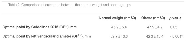
*as measured from the distal end of the sternum
Bottom Line: Radiographically, the location on the sternum that corresponds to optimal compression of the LV is more cranial in obese patients than in non-obese patients. It remains to be seen whether the recommendations for hand placement in CPR should be adjusted, but we may want to consider staying within 4cm of the bottom of the sternum in patients of normal weight.
Show References
- The Surviving Sepsis Campaign recently republished the 2018 update to their guidelines, namely, the recommendation that physicians initiate treatment measures using a "1-Hour Bundle" rather than the 3 and 6-hour bundles previously recommended:

- Also included was the level of evidence for each bundle component. There was no additional evidence provided to support the within-one-hour recommendation.

- There has been no well-designed, randomized trial to demonstrate benefits to administration of the various bundle components at specific time points. There are observational studies that show benefits to early protocolized therapy, including a restrospective study by Seymour et al. that showed benefits to earlier administration of antibiotics (but notably, not IV fluid administration), primarily in patients with septic shock requiring pressors.2
- There have been a variety of studies demonstrating harm with unecessary IV fluid administration,3-5 and inappropriate antibiotic use puts patients at risk for C.difficile colitis, drug reactions, and promotes drug-resistant organisms. Studies to date do not examine adverse events in patients initially treated for sepsis who do not end up being septic.
Take Home Points:
- Early recognition of sepsis is crucial to initiating necessary therapies and improving outcomes.
- Patients with sepsis and septic shock benefit from early appropriate antibiotics, source control, and appropriate resuscitation.
- Empiric treatment of all-comers with possible sepsis with broad spectrum antibiotics and 30ml/kg of IV fluids, in order to meet a 1-hour deadline, has definite potential for harm.
Show References
Precedex (dexmedetomidine) is a selective alpha-2 adrenergic receptor agonist used as a sedative.
It is unique among sedatives typically used in the ICU in that it lacks GABA activity and lacks anticholinergic activity.
Previous studies have shown significant positive changes in sleep patterns in critically ill patients sedated with dexmedetomidine:
-improved sleep efficiency – decreased sleep fragmentation, decreased stage 1 sleep, increased stage 2 sleep
-improved distribution of sleep (with more than ¾ sleep occurring at night)
Given importance of sleep and preservation of day-night cycles/ circadian rhythms in prevention of delirium, a recent randomized controlled trial evaluated dexmedetomidine's effect on delirium.
100 delirium-free critically ill adults receiving sedatives were randomized to receive nocturnal (21:30-06:15) IV dexmedetomidine (titrated to RASS -1 or max 0.7 mcg/kg/hr) OR placebo until ICU discharge.
80% of patients in the dexmedetomidine group remained delirium-free vs 54% in the placebo group.
There was no difference in the incidence of hypotension, bradycardia, or both between groups.
Show References
DOACs and the Critically Ill
- The use of DOACs for the prevention of stroke and venous thromboembolism is increasing.
- Though DOACs may be non-inferior to warfarin for these indications, it is important to consider the following pearls on DOACs in the critically ill patient:
- Acute kidney injury can double the half-life of dabigatran to more than 30 hours
- Hepatic failure can markedly increase the half-life of the factor Xa inhibitors
- PT, aPTT, and INR may not accurately assess the risk of bleeding. Use dilute thrombin time (TT), ecarin clotting time (ECT), or TEG/ROTEM to assess coagulopathy
- Can consider PCC (25 to 50 IU/kg) for life-threatening hemorrhage. The evidence supporting this recommendation is not robust.