
Search
Subscribe to daily UMEM Pearls (FREE!) via:
81-100 of 366 results with category "Cardiology"
Question
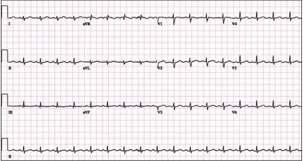
A 48 year-old female presents to the ED with progressive dyspnea and chest discomfort over the past 3 months. HR = 105, BP = 100/60 mmHg, with mild JVD on exam. Her ECG is shown below. What ECG abnormalites are present? What does your differential diagnosis include? What is the best initial diagnostic test?
Show Answer
Show References
Attachments
- 1310132023_ECGs_with_small_QRS_voltages.pdf (578 Kb)
- 1310132023_Low_QRS_voltage_and_its_causes.pdf (108 Kb)
Acute Aortic Syndromes
Classically, aortic dissection is considered the primary culprit in patients with chest pain that radiates to the back (aortic pain) or chest pain combined with ischemia (cerebral, cardiac, peripheral), syncope, or cardiac arrest. However, it should not be your only concern: the rate of aortic rupture is much higher in penetrating atheromatous ulcer (42%) and intramural hematoma (35%) than in aortic dissection (types A 7.5% and type B 4.1%).
Chest pain with concomitant ischemic symptoms and acute decompensation should prompt consideration of several etiologies under the umbrella of aortic syndromes and not limited to dissection :
- Penetrating atheromatous ulcer - rupture of an atheromatous plaque through the internal elastic lamina, with subsequent localized medial disruption and potential dissection, pseudoaneurysm formation, or free rupture
- Intramural hematoma - rupture of the vasa vasorum or hemorrhage within an atherosclerotic plaque followed by aortic wall infarct
- Aortic dissection- an intimal tear with resultant propagation within the middle third of the medial layer of the aorta
- Aneurysm leak or rupture - progressive vessel dilation and increased wall tension
- Traumatic transection - rapid deceleration forces or direct trauma, commonly shearing distal to left subclavian artery at aortic isthmus where the aorta is fixed by ligamentum arteriosum
Show References
The primary goal in management of STEMI is rapid coronary revascularization. STEMI's are occasionally complicated by ventricular fibrillation (VF) arrest. High quality chest compressions and early defibrillation will improve survival. But what can be done in cases where conventional ACLS measures fail and patients have shock-refractory VF?
Some have suggested that emergent PCI with ongoing CPR en route may be beneficial. This option may be considered in close consultation with cardiology if the arrest is thought to be driven by ongoing ischemia and infarction. However, definitive data is lacking and this has only been described in a handful of case reports.
There may also be a role for venoarterial ECMO to aid in perfusion of vital organs and limit the risk of multisystem organ failure. The ECMO circuit can also help facilitate therapeutic hypothermia after the culprit vessel(s) is revascularized and rhythm is restored.
Chances for survival are highest in younger patients, those that do not have chronic illnesses, and those who received immediate CPR after arrest.
Summary:
Consider emergent consultation for salvage PCI and ECMO in select cases of shock-refractory ventricular fibrillation associated with STEMI
Want more emergency cardiology pearls? Follow me @alifarzadmd
Show References
Attachments
Is RBBB More Indicative of Large Anteroseptal MI?
- Conventionally a new onset left bundle branch (LBBB) with acute myocardial infarction (MI) is associated with a massive MI
- Proximal left anterior descending artery (LAD) septal perforators perfuse the right bundle branch and the anterior fascicle of the left bundle branch ~90% of cases
- The right coronary artery (RCA) perfuses the posterior fascicle of the left bundle branch ~90% of cases
- Given the anatomy, a LAD occlusion should cause RBBB and/or LAFB; both a proximal LAD and RCA occlusion would be required for MI to cause LBBB
- A recent cohort study analyzed 233 patients to evaluate if RBBB or LBBB was associated with a large anteroseptal scar:
- RBBB was associated with larger scar size (24% vs. 6.5%; p<0.0001)
- RBBB was more indicative of ischemic heart disease (79% vs. 29%; p<0.0001)
- Based on this preliminary data RBBB may have a stronger association with ischemia and anteroseptal scarring than LBBB (*limitations - small cohort of cardiomyopathy patients with an EF<35%, further study is required)
Show References
Colchicine is known to be effective in treatment of recurrent pericarditis, but until recently its efficacy during the first attack of acute pericarditis has been uncertain.
A recent multicenter, double-blinded, RCT of patients with acute pericarditis found colchicine to be effective in reducing the rate of incessant or recurrent pericarditis (primary outcome), as well as the rate of hospitalization. Here are some highlights:
- 240 patients with acute pericarditis received conventional therapy (aspirin or ibuprofen), half of them were randomized to also get colchicine, the other half to placebo for 3 months
- Incessant or recurrent pericarditis: 16% in the colchicine group versus 37% in the control group (relative risk reduction=0.56; CI 0.30-0.72; NNT =4; p < 0.001)
- Symptom persistence at 72 hours, recurrences per patient, and hospitalization rate were all significantly reduced in the colchicine group
- There were no significant differences in adverse effects or discontinuation of the study drugs
Bottom-line:
Colchicine is a safe and effective drug for the treatment of acute pericarditis. Consider adding colchicine to conventional therapies to reduce duration of symptoms, recurrences, and rate of hospitalization.
Show References
Attachments
- In 1936 early repolarization (ER) was 1st described as ST-segment elevation in the absence of coronary artery disease, typically viewed as a benign ECG finding (BER) not association with increased cardiovascular mortality
- Classically the prevalence of BER tends to be associated with young athletes, male sex, and black race
- Recent data from Haissaguerre et al. and Tikkanen et al. suggest that certain subtypes of ER may be associated with a predisposition for malignant arrhythmias and sudden cardiac death (SCD)
- Although ER has various definitions contingent on the author, it consists of two components:
- 1.) Prominent J wave
- 2.) ST-segment elevation
- This article (9/13 JACC) focuses on the analysis and importance of the ST-segment contour and its possible relation to “malignant” repolarization
- Several studies (subgroup analysis) have found that a rapidly ascending ST-segment blending with the T-wave (Figures: A & C) confers BER, whereas a flat, horizontal, or even descending ST-segment (Figures: B & D) prior to the T-wave has potential to be malignant
*Please see the attachment below for Figures A-D
Show References
Attachments
Adult ED patients are commonly found to have markedly elevated blood pressures (>160/100) without any signs or symptoms of acute organ injury (ie, cardiovascular, renal, or neurological).
A recently revised ACEP clinical policy aims to guide emergency physicians in the evaluation and management of such patients.
They make the following recommendations (Level C):
- Routine screening tests (ie, CXR, ECG, UA, BMP) do not reduce adverse outcomes and are not required from the ED.
- Initiation of medical treatment does not reduce adverse outcomes and is not required in the ED.
- Patients with persistently elevated blood pressure should be referred for primary care follow-up.
- In select patient populations (eg. poor access to care), a screening creatinine level may identify renal injury that may alter disposition.
- If medication is started in the ED, the goal should be to facilitate gradual long-term control. Rapidly lowering blood pressure may be harmful.
Bottom-line:
There's little evidence to guide the decision of which patients with markedly elevated blood pressures to test or treat in the ED. This new clinical policy suggests that routine screening and treatment is not required. Asymptomatic patients should be referred for close follow-up, but consider a BMP in patients with poor follow up.
Want more emergency cardiology pearls? Follow me @alifarzadmd
Show References
Attachments
- 1st generation drug-eluting stents (DES) have been shown to reduce restenosis and target vessel revascularizations (TVR) compared with bare-metal stents (BMS) in patients with STEMI
- 1st generation DES have also been associated with increased rates of very late stent thrombosis (ST), raising concerns over the safety of these devices in patients with STEMI, who compared to patients with stable coronary artery disease, have greater rates of ST due to heightened platelet activation and the presence of thrombus
- The most important finding in this study is the significantly reduced risk of 1-year cardiac death, MI, and ST with CoCr-EES (cobalt-chromium everolimus eluting stent) compared to BMS
- The observed reduction in MI, ST, and composite cardiac death rates with CoCr-EES compared to BMS is consistent with experimental data suggesting that stents covered by fluorinated polymers are less thrombogenic than even BMS
Show References
- Classically MVP is considered a benign diagnosis associated w/palpitations, atypical chest pain, dyspnea, and carries a low risk of complications
- A recent study investigated MVP and its association w/ventricular arrhythmias in a cohort of unexplained out-of-hospital cardiac arrest (OHCA)
- A small subset of patients w/MVP experienced life threatening arrhythmias coined "malignant" MVP
- Malignant MVP was most often associated w/female sex, bileaflet valve, and frequent complex ventricular ectopic activity
Show References
Tight glycemic control (HbA1C<7%) has previously been recommended in CAD based on data from the United Kingdom Prospective Diabetes Study (UKPDS)
A recent study (JACC) evaluated the relationship between glycemic control, cardiovascular disease (CVD) risk, and all-cause mortality
Patients with a mean HbA1C 7-7.4% were compared to those with mean HbA1C <6%; tight glycemic control had a 68% increased risk of CVD hospitalization
Lenient HbA1C>8.5% also had significantly higher risk
CVD risk and all-cause mortality is greater with both aggressive and lax glycemic control and the optimal reference range may lie between 7-7.4%
Show References
- A recent meta–analysis of 12 studies (6,538 patients with 1,824 ROSC) assessed the quality of cardiopulmonary resuscitation (CPR) using either manual vs. mechanical (load-distributing or piston-driven) compressions in out-of-hospital cardiac arrest
- Compared w/manual CPR, load-distributing band CPR had significantly greater odds of ROSC (odds ratio, 1.62 and p<0.001)
- The treatment effect for piston-driven CPR was similar to manual CPR
- The difference in percentages of ROSC rates from CPR was 8.3% for load-distributing band CPR and 5.2% for piston-driven CPR
- Compared with manual CPR, combining both mechanical CPR devices produced a significant treatment effect in favor of higher odds of ROSC with mechanical CPR devices (odds ratio, 1.53 and p<0.001)
Show References
Radiation therapy is frequently utilized in the management of numerous thoracic malignancies
Cardiovascular disease is now the leading cause of nonmalignancy death in radiation-treated cancer survivors
The spectrum of radiation-induced cardiac disease is broad
The relative risk of CAD, CHF, pericardial/valvular disease, and conduction abnormalities is particularly increased
Early identification of potential cardiac complications w/cardiac MR and echocardiography provides an opportunity for regular assessment and potentially improved long term mortality
Show References
- Typically the normal ECG shows progression of T-wave size across the precordial leads & the T-wave in V1 is inverted or flat
- A large upright T-wave in V1 can be considered normal when there is high voltage/LVH or LBBB
- A new upright T-wave in V1 can be indicative of significant atherosclerotic disease
- If the T-wave in V1 is larger than the T-wave in V6 have a high suspicion for myocardial disease
- A new tall upright T-wave in V1 has ~84% specificity for ischemic heart disease (Barthwal)
Show References
- Statin therapy significantly reduces the risk for thrombotic events
- A recent study sought to determine the impact of short-term intensive statin therapy on intracoronary plaque lipid content
- 87 patients with multivessel CAD undergoing percutaneous coronary intervention and at least 1 other severely obstructive were randomized to intensive (rosuvastatin40 mg daily) or standard-of-care lipid-lowering therapy
- Upon follow-up, median reduction (95% confidence interval) was significantly greater in the intensive versus standard group ( p=0.01)
- Short-term intensive statin therapy in small trials reduces lipid content in obstructive lesions and further large studies with longer follow-up are warranted
Show References
Bifascicular block
- Right bundle branch block (RBBB) + left anterior fascicular block (LAFB)
- RBBB + left posterior fascicular block (LPFB)
- Complete left bundle branch block (LBBB)
Incomplete Trifascicular block
- Bifascicular block w/1st degree AV block *classically referred to as “trifascicular block”*
- Bifascicular block w/2nd degree AV block
- Alternating LBBB + RBBB
Complete Trifascicular block
- Bifascicular block w/3rd degree AV block
Show References
Stanford type A (proximal) aortic dissection accounts for ~60% of all aortic dissections
Classic treatment includes direct surgical replacement of the ascending aorta w/prosthetic graft (+/- AV aortic repair/replacement)
~20-30% of these patients (*institutional dependent) are considered poor candidates for surgery and receive only medical management, which innately results in substandard outcomes
In this study those who were considered poor candidates for surgical repair underwent novel endovascular treatment
Endovascular repair in this study was considered both appropriate and improved traditional medical outcomes in patients who were considered poor candidates
Show References
- Syncope is a sudden lack of blood supply to the brain typically caused by a problem in the regulation of blood pressure or a problem with the heart
- Syncope can be broadly classified in 3 categories neural reflex (~60%), orthostatic (~15%), and cardiac (~15%)
- >Even in the absence of a firm diagnosis of cardiac syncope, the presence of known structural heart disease (CAD) or evidence a primary electrical disorder is associated with a poor prognosis
- Cardiac causes of syncope can also be divided into 3 categories: structural heart disease, obstructive lesions, and arrhythmogenic potential
- Structural: Ischemic heart disease, dilated cardiomyopathy, ARVD,
- Obstructive: HCM, aortic/mitral stenosis, atrial myxoma, pulmonary HTN, PE, tamponade
-
- Brady: AV block, sick sinus, sinus arrest/pause
- Tachy: SVT (AVNRT/AVRT), accessory pathways (WPW), or primary arrhythmias (LQTS, SQTS, CPVT, Brugada)
Show References
- Mirror-image dextrocardia is the most common form of cardiac malposition and is commonly associated with situs inversus of the abdominal organs
- The anatomic right ventricle is anterior to the left ventricle and the aortic arch curves to the right and posteriorly
- 25% percent of these patients will have associated sinusitis and bronchiactasis (Kartagener’s syndrome)
- ECG changes associated with dextrocardia include:
- Right-axis deviation
- Global negativity in leads I and aVL (negative QRS w/inverted P and T waves)
- Lead aVR similar to the normal aVL (positive QRS)
- Absent R wave progression in precordial leads/dominant S waves
Show References
- MI without obstructive CAD is common, occurring in 5–10% of patients w/woman most commonly affected
- Mechanisms for MI without obstructive CAD include vasospasm, embolism, myocarditis, dissection, tako-tsubo, and occult plaque rupture
- Recent studies have applied cardiac MRI (CMR) with intravascular ultrasound (IVUS) to determine the mechanism of MI without obstructive CAD
- In this study plaque disruption frequently occurred when the angiogram was normal or showed minimal atherosclerosis; Plaque rupture was demonstrated on IVUS in ~40% of women studied
- IVUS and CMR identified the potential mechanism of MI in 70%
- Consider theses adjunctive tools in the assessment of all patients with a clinical syndrome of MI who do not have obstructive CAD at angiography
Show References
- The ADAPT (2-Hour Accelerated Diagnostic Protocol to Assess Patients With Chest Pain Symptoms Using Contemporary Troponins as the Only Biomarker) trial was a prospective observational validation study designed to assess a predefined ADP (Accelerated Diagnostic Protocol)
- A low risk patient in this ADP was defined by TIMI 0, ECG w/no ischemic changes, and negative troponin at 0-and 2-hours after presentation
- Primary endpoint was assessment of any major adverse cardiac event (MACE)
- Of 1,975 patients enrolled, 302 (15.3%) had a MACE
- ADP classified 392 patients (20%) as low risk and only 1 (0.25%) had a MACE
- ADP had a sen 99.7%, NPV 99.7%, spec 23.4%, and PPV 19.0%
- Despite ADP identifying patients as low risk for MACE standard of care still requires rapid early outpatient follow-up or further inpatient testing