Core Facilities
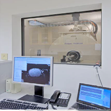
7 Tesla Biospec Avance III MRI Scanner
Description: Small animal scanner, 30cm open bore
Manufacturer: Bruker Corporation
Click here for manufacturer's website
The scanner is a part of the Center for Innovative Biomedical Resources (CIBR) >
Details
Please select a section for more details:
Capabilities
7 T Bruker BioSpec® 70/30 USR
Software Version
Paravision 6.0.1
Hardware Version
7.0 T Bruker Biospin (BioSpec® 70/30 USR)
The instrument is based on the AVANCE III MRI scanner architecture.
Magnet BioSpec 70/30 USR Technical Details:
Field Strength: 7T
Bore Diameter: 30cm
Zero Helium boil-off technology: Yes
Gradient Coils
System gradient/shim set B-GA20S
B-GA20S Gradient Specifications:
Inner diameter: 205mm
Gradient Strength: 200mT/m with 200A, 300V
Max. linear slew rate: 640T/m/s with 200A,300V
B-GA20S Shim Specifications:
X, Y, Z: 2500Hz/cm
Gradient/Shim Insert B-GA12S
B-GA20S Gradient Specifications:
Inner diameter: 116 mm
Gradient Strength: 400 mT/m with 200 A, 300 V
Max. linear slew rate: 3400 T/m/s with 200 A,300 V
B-GA12S Shim Specifications:
X, Y, Z: 5700 Hz/cm with 200 A
Z2: 740 Hz/cm2 at 3 A
XZ, YZ: 2100 Hz/cm2 at 3 A
X2-Y2, 2XY: 1000 Hz/cm2 at 3 A
RF-System
The system has four transmit channels and four receive channels and is capable of parallel imaging both on the transmit and receive side. The system comes with both narrow band operating at the proton/fluorine frequencies and a broad band covering the lower frequency with capability of imaging Na-23, C-11, P-31 and Li-7.
Types of Coils
- Double tuned 20mm surface coils operating at the proton/phosphorus frequencies
- Double tuned 20mm surface coils operating at the proton/carbon frequencies
- Rat Brain Array Coil – 4 Channels
- 112mm OD/ 86mm ID volume coil
- 112mm OD/ 72mm ID volume coil
- 1H Rat Head / Mouse Body Volume Coil - 40 mm
- 1H/19F Rat Head / Mouse Body Volume Coil - 40 mm
QA/QC phantoms
2 QA phantoms in difference sizes for 1H imaging
Contact
Rao Gullapalli, PhD
Phone: 410-706-2694
rgullapalli@som.umaryland.edu
Su Xu, PhD
Phone: 410-706-5419
sxu@umm.edu
Template for NIH "Facilities and Other Resources"
Please check with your collaborator before using this template, to make sure the content is accurate and up to date.
The University of Maryland houses the Core for Translational Research in Imaging @ Maryland (C-TRIM) which is operated by faculty associated with the Center for Advanced Imaging Research (CAIR) within the University of Maryland School of Medicine’s Department of Diagnostic Radiology & Nuclear Medicine. Within the core, both animal and human imaging facilities are available.
Core for Translational Research in Imaging @ Maryland (C-TRIM): The facilities of C-TRIM are located in two buildings. One is located in Howard Hall 6th floor where the 7 Tesla Bruker small Animal MRI, Siemens Inveon microPET/CT, SPECT, Xenogen, MR guided Focused Ultrasound (MRgFUS), fluorescence and bioluminescence imaging and wet lab for tissue processing are available. The other location is in the basement of the new Health Sciences Research Facility III (HSFIII) building where a 9.4 Tesla Bruker system is available. This location also hosts the human imaging arm of C-TRIM where a research dedicated 3.0 Tesla PRISMA FIT scanner and a state-of-the-art Siemens 3.0 Tesla mMR biograph system (combined PET/MR) are located. C-TRIM provides cross-sectional imaging services to various investigators on campus.
Animal MRI at C-TRIM: Currently the core houses a NCRR funded 7 Tesla 30 cm MRI scanner from Bruker Biospin that operates on Avance VI software system. The location is also conveniently close to the animal imaging facilities. Two sets of gradient coils are available to facilitate optimal imaging of larger and smaller rodents operating at slew rates of 800 T/m/s with a peak gradient strength of 200 mT/m and the second one operating at a slew rate of 5000 T/m/s with a peak gradient of 400 mT/m respectively. The scanner has multi-nuclear capabilities. Parallel imaging is available with 4-receive and 4 transmit channels available with the system. Several RF coils, including P-31, C-13 and Na-23 are available to fit the anatomy of various rodents. Two volumetric quadrature coils, 30mm and 40 mm diameter respectively are available to accommodate various size animals. In addition, a 4 channel receive coil is available for optimal reception of signals regardless of the transmit coil used. A large animal body coil is also available to image with the larger diameter gradient. Physiology monitoring systems are available to monitor the animals along with the safe delivery of anesthesia.
Along with the 7.0 Tesla scanner, we recently acquired a 9.4 Tesla 30 cm system which is located in HSFIII. This system also has two gradient coils to accommodate both large and small rodents. For relatively large rodents, there is a 20 cm gradient coil operating at 640T/m/s with a peak gradient strength of 20mT/m. The smaller bore gradient operates at a max slew rate of 3,440 T/ms with a peak gradient of 440 mT/m. Similar coils as for the 7 Tesla are available and this system also has broadband capability. In addition, the system is equipped with a 2 channel cryo-coil for mouse imaging. The system has the same capabilities as the 7.0 Tesla and more, including a cryocoil for mouse imaging.
Two SurgiVet MR-conditional animal anesthesia systems and an SA Instruments model 1030 gating and monitoring system for use with a clinical MR scanner are available for delivery of isoflurane anesthesia and to monitor physiological parameters (ECG, respiration, temperature, heart rate, and blood oxygen saturation).