Core Facilities
3 Tesla Prisma MRI Scanner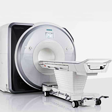
Description: State-of-the-art human whole body scanner, 60cm open bore
Manufacturer: Siemens Healthineers
Click here for manufacturer's website
Details
Please select a section for more details:
Capabilities
- Brain functional MRI
- Cardiac function
- High-angular diffusion tensor imaging (DTI)
- Cerebral blood perfusion (non contrast based)
- GABA editing
- Whole-brain spectroscopic imaging (EPSI)
- Pediatric and neonatal imaging coils
- 100 Kilowatt uninterruptible power supply (UPS)
Specifications
- 60cm open bore
- Parallel imaging with 64 receive channels
- 64 channel head RF coil
- XR 80/200 gradient coil (80mT/m strength on each axis, 200mT/m/ms rise time)
- TIM 4G receive architecture
- Software version VE11C
Contact
Rao Gullapalli, PhD
Phone: 410-706-2694
rgullapalli@som.umaryland.edu
Jiachen Zhuo, PhD
Phone: 410-706-2697
jzhuo@som.umaryland.edu
Thomas Ernst, PhD
Phone: 410-706-1045
ternst@som.umaryland.edu
Peripheral equipment
- Rear-projection system for visual presentation
- MR-compatible earphones
- Handheld button response boxes
- Mock scanner
- Animal RF coils
- Camera-based KinecCor motion tracking system
Template for NIH "Facilities and Other Resources"
This is a template for the 3 Tesla Prisma MRI Scanner, which can be used in the "Facilities and Other Resources" page of NIH applications. Before using this template, please contact your collaborator to ensure the information is correct and up to date.
"The Maryland Center for Advanced Imaging Research (MCAIR) houses a research dedicated 3.0 Tesla Siemens Prisma scanner that is equipped with a state of the art gradient and computer system. The Prisma has state of the art parallel imaging technology with 64 receive channels, and a 64-channel head radio-frequency (RF) coil. Several customized RF coils are also available that allow optimal imaging of small and large animals. The Prisma scanner has audio-visual presentation (rear-projection) to facilitate functional MRI protocols, and handheld button response boxes are present for the study participants to provide response during a functional paradigm. Both E-Prime and Presentation programs are available to the users to design their functional paradigms. A mock scanner is available for research participant training.
The scanning room has been designed with optimum RF shielding. All electronic equipment brought into the scan room is passed through a penetration panel which is conveniently located at the scanner console. The Center also features power conditioning to enhance MRI temporal stability. Preventive maintenance that involves measurement of temporal stability and noise characteristics are done on a monthly basis above and beyond what is performed by the vendors of the respective machines. A 100 Kilowatt uninterruptible power supply (UPS) provides continuous conditioning of the electrical power for the scanners, shielding the gradient and radio-frequency amplifiers from any fluctuations in building power.
A strong research relationship exists between MCAIR and scientists from Siemens Healthineers, which facilitates progress on ongoing research projects. Several MR physicists are trained on the use of the IDEA pulse sequence environment and the ICE image reconstruction environment available on the Siemens scanner."