
CONTACT
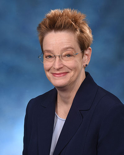
Christine Brantner, PhD
Director
cbrantner@umaryland.edu
LOCATION
Room 696, John Eager Howard Hall
660 West Redwood Street
Baltimore, MD 21201
HOURS
Monday through Friday
9 a.m. - 5 p.m.
PHONE
Office: 410-706-7992
Email: Coreimaging@umaryland.edu
About Us
The Electron Microscopy Core Imaging Facility (EMCIF) is part of the Center for Innovative Biomedical Resources (CIBR) and is located on the 6th floor of Howard Hall. It is jointly sponsored by the University of Maryland School of Medicine, University of Maryland School of Dentistry, and the University of Maryland Marlene and Stewart Greenbaum Comprehensive Cancer Center.
EMCIF provides electron microscopy-related research and imaging services to all faculty and staff of the University of Maryland campus and the academic and industrial community in the Washington D.C. and Baltimore areas. All services, instrumentation, and training courses are available to anyone, academic or private users, with an interest in electron microscopy.
EMCIF services are managed by iLab core facility management program. New users must register in iLab to access instrument and EMCIF services. Follow the quick links below for iLab registration and instructions. Please contact EMCIF to inquire about our services.
Core Services
- Conventional TEM sample preparation, including embedding in various types of resin and ultrathin sectioning
- Conventional SEM sample preparation, including chemical dehydration, critical point drying and sputter coating
- Cryo-sample preparation for both TEM and SEM, including high pressure freezing, freeze substitution, cryo-ultramicrotomy, plunge freezing and freeze fracture
- Immuno electron microscopy using pre-embedding, post embedding or Takuyasu methods
- Negative staining of purified macromolecular complexes, bacteria, viruses, liposomes, nanoparticles, or viral like particles (VLP)
- Advanced microscopy techniques, such as cryoEM, correlative LM/EM (CLEM) and 3D EM
- Electron microscopes imaging for trained or novice users
- Advanced consultation and training of electron microscopy related techniques and equipment usage
- Annual Current Electron Microscopy Techniques workshop
- Annual Ultramicrotomy Minicourse
- Instrument demonstration
iLab
Getting Started with iLabs
For postdocs, students, research staff & other non-Principle Investigators.
Approving Members to Your iLab (For PIs)
Approving members to your iLab For Principle Investigators
This report explains how to reconcile expenses accurately
EMC Core Instructions
How to order services and schedule equipment with Electron Microscopy Core