
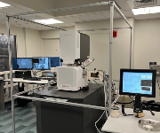
 The FEI Apreo 2 S combines high- and low-voltage ultra-high resolution capabilities with a low vacuum mode for non-conductive material. The electron detectors are optimized for fast imaging and easy collection of all available signals.
The Apreo 2 S can operate in traditional high vacuum mode, high vacuum with immersion mode and beam deceleration as well as low vacuum mode for non-conductive specimens that do not tolerate normal coating procedures.
Features & Specifications
SEM Imaging: Traditional high vacuum secondary electron imaging.
Large Area SEM Imaging: Using MAPS software, many images are acquired and stitched together in large format as a project. This capability allows larger resin blockfaces than TEM imaging and with inverted image contrast, the images look like they were generated with a TEM (no grid bars).
Cryo SEM Imaging: Beam sensitive samples can be frozen in liquid nitrogen slush and imaged on a temperature-controlled stage to preserve structure. Example: hydrogels.
Volumescope 3D Imaging: Using a special stage with a diamond knife installed, the sample blockface can be imaged and sectioned repeatedly to obtain a stack of 2D images that can be reconstructed in 3D.
Electron Optics: Field emission gun (Schottky) with SmartAlign technology. Voltage: 200 eV to 30 keV. Beam Current: 1 pA to 50 nA. Resolution: high vacuum, field free mode = 1.2 nm at 1 kV; high vacuum, immersion mode = 0.9 nm at 1 kV.
Vacuum: Oil-free vacuum system, high vacuum mode, low vacuum mode with water vapor added to the chamber for non-conductive samples that will not tolerate coating.
Sample Navigation: High precision specimen goniometer with 110 mm travel in x and y, 5-axes motorized x-y-z-tilt-rotate stage, unique standard specimen holder with labeled positions Navigation camera allows for double-click-to-center, stores and recalls positions, compucentric rotation and tilt.
Scanning System: High-resolution digital scanning engine controlled from the User Interface, selectable pixel density, dwell time 25 ns/pixel to 25 ms/pixel, electronic scan rotation.
Detection: High-vacuum secondary electron detector (Everhart-Thornley SED), low vacuum SED (LVD), Trinity detection system: T1 – segmented, lower in-lens detector, T2 and T3, STEM detector, Lo Vac Directional backscatter detector, IR-CCD, and Nav-Cam color optical camera for top-down images for navigation.
Imaging: FLASH technology which automatically performs focus, lens alignment and stigmator alignment in seconds; images can be live, averaged or integrated; drift corrected frame integration; images are saved as tiff.
Grant Citation
Any work performed on this instrument that is used in a publication needs to acknowledge the granting source that allowed UMB to obtain this microscope. Please use the following statement in your Acknowledgements:
“This work was supported by the National Institutes of Health (NIH) S10 [1S10OD030274-01].”