
Nikon W1 Spinning Disk
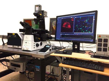 Nikon W1 spinning disk
- High-speed spinning disk confocal on Nikon Ti2 inverted microscope, designed for rapid fluorescence imaging.
- Provides enhanced signal-to-noise ratio, ultra-wide field of view, and improved light efficiency.
- Enables gentle imaging with reduced phototoxicity and photobleaching, ideal for live samples.
- Equipped with a stage-top incubation chamber for imaging live samples.
- Motorized stage and software for automated tiling, stitching, and reconstruction
- 7 laser lines for multicolor imaging (list them out)
- Dual Hamamatsu sCMOS camera for simultaneous two-channel imaging.
- Availability of two pinhole sizes (50um and 25um) to cover a wider range of objective magnifications.
- Equipped with Digital Micromirror Device (DMD) for high-speed, patterned photo-stimulation, enabling precise optogenetics, photo-activation, and FRAP (Fluorescence Recovery After Photobleaching).
- Availability of TIRF optics to enable high-contrast, surface-specific imaging of samples within 100-200 nm of the coverslip, minimizing background fluorescence.
- NIS-Elements General Analysis 3 (GA3) to create customized, automated imaging and quantification workflows.
Nikon A1R + X1 Spinning Disk Microscope
Nikon A1R + X1 Spinning Disk Microscope
The Nikon A1R + W1 Spinning Disk combines the flexibility of a point-scanning confocal microscope with the speed and sensitivity of a spinning disk system. The A1R resonant scanner enables high-speed confocal imaging with excellent optical sectioning and spectral imaging capabilities, while the W1 spinning disk attachment provides rapid, low-phototoxicity imaging ideally suited for live-cell and long-term imaging applications. The system supports fast multi-channel acquisition, z-stack and 3D imaging, time-lapse imaging, and imaging of sensitive biological samples with reduced photobleaching.
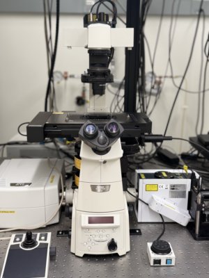
- High-speed spinning disk confocal on Nikon Ti2 inverted microscope, designed for rapid fluorescence imaging.
- Enables gentle imaging with reduced phototoxicity and photobleaching, ideal for live samples.
- Motorized stage and software for automated tiling, stitching, and reconstruction
- 6 laser lines for multicolor imaging (405, 445, 488, 514, 561, 640).
- Dual EMCCD camera for simultaneous two-channel imaging **
- 4 PMTs for confocal imaging with fast resonant scanners.
- Separate galvanometer scanner and 405 laser (50 mW) for photo-bleaching/uncaging.
Applications
- Live-cell imaging of dynamic processes (cell division, migration, tracking)
- Calcium signalling and other fast dynamic events.
- Large-area tile scans of sectioned tissue samples.
- High-content screening for automated, high-throughput testing and cellular studies.
- Specialized photobleaching and photoactivation, such as FRAP, photo-switching, and uncaging, using a 405 nm laser.