
Nikon A1 Laser Confocal
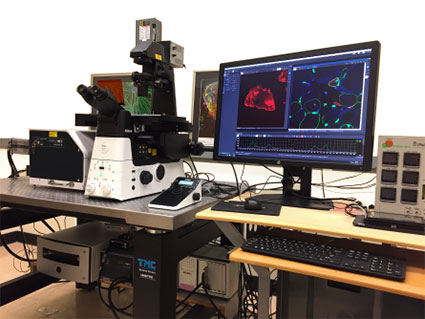
Nikon A1 Laser Confocal Microscope
The Nikon A1 Confocal is a versatile laser scanning confocal microscope designed for high-resolution fluorescence imaging of fixed and live biological samples. The system supports multi-channel imaging, optical sectioning, z-stack acquisition, and 3D reconstruction, enabling detailed visualization of cells, tissues, and subcellular structures. With sensitive detectors and flexible imaging modalities, the Nikon A1 is well-suited for a wide range of applications, including protein localization, co-localization analysis, and quantitative fluorescence imaging.
- Inverted microscope on a Nikon Ti2 stand.
- Equipped with 405, 488, 561, and 647nm laser lines.
- 4 high-sensitivity detectors for multicolor imaging.
- Excellent for immunocytochemistry with advanced tiling and stitching capabilities.
Zeiss 710 NLO & Zeiss 7MP
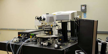 Zeiss 710 NLO & Zeiss 7MP
- Two separate, upright microscope systems equipped with multiphoton excitation for imaging live cells, slices and whole animals
- 710 NLO equipped with a confocal LSM and spectral detection
- 7MP equipped with two lasers for imaging a very broad range of fluorophores.
- Each equipped with 4 detectors for high-sensitivity multicolor imaging
- 7MP is ideal configuration for in vivo imaging
Applications:
- Optical sectioning of fluorescent tissue using confocal or multiphoton imaging
- Tile scanning and stitching of composite images
- 3D reconstruction of biological tissue/cells
- In vivo time-lapse imaging
Zeiss 5Live & Zeiss 510
Zeiss 5Live & Zeiss 510
- Point-scanning and slit-scanning confocal microscope
- Fast acquisition frame rates for studying dynamic cellular processes at physiological temperatures
- Dual scan heads (5Live) allow simultaneous imaging and optical manipulation
- Excitation (488, 543, 560, 633, Ti:Sapphire laser)
Abberior Facility Line STED Microscope
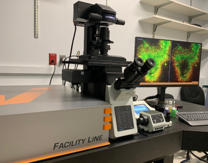 Abberior Facility Line STED Microscope
- Super-resolution STED microscope capable of lateral resolution ~40 nm and axial resolution ~80 nm.
- Suitable for super-resolution imaging in cultured cells, tissue slices, and other preparations.
- Fluorescence Lifetime Imaging (FLIM) for FRET and other new measurements.
- Core hardware for image acquisition
-
- Laser lines: 405, 485, 561 and 640 nm for imaging and a pulsed STED laser at 775nm.
- Detection via 3 photon-counting APDs and 1 Matrix detector for effective removal of background signals.
- Inverted Olympus IX83 with 10x (dry), 20x (dry), 40x (dry), 60x (silicone oil) and 60x (oil) objectives for a broad range of applications, motorized stage, and Z drift compensation.
-
- Capable of imaging fixed and live samples.
-
- Okolab stage-top incubator for temperature and CO2 control for live-cell imaging.
- A range of sample holders is available.
- STED-capable live-cell stains are available for a variety of intracellular targets.
-
- Unique accessories to increase resolution, decrease photobleaching, and:
-
- Adaptive optics to correct for spherical aberrations and compensate for sample-induced aberrations.
- Adaptive Illumination that facilitates high resolution and low-light gentle imaging of live-cells.
- Novel approaches called DyMIN and RESCue that reduce the total light power delivered to the sample during image scanning.
-
Applications
- Nanoscale Super-Resolution Imaging (STED) facilitating resolution down to 30–50 nm. Enables visualization of sub-diffraction structures like cytoskeleton (actin, microtubules), synaptic vesicles and organelle sub-compartments.
- Time-Resolved & Lifetime-Enhanced Imaging (Time-Bin / Gating) improves resolution and contrast by filtering the background, enabling lifetime-based separation of signals, and also can reduce autofluorescence and enhance signal specificity.
- Deep Tissue & Aberration-Corrected Imaging using the adaptive optics.
- Live-Cell STED Imaging (Advanced Applications)
Zeiss LSM 880 with Airyscan
Zeiss LSM 880 with Airyscan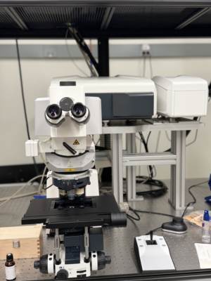
- Point scanning confocal on an upright Zeiss Examiner microscope stand.
- Offers high sensitivity, enhanced resolution in x, y and z and high image-acquisition speed.
- Equipped with Zeiss Airyscan and Fast Airyscan detector, which can improve the signal to noise ratio (4-8x) and increase resolution 1.7x
- Definite Focus to prevent focal drift.
- Equipped with 7 laser lines (355, 458/488/514 nm (Argon), 561 nm, 594 nm, and 633 nm) and a motorized stage and software for automated tiling and stitching.
- A 34-channel GaAsP-PMT array detector enables precise spectral imaging.
- Wide range of objective lenses to meet diverse imaging needs in biological research.
Applications
- Superresolution Imaging with the Airyscan provides a lateral resolution of about 140nm and an axial resolution of 400nm.
- Tile scanning of large tissue section samples.
Nikon A1R + X1 Spinning Disk Microscope
Nikon A1R + X1 Spinning Disk Microscope
The Nikon A1R + W1 Spinning Disk combines the flexibility of a point-scanning confocal microscope with the speed and sensitivity of a spinning disk system. The A1R resonant scanner enables high-speed confocal imaging with excellent optical sectioning and spectral imaging capabilities, while the W1 spinning disk attachment provides rapid, low-phototoxicity imaging ideally suited for live-cell and long-term imaging applications. The system supports fast multi-channel acquisition, z-stack and 3D imaging, time-lapse imaging, and imaging of sensitive biological samples with reduced photobleaching.
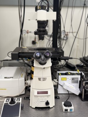
- High-speed spinning disk confocal on Nikon Ti2 inverted microscope, designed for rapid fluorescence imaging.
- Enables gentle imaging with reduced phototoxicity and photobleaching, ideal for live samples.
- Motorized stage and software for automated tiling, stitching, and reconstruction
- 6 laser lines for multicolor imaging (405, 445, 488, 514, 561, 640).
- Dual EMCCD camera for simultaneous two-channel imaging **
- 4 PMTs for confocal imaging with fast resonant scanners.
- Separate galvanometer scanner and 405 laser (50 mW) for photo-bleaching/uncaging.
Applications
- Live-cell imaging of dynamic processes (cell division, migration, tracking)
- Calcium signalling and other fast dynamic events.
- Large-area tile scans of sectioned tissue samples.
- High-content screening for automated, high-throughput testing and cellular studies.
- Specialized photobleaching and photoactivation, such as FRAP, photo-switching, and uncaging, using a 405 nm laser.
Nikon NSTORM
Nikon NSTORM
The Nikon NSTORM microscope can perform STORM, DNA-PAINT, and TIRF imaging along with correlative confocal and super-resolution imaging. This enables colocalization and protein interaction studies at the nanometer scale.
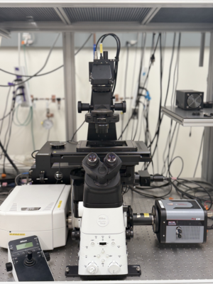
- Combined resonant galvanometer point scanning confocal and Nikon NSTORM Single molecule localization microscope capable of ~20nm resolution.
- Inverted Ti2 stand with 10, 20X dry, 10X LWD Water immersion, and 40X, 60X immersion Oil and 100X Silicone objectives.
- Motorized stage and software for automated tiling and stitching.
- 6 laser lines: 405, 445, 488, 514, 561, and 647.
- Princeton Instruments ProEM camera.
Applications
- Single-Molecule Localization (STORM/PALM) achieves ~20–30 nm resolution and resolves structures beyond the diffraction limit. Ideal for nanoscale organization of proteins and complexes.
- Membrane & Surface Imaging (TIRF/STORM), facilitating high-contrast imaging at the cell membrane. Aids in the study of receptor dynamics, endocytosis/exocytosis, and/or cell adhesion complexes.
- 3D Localization Microscopy: Axial (z-axis) resolution with specialized optics enabling 3D reconstruction of nanoscale structures.