
CONTACT:

Thomas Blanpied, PhD
Director
Department of Physiology
Room 5-015, Bressler Research Building
tblanpied@som.umaryland.edu
410-706-4769
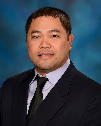
Joseph Ryan H. Mauban, PhD
Confocal Core Manager
Room 523, Howard Hall
jmauban@umaryland.edu
410-706-6170

Shilpa Dilip Kumar, PhD
Assistant Professor
Room 584, Howard Hall
SDKumar@som.umaryland.edu
410-706-0859

Yajie (Kevin) Liang, MB, PhD
Assistant Professor
Department of Diagnostic Radiology and Nuclear Medicine
HSF III, Room L127 Y
yajie.liang@som.umaryland.edu
LOCATION:
Room 610, Health Sciences Facility l
685 West Baltimore Street
Baltimore, MD 21201
HOURS:
Monday through Friday
7:00am - 5:00pm
PHONE:
410-706-6170
Mission
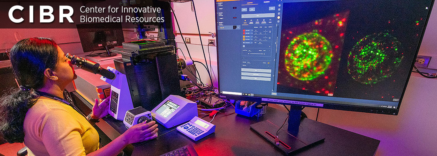
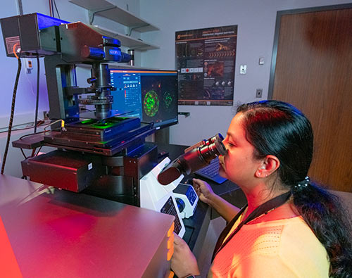
The mission of the Confocal Microscopy Core is to provide researchers with access to state-of-the-art optical imaging technologies, expert technical support, and advanced image analysis resources that enable high-quality, reproducible biological research. The Core is committed to supporting a broad spectrum of scientific applications by offering cutting-edge microscopy platforms, and by providing comprehensive training in experimental design, sample preparation, image acquisition, image processing, and quantitative analysis to empower users to generate meaningful and publication-quality data. The Core strives to foster innovation, collaboration, and scientific rigor by assisting investigators in selecting appropriate imaging approaches, optimizing workflows, and implementing reproducible image analysis strategies. Through education, consultation, and access to advanced technologies, the Confocal Microscopy Core serves as a collaborative resource that accelerates discovery across diverse areas of biomedical and biological research.
Services
The Core also includes a dedicated cell culture room equipped with an incubator, culture hood, and widefield fluorescence microscope to support the preparation and maintenance of cultured and live-cell samples prior to imaging experiments. In addition, the space is equipped to support the preparation of live animal samples for imaging applications.
Learn More
Equipment
Explore the advanced imaging systems available within the Confocal Microscopy Core, including confocal, spinning disk, super-resolution, lightsheet, and multiphoton microscopes. Detailed specifications and imaging capabilities are provided for each instrument.
Applications
Discover the wide range of biological applications supported by the Core, from live-cell and tissue imaging to super-resolution, developmental biology, neuroscience, and quantitative fluorescence imaging.
Image Analysis
Learn about the image processing, visualization, and quantitative analysis tools available through the Core, including commercial and open-source software platforms for multidimensional microscopy data analysis.
Training and Core Policies
Access information on user training, instrument access requirements, scheduling procedures, data management, and facility policies designed to ensure safe and efficient use of Core resources.
Pricing
View the current hourly rates and service fees for microscope usage, training, assisted imaging sessions, and image analysis resources available through the Core.
Courses and Workshops
Explore educational opportunities offered by the Core, including hands-on bioimage analysis workshops, lecture series, and specialized training sessions in microscopy and quantitative imaging.
Learning Resources
Browse protocols and educational materials to support microscopy experiments, image acquisition, and reproducible image analysis workflows.
News and Announcements
Stay updated on new instrumentation, software availability, workshops, policy updates, maintenance schedules, and other important announcements from the Confocal Microscopy Core.