
The Confocal Core provides comprehensive image analysis services to help researchers transform complex imaging data into meaningful, quantitative insights. We offer access to powerful commercial platforms such as Bitplane Imaris, Zeiss Arivis, and Nikon NIS-Elements for advanced visualization, segmentation, and 3D/4D/5D analysis. In addition, we support a wide range of open-source tools, including FIJI and Cellpose, enabling flexible and customizable workflows. With expert guidance and training, our core ensures rigorous, reproducible, and efficient analysis tailored to diverse research applications.
Please reach out to Confocal Core staff for any questions related to data analysis and quantitative image processing. Whether you need help with image interpretation, segmentation, colocalization, or selecting the most appropriate analysis workflow for your project, the team can provide guidance and support to generate accurate, reproducible, and publication-quality quantitative data from imaging experiments.
Bitplane Imaris
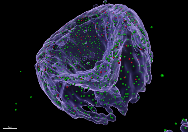
Imaris is a powerful, user-friendly platform designed for advanced visualization, analysis, and interpretation of 3D and 4D microscopy datasets. Widely used in life sciences, it enables researchers to explore complex biological structures and dynamic processes with high precision.
Imaris offers intuitive tools for 3D rendering, surface and spot detection, cell tracking, and colocalization analysis, making it especially valuable for studying cells, tissues, and subcellular structures over time. Its interactive interface allows users to seamlessly move between visualization and quantitative analysis, helping extract meaningful biological insights from large imaging datasets.
The software also supports automation and reproducibility through customizable workflows and batch processing.
Zeiss Arivis
Arivis is a powerful platform specifically well-suited for the analysis of large-scale datasets generated by light-sheet microscopy. Designed to handle terabyte-sized images, it enables smooth, high-performance visualization and exploration of whole organs, embryos, or cleared tissue samples without compromising resolution.
For light-sheet applications, Arivis excels in fast 3D and 4D rendering, allowing users to navigate complex volumetric datasets and identify structures of interest with ease. Its advanced tools support segmentation, object detection, and quantitative analysis across large fields of view, making it ideal for studying spatial relationships and biological organization at scale.
Nikon NIS-Elements
NIS-Elements offers a wide range of tools for image processing, segmentation, object tracking, and colocalization analysis. The software includes modules for AI-based analysis and high-content screening, allowing users to handle large datasets and extract meaningful biological information. With its intuitive interface and seamless integration with Nikon hardware, NIS-Elements is a versatile solution for both image acquisition and downstream analysis in microscopy workflows.
Fiji/ ImageJ
FIJI (Fiji, “Fiji Is Just ImageJ”) is a widely used, open-source image processing platform built on ImageJ and specifically tailored for biological and biomedical image analysis. It provides a flexible and extensible environment for handling a broad range of microscopy data, from simple 2D images to complex multidimensional datasets.