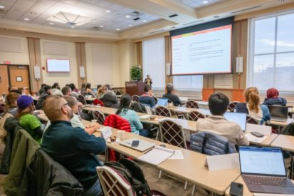
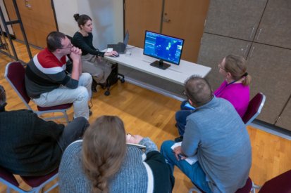
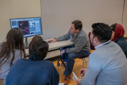
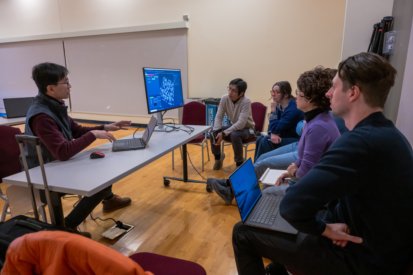
The Confocal Core is committed to supporting researchers not only through access to advanced imaging instrumentation, but also through education and training in modern microscopy and quantitative image analysis. To help users develop the skills needed for rigorous and reproducible imaging workflows, the Core offers a variety of educational programs throughout the year. These include an annual hands-on Bioimage Analysis Workshop covering topics such as image processing, segmentation, quantification, and visualization using both commercial and open-source software platforms and a recurring lecture series in Bioimage analysis. These courses and workshops are designed for users of all experience levels and aim to empower researchers to extract meaningful, reproducible, and publication-quality data from their imaging experiments.
Nanocourse: MINDFLUX—Theory and applications of MINFLUX microscopy (Fall 2026)

The use of light microscopy to measure the dynamics and interactions of single molecules in cells has developed remarkably quickly in recent years. The latest and greatest fluorescence microscopy method in this area, called MINFLUX, achieves unparalleled resolution both in images and in tracking the motion of single molecules, exceeding 2 nm localization precision—far less than the size of a protein. With these capabilities, MINFLUX imaging provides a bridge between the spatial resolutions of structural biology (such as NMR or cryoelectron tomography) and confocal fluorescence imaging (such as STED or spinning disk microscopy). In just the few years since its development, MINFLUX has already been used for nanometer-resolution measurement of protein organization at the plasma membrane and in organelles, quantifying stoichiometry in protein complexes, visualizing protein arrangement in single viral particles, measuring locus-dependent chromatic dynamics, tracking the diffusion of single lipid molecules, and even measuring the motion of single protein domains during ion channel opening.
A MINFLUX microscope will be coming to UMB this year, providing exciting opportunities for deciphering molecular actions in cells, dissecting macromolecular disorder in human disease, and accelerating drug discovery. This course will discuss the unique strengths of MINFLUX imaging, the
technological achievements that make it work, and biological applications. The course will principally use readings of the primary literature from neuroscience, biochemistry, virology, and cancer biology. The intended audience is advanced graduate students, but postdocs and faculty should feel free to contact the course directors for discussion about audit options.
Beyond the Microscope: Microscopy Image Analysis Made Practical - a recurring lecture series and hands-on classes in Bioimage Analysis
The Confocal Microscopy Core hosts a recurring Image Analysis Lecture Series four times per year, featuring lectures and hands-on sessions on emerging analysis methods, best practices in quantitative microscopy, and workflow development for diverse biological applications. The course is designed for students and researchers at the University of Maryland, School of Medicine. This recurring set of events will provide both foundational and advanced hands-on training in quantitative image analysis, with a strong focus on practical tools and workflows.
Highlights:
- Introduction to Image Analysis Concepts
- ImageJ/Fiji Basics
- Segmentation Techniques (Basic & Advanced)
- Additional Key Tools & Methods in Image Analysis
- Introduction to Bitplane Imaris
The lecture series is designed for all researchers (faculty, students, post-docs, and staff) who are interested in using ImageJ/Fiji for image analysis. The classes are open to participants who are actively acquiring data on the microscopes and are interested in learning ImageJ for future analysis projects. The topics covered during the class will include the basics of ImageJ for digital image processing; advanced topics like segmentation with classical and AI-based methods, colocalization analysis, and tracking and automation using ImageJ macros. Experience with ImageJ is not required. Attendees are encouraged to bring their own microscopy data, if they have any. After the meeting, we will have time to discuss individual questions concerning image analysis using ImageJ/Fiji on the participant's own data.
UM-MIND X-CHANGE/UMGCCC/CIBR Bioimage Analysis Workshop

The University of Maryland - Medicine Institute for Neuroscience Discovery (UM-MIND), together with the University of Maryland Greenebaum Comprehensive Cancer Center (UMGCCC) and the Center for Innovative Biomedical Resources (CIBR), annually conducts a Bioimage Analysis Workshop. Trainees, faculty, and staff at all levels at the University of Maryland School of Medicine and with UM-MIND’s partner institutions at the University of Maryland, Baltimore County (UMBC), and Morgan State University (MSU) through the MIND X-CHANGE may apply to attend. Although the workshop is designed primarily for neuroscience and cancer researchers, researchers from all departments are encouraged to participate, as many image analysis approaches and applications are broadly applicable across diverse areas of biological research.
This workshop will introduce biologists to the fundamentals of image analysis and provide practical strategies for efficient image processing and quantitative data analysis through a combination of instructional lectures and hands-on training sessions. Participants will gain exposure to both open-source platforms such as FIJI and Cellpose, as well as commercial image analysis solutions from Nikon, Bitplane, Leica Microsystems, and Thermo Fisher Scientific, with demonstrations and guidance provided by specialists from the respective companies.
Please contact the Core staff if you are interested in learning more about, or participating in, any of the courses and workshops offered through the Confocal Microscopy Core.