.jpg)
CONTACT:

Co-Director
jstains@som.umaryland.edu
Co-Director
cward@som.umaryland.edu
LOCATION:
Howard Hall, 5th Floor
660 West Redwood Street
Baltimore, MD 21201
HOURS:
Monday through Friday
8:03am - 5:00pm
EMAIL:
ABOUT US
The Musculoskeletal Physiology Service Center provides comprehensive outcome measures to assess bone and muscle structure, function, and physiology in preclinical models and training and assistance in the holistic assessment of in vivo and ex vivo musculoskeletal phenotypes. The Center seeks to expand the research programs of UMB investigators by way of providing anyone with a musculoskeletal phenotype the ability to collaborate with experts to explore said phenotype.
The services provided are available to all UMB researchers and extramural users on a fee-for-service basis.
SERVICES
Our Musculoskeletal Physiology Service Center offers a comprehensive range of specialized services:
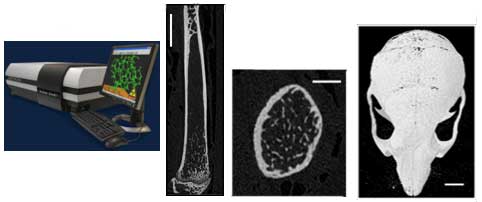
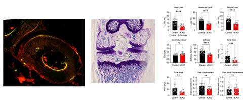
Microcomputed Tomography
Ex vivo/specimen quantification of bone microarchitecture and mineralization using the Skyscan 1172 microCT instrument. Soft tissue imaging with contrasting agents can also be performed.
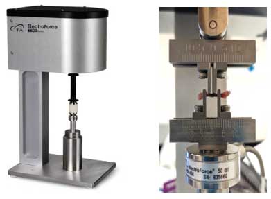
Bone Loading
Applies axial loads to bones in anesthetized mice to stimulate new bone formation, using the Bose ElectroForce 3200 instrument.
Mechanical Property Testing
Investigates bone functional properties using four-point bending and biomechanical properties of materials. In addition to testing bone, we can test other biomaterials in compression or tension including tendon and 3D fabricated devices.
Static and Dynamic Histomorphometry
Analyzes bone histomorphometry, including osteoblast and osteoclast numbers. Bioquant software, data.
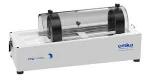
Electrocardiogram (ECG)
The EMKA ECG Tunnel enables the non-invasive assessment of the ECG in conscious restrained mice. Two tunnels available for simultaneous assessment in two mice.
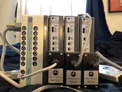
BioPaq
The BioPaq system enables ECG assessment in anesthetized mice using wire electrodes (up to 4 mice simultaneously).
Gait Dynamics
The DigiGait from Mouse Specifics performs ventral plane videography of mice walking on a transparent treadmill belt. A high-speed, high-resolution digital video camera continuously images the underside of the walking mouse. The software generates digital paw prints and dynamic gait signals, providing a temporal record of paw placement. More than 20 gait indices are reported from the analyses that are related to specific functional gains or losses.
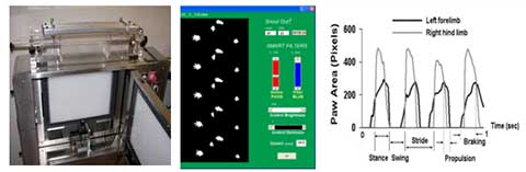
(Left panel) Digital paw prints based on image analysis (Middle panel). The positions of the paws in space over time are determined to generate dynamic gait signals for each limb from which gait indices are automatically reported. (Right panel)
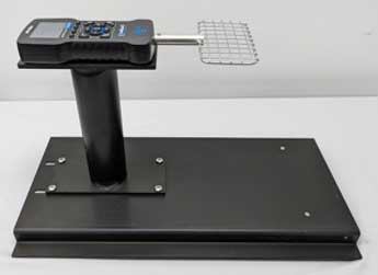
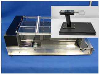
Grip Strength
The Columbus Grip Strength Meter (see pic) enables the quantification (force in grams/pounds) of grip strength force and its fatiguability in mice or rats. This indirect measure of gross neuromuscular performance effectively quantifies dysfunction in murine models of motor neuron and muscle disease.
Inverted Grid
A timed measure of a mouses ability to hang inverted on a wire grid (time in seconds). This indirect measure of gross neuromuscular performance effectively quantifies dysfunction in several murine models of motor neuron and muscle disease.
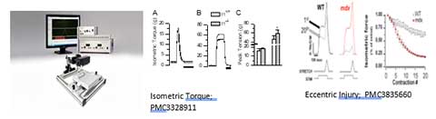
Nerve-evoked Muscle Function
The Aurora scientific 3-in-1 (Left panel) enables neuromuscular performance in the anesthetized mouse or rat that eliminates the influence of behavior or motivation. These assays are performed without surgical isolation of the muscle or alteration of the neural or vascular supply, and thus examine the function of muscle in its near native state. This approach also allows the assessment of nerve evoked isometric torque (Middle panel) or controlled induction of muscle injury (i.e., via eccentric contractions), which has proved to be a valuable assay in studies of muscular dystrophies and aging.
Rotarod
The RotaRod (Hugo Basil) assesses motor function and coordination, or fatigue, in rodents by measuring the time an animal stays on a rotating rod before falling.
Treadmill Exercise
The Columbus Exer6 is a 6 lane rodent treadmill enabling forced exercise training or quantification of exercise capacity in mice (6 simultaneously) or rats (6 or 3 depending on size). Most rats and mice are amenable to this test with proper acclimation to the treadmill.
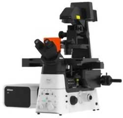
Fluorescence Imaging
Nikon Ti2 inverted microscope equipped with AX confocal (4 color) and wide-field fluorescence (PCO camera and 4 color LED light source). This system supports musculoskeletal investigators work at the tissue and cellular level.
PUBLICATIONS
Gould NR, Williams KM, Joca HC, Torre OM, Lyons JS, Leser JM, Srikanth MP, Hughes M, Khairallah RJ, Feldman RA, Ward CW, Stains JP. Disparate bone anabolic cues activate bone formation by regulating the rapid lysosomal degradation of sclerostin protein. Elife. 2021 Mar 29;10:e64393. doi: 10.7554/eLife.64393. PMID: 33779549; PMCID: PMC8032393.
Aljohani H, Senbanjo LT, Al Qranei M, Stains JP, Chellaiah MA. Methylsulfonylmethane Increases the Alveolar Bone Density of Mandibles in Aging Female Mice. Front Physiol. 2021 Oct 4;12:708905. doi: 10.3389/fphys.2021.708905. PMID: 34671266; PMCID: PMC8521043.
Balenga N, Koh J, Azimzadeh P, Hogue J, Gabr M, Stains JP, Olson JA Jr. Parathyroid-Targeted Overexpression of Regulator of G-Protein Signaling 5 (RGS5) Causes Hyperparathyroidism in Transgenic Mice. J Bone Miner Res. 2019 May;34(5):955-963. doi: 10.1002/jbmr.3674. Epub 2019 Feb 28. PMID: 30690792; PMCID: PMC8210536.
Long KK, O'Shea KM, Khairallah RJ, Howell K, Paushkin S, Chen KS, Cote SM, Webster MT, Stains JP, Treece E, Buckler A, Donovan A. Specific inhibition of myostatin activation is beneficial in mouse models of SMA therapy. Hum Mol Genet. 2019 Apr 1;28(7):1076-1089. doi: 10.1093/hmg/ddy382. PMID: 30481286; PMCID: PMC6423420.
Choi JY, Lai JK, Xiong ZM, Ren M, Moorer MC, Stains JP, Cao K. Diminished Canonical β-Catenin Signaling During Osteoblast Differentiation Contributes to Osteopenia in Progeria. J Bone Miner Res. 2018 Nov;33(11):2059-2070. doi: 10.1002/jbmr.3549. Epub 2018 Aug 1. PMID: 30001457; PMCID: PMC7739562.