CONTACT:

Thomas M. Ernst, Dr. rer. nat.
Director
thomas.ernst@umm.edu
(410) 706-1045

Su Xu, PhD
Manager
sxu@umm.edu
410-706-6384
Jiachen Zhuo
Manager
jzhuo@umm.edu
410-706-2697

Mark Smith, PhD
PET/CT Imaging Manager
mark.smith@som.umaryland.edu
410-328-1320
LOCATION:
Room 645, John Eager Howard Hall
660 West Redwood Street
Baltimore, MD 21201
HOURS:
Monday through Friday
7:00am - 5:00pm
PHONE:
Phone: 410-706-CTRM (2876)
Fax: 410-328-5937
MISSION:
To foster a collaborative environment that facilitates novel innovations in imaging and image guided therapeutics that can be translated to the clinic.
SERVICES:
The staff of C-TRIM provides consultation on all imaging related research. Assistance is available for the design of experiments and to optimize imaging techniques. Image processing and analysis expertise is available within the core and training is provided upon request. The core also conducts an annual retreat where specific areas of research are highlighted. One aspect of the core is to develop new diagnostic imaging technologies and to develop image guided therapeutic interventions to remain at the state-of-the-art.
INSTRUMENTATION:
Bruker BiospecAvance III 7 Tesla and 9.4 T Small Animal MRI Scanners
- High-resolution qualitative and quantitative assessment of structure and function for CNS and various body applications
- Multi-nuclear spectroscopy (H-1, C-13, Na-23, P-31, F-19 etc)
- High-resolution Diffusion Tensor Imaging for detecting microstructural and cellular changes
- Vascular studies, cerebral blood flow, cardiac function analysis
- H-1 MRI CryProbeTM 2 Element Array kit for mice on 9.4 T scanner which provides a remarkable SNR gain compared to regular MRI coils operating at room temperature
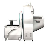 Siemens Inveon Small Animal PET- CT Imaging System
- Dockable PET-CT for combined anatomic and functional imaging
- High specificity radionuclide uptake
- Metabolic imaging
- High resolution system, (50 μ m for CT and 1.2 mm for PET) with extended FOV
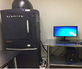 Xenogen IVIS Spectrum Optical in vivo Imaging System
- Rapid whole-body optical images of mice, rats or rabbits
- Wide range of fluorescence excitation and emission filters
- Wide array of molecular biology assays including GFP and luciferase
- Measures proteasome activity, monitor tumor growth, drug efficacy
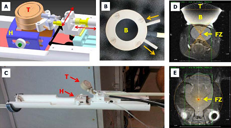
MR-Guided Focused Ultrasound (MRgFUS) System
- Integrated with MR for image guidance
- Tumor ablation studies, blood brain barrier disruption and neuromodulation
CORE APPLICATIONS INCLUDE:
- High-resolution anatomic imaging for CNS and body applications (MR/CT)
- Tumor kinetics using receptor specific exogenous agents (MR/PET)
- Multi-nuclear MR spectroscopy (H-1, C-13, Na-23, P-31, F-19 etc.)
- Metabolomic studies (MR/PET)
- High-resolution Diffusion Tensor Imaging for detecting microstructural and cellular changes (MR)
- Vascular studies (CT/MRI)
- Cerebral blood flow studies using endogenous contrast (MR)
- Cardiac functional analysis (MR/PET/CT)
- Investigation of Blood-Brain Barrier disruption for various particle delivery (MR/MRgFUS)
- Neuromodulation using low energy ultrasound (MRgFUS)
- Ablative image guided surgery
- Focal image guided body and neuro thermal therapy applications. (MRgFUS)
- Bone density measurements (CT)
- Cardiac metabolism (MR/PET)
- Musculoskeletal studies (MR/CT)
- Detection of novel fluorophores (Xenogen)
- GFP and Luciferase imaging (Xenogen)
- Monitoring tumor growth (CT/MR/PET/Xenogen)