Flow & Mass Cytometry Facility
MISSION:
To ensure that University of Maryland investigators have access to flow cytometry and mass cytometry services for their research. A facility with dedicated operators ensures well-performing instruments and optimal results with a minimal outlay of expenses. Established in 1991, this facility has state-of-the art equipment and a highly-trained and experienced staff.
SERVICES:
Multichromatic flow cytometry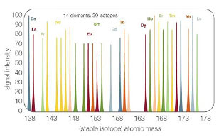
Including markers for:
- Lineage
- Maturation
- Activation
- Homing
- Intracellular cytokines
- Cell sorting (up to 6-way)
based on GFP and/or multichromatic staining - Mass Cytometry (>60 parameters)
- Serum/supernatant cytokine levels using bead kits (e.g. BD Pharmingen CBA kit)
- Cell cycle analysis (PI, DAPI)
- Cell proliferation (CFSE, PCNA, BrdU and Ki67)
- Apoptosis (Annexin V vs. PI; TUNEL; subG0/G1 peak analysis)
- Green fluorescence protein (GFP) (eukaryotic and prokaryotic)
- Advice with experimental design and data analysis
INSTRUMENATION:
BD LSR II Flow Cytometer:
- 4 lasers: 407, 488, 552, and 641 nm
- 16 parameters (14 colors plus forward and side scatter)
Beckman Coulter MoFlo Astrios Cell Sorter
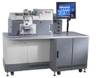
- 4 lasers: 355, 407, 488, and 641 nm
- 21 parameters (19 colors plus forward and side scatter)
- Up to 6-way high speed sorting
- CyCLONE single cell sorting
Fluidigm CyTOF Mass Cytometer
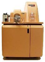
- >35 parameters based on mass spectrometry detection of metal isotope- labeled antibody staining
- No need for single color controls or fluorescence compensation
Fluidigm Helios Mass Cytometer
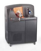
- >60 parameters based on mass spectrometry detection of metal isotope- labeled antibody staining
- No need for single color controls or fluorescence compensation
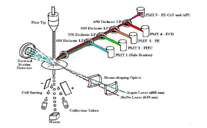
Principles of Flow Cytometry
Fluidics
- Cells in a single-cell suspension
- Flow in a single file through
Optics
- An illuminated volume where they
- Scatter light and emit fluorescence
- That is filtered, collected and
Electronics
- Converted to digital values
- That are stored on a computer
- And put through software for analysis
Revised from Dr. Robert Murphy, Carnegie Mellon University, Pittsburgh, PA
Principles of Mass Cytrometry
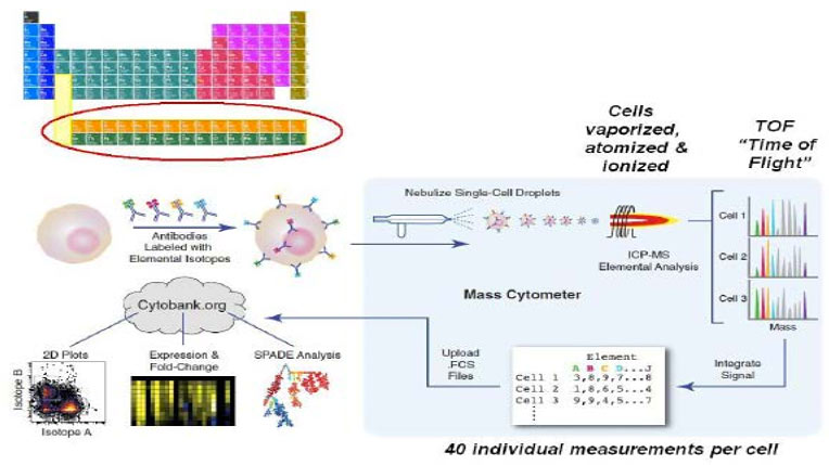
Bendall & Simonds et al., Science 332, 687 (2011) www.cytobank.org/nolanlab

