Search
1-8 of 8 results with category "Ophthamology"
Prior research has shown that EPs can accurately detect ocular pathology using POCUS.
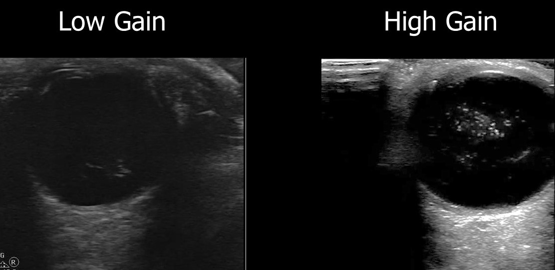
A recent retrospective review looked at how ultrasound image gain levels impacted the accuracy of POCUS for detection of retinal detachment, retinal hemorrhage and posterior vitreous detachment.
They included 383 ED patients who received ocular POCUS and ophthalmology consultations.
Conclusions: The authors found that increasing the gain for ocular POCUS images allowed for increased sensitivity.
Here is an example of a vitreous hemorrhage on ocular POCUS using low gain and high gain.
Show References
All to often we see children that are sent to the ED for "Pink Eye" as the school nurse will not allow them back into class unless they are treated with antibiotics. A recent study out of New York identified 4 factors that are associated with low risk (<8% chance) of bacterial (culture postive) conjunctivitis. They are:
- age 6 years
- presentation during April through November
- watery or no discharge
- no glued eye in the morning
An editorial in journal watch comments that if this study can be replicated in other geographic areas we could change the practice of prescribing antibiotics that are not necessary.
Show References
Iritis is a common diagnosis in the ED, but did you know it was actually a subset of Uveitis.
Uveitis is an inflammation of one or all parts of the uveal tract which consists of the iris, the ciliary body, and the choroid.
The subsets of uveitis are:
- anterior
- confined to the iris and anterior chamber -- iritis
- confined to the iris, anterior chamber, and ciliary body -- iridocyclitis.
- Posterior uveitis -- choroiditis and chorioretinitis, is uncommon, with the exception of cytomegalovirus (CMV) retinitis in patients with AIDS.
Treatment of iritis and uveitis next week.
Show References
Some of the causes of acute vision loss are:
- Cardiac Causes include:
- Emboli -- causes can be atherosclerotic plagues, atrial fibrillation, endocarditis.
- Arteritis
- Dissection
- Hematologic causes
- Hypercoaguable state
- Hyperviscosity
- Anemia
- Ocular Causes
- Angle-closure glaucoma
- Papilledema/neoplasm: Intracranial hypertension
- Intraocular foreign bodies:
- Central retinal artery occlusion
- Anterior ischemic optic neuropathy
- Ruptured globe
- Miscellaneous
- Migraine
- Hysteria
- Drugs (i.e.: viagra and its counterparts)
Show References
Vision loss whether acute or chronic is a common presenting complaint to the ED. This will be the first in a series of pearls on the subject. This pearl will address the nomenclature used by ophthalmology based on the length of vision loss.
• Transient visual obscuration - Episodes lasting seconds. Usually associated with papilledema and increased intracranial pressure.
• Amaurosis fugax - Brief, fleeting attack of monocular partial or total blindness that lasts seconds to minutes
• Transient monocular visual loss or transient monocular blindness - A more persistent vision loss that lasts minutes or longer
• Transient bilateral visual loss - Episodes affecting one or both eyes or both cerebral hemispheres and causing visual loss
• Ocular infarction - Persistent ischemic damage to the eye, resulting in permanent vision loss
Show References
MEWDS (Multiple Evanescent White Dot Syndrome)
- A rare, unilateral, self-limiting inflammatory disease
- Afflicts young women more than men in a 4:1 ratio.
- Patients typically present complaining of
- Sudden, painless, monocular decline in central acuity
- Photopsias-- def. appearance as of sparks or flashes in retinal irritation
- Dyschromatopsia-- def> disorder of color vision
- Central/paracentral scotomas
- Visual acuity usually in the 20/40-20/400
- Fluorescein angiography of active lesions typically demonstrate a "wreath-like" hyperfluorescence of the white dots
- Disease is usually self limited (resolves in weeks to months) with an excellent prognosis.
- There are no known treatment options.