Search
261-280 of 465 results with category "Orthopedics"
Back pain accounts for more than 2.6 million visits
30% of ED patients receive X-rays as part of their evaluation
Imaging can be avoided in a majority of these patients by focusing on high risk (red flags) findings in the history and physical exam.
Patients who can identify a an acute inciting event without direct trauma likely have a MSK source of pain.
Imaging rarely alters management
Attempt to avoid imaging in patients with nonspecific lower back pain of less than 6 weeks duration, with a normal neurologic exam and without high risk findings (fever, cancer, IVDA, bowel or bladder incontinence, age greater than 70, saddle anesthesia, etc)
Patients with radiculopathy (sciatica) and are otherwise similar to the above also do not require emergent imaging
Some radiology pearls concerning ankle pain and fractures courtesy of David Bostick and Michael Abraham
Maisonneuve fracture – fracture of the medial malleolus with disruption of the tibiofibular syndesmosis with associated fracture of the proximal fibular shaft (http://radiopaedia.org/articles/maisonneuve-fracture)
When to look for high fibular fracture
- Isolated fracture of medial malleolus
- Isolated fracture of malleolus tertius without fracture on the lateral side
- Any painful swelling or hematoma on medial side without a fracture on x-ray
Always look for avulsion fracture of 5th metatarsal styloid in patients with ankle pain and
no obvious fractures
Dans-Weber Classification – for lateral malleolar fractures (http://radiopaedia.org/articles/ankle-fracture-classification-weber)
- Type A – fracture below ankle joint
- Type B – at level of joint with tibifibular joint intact
- Type C – fracture above joint with tears syndesmotic joint
Patellar tendonitis aka jumpers knee
Activity related knee pain due to degenerative, micro injury rather than an inflammatory process
Up to 20% in jumping athletes
Anterior knee pain during or after activity
Bassett Sign:
a) Tenderness to palpation with knee in full extension (patellar tendon relaxed)
b) No tenderness with knee in flexion (patellar tendon tight)
Is there any benefit to the use of prednisone in the treatment of lower back pain? One study showed that about 5% of patients receive prednisone for the treatment of their low back pain, but does it work.
A recent study by Eskin et al published in the Journal of Emergency Medicine looked at this question. They conducted a randomized controlled trial of 18-55 year olds with moderately severe low back. Patients were randomized to receive prednisone 50mg for 5 days or placebo.
The study enrolled a total of 79 patients, and 12 were lost to follow up. At followup there was no difference in their pain, or in them resuming normal activities, returning to work, or days lost from work. To make matters worse more patients in the prednisone group sought additional medical treatment 40% versus 18%.
Conclusion: With the results of this study we should continue the treatment of low back pain with non-steroidials, muscle relaxants and exercise. There does not appear to be any role for steroids in the treatment of these patients.
Show References
Return to Play After Infectious Mononucleosis (IM)
-Long incubation period make it difficult to determine source or onset
Presentation often atypical with nothing more than fatigue, decreased energy or decreased athletic performance.
DDX: Herpes simplex, HIV, CMV, toxo and strep (simultaneous infection may be seen in up to 30%)
Classic 3 to 5 day prodromal period (malaise, fatigue, anorexia)
Symptoms then progress into the classic “triad” of IM
Fever, pharyngitis, lymphadenopathy (esp. posterior cervical nodes)
May also have posterior palantine petechiae ( of cases), jaundice, exudative pharyngitis, rash and splenomegaly)
Rash (10% to 40%), transient, generalized maculopapular, petechial or urticarial)
Most commonly seen in patients treated with PCN antibiotics
Splenomegaly is an important complication in the athletic population
Mononucleosis makes the spleen susceptible to rupture (traumatic or spontaneous)
- Lymphocytic proliferation enlarges the spleen beyond protection from the ribs
- Physical examination has been shown to be unreliable for determining splenomegaly
- Highest risk is in the first 21 days (rare after 28 days)
Ultrasound is the modality of choice
-Splenomegaly peaks at 2 to 3 weeks and resolves in the majority between 4 to 6 weeks
Return to play is generally allowed after 4 weeks from diagnosis in the absence of splenomegaly and resolution of symptoms.
Cervical Cord Neuropraxia (CCN)
A concussion of the spinal cord as a result of an on-field collision.
A transient motor and/or sensory disturbance, lasting less than 24 hours.
A distinct and separate entity from spinal cord injury resulting in quadriplegia
Incidence 7.3 per 10,000 athletes
Approx. 50% of players experiencing CCN who return to play, have a second episode
The risk of this second episode is inversely proportional to the size of the cervical bony canal
Athletes with narrow canal diameter are more likely to have a 2nd episode
Those with normal canal diameter (14 mm on MRI) have a 5% risk
Those with a narrow canal (9 mm or less)) have a greater than 50% risk.
Whether repeat episodes lead to permanent spinal cord injury is unknown
Show References
Football helmets
A review of head and neck injuries from football from 1959 to 1963 found the rates of intracranial hemorrhage /intracranial death were 2-3X higher than the rates of cervical spine fracture/dislocation or cervical quadriplegia. In contrast, a study of football injuries from 1971 to 1975, revealed a dramatic reversal in rates. Cervical injuries now exceeded the rate of ICH by 2-4X.
A 66% reduction in ICH
A 42% reduction in craniocerebral deaths
A 204% increase in cervical spine fractures and dislocations
The shift was attributed to the modern football helmet, whose superior protection promoted “spearing” (headfirst tackling technique). Spearing involves hitting with the crown of the helmet leading to axial loading of the spine. Spearing accounted for 52% of the quadriplegia injuries from 1971 to 1975. Research by Joesph Torg, M.D., resulted in rule changes that led to an immediate 50% reduction in quadriplegia in NCAA football.
As a parent, coach or team physician, teach and enforce proper form and protect our young athletes.
Show References
Some quick facts about Knee Injuries:
- The most common cause of acute traumatic hemarthrosis of the knee is an anterior cruciate ligament tear.
- Most patients with an ACL injury will give a history of immediate pain, disability, knee swelling and audible pop.
- The most common ligament injuried in the knee is the medial collateral ligament.
- Patella dislocations
- Usually lateral dislocations and often spontaneous reduce.
- Hyperextend the knee to make the reduction easier.
- Dislocation of the knee:
- Anterior is the most common and usually secondary to hyperextension
- Popliteal artery injury is commonly seen and must be looked for. Easy bedside test is Ankle Brachial Index.
- Normal pulses do NOT exclude a vascular injury.
- Patients should be monitored for vascular complications and compartment syndrome.
- Vascular injuries due to knee dislocation are associated with a high rate of amputation, which markedly increases if not repaired within 6-8 hours.
When examining a knee for a meniscal injury the commonly described tests are the McMurray Test and Apley Test. However, these tests have sensitivities of 48-68% and 41% respectfully, and specificities of 86-94% and 86-93% respectfully. Depending on whether you are looking at the medical or lateral meniscus.
The Thessaly Test that was first described in 2005 can be performed with knee in either 5 or 20 degrees of flexion and has a senstivity of 89-92% and specificity of 96-97% when performed in 20 degrees flexion. The test also tends to be easier to perform.
To perform the test:
- Stand on affected leg only with the other leg held up in the air. The examiner holds hands for balance.
- Flex knee to be test to 20 degrees, while the other leg is held in the air
- Internally and Externally Rotate Knee
- Positive test is pain at medial or lateral joint line with possible locking/catching sensation
Essentially you and your patient will look like you are doing the twist as they rotate their knee with you holding their hands.
A video of the technique can be found at http://youtu.be/R3oXDvagnic
Show References
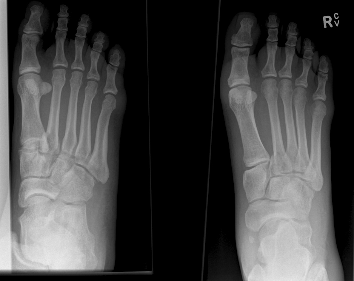
Lisfranc Fracture:
Typically consists of a fracture of the base of the second metatarsal and dislocation, though it can also be associated with fractures of a cuboid. Common current mechanism of injury is when a person steps into a hole and twists the foot. The original mechanism of injury that was described was when a horseman would fall of their horse with their foot still trapped in a stirrup.
Diagnosis should be considered if patient has difficultly weight bearing with pain on palpation over the 2nd and 3rd metacarpal head with an appropriate mechanism.
Pearls:
- Fracture findings on plain films may be subtle.
- If in doubt obtain weight bearing AP views of the foot to demonstrate dislocation/fracture.
- If weight bearing films are negative and you are still suspicious consider a CT scan of the foot.
Risk Modifiers for Concussion and Prolonged Recovery
A history of prior concussion is a risk factor for future concussion (>2x risk).
For individual sports, boxing has the highest risk.
For team sports, football, ice hockey and rugby have the highest risk.
Women’s soccer confers the highest risk for female athletes.
Younger age confers increased risk.
Female sex confers higher risk when comparing similar sports with similar rules.
Those with migraine headaches may be at increased risk.
Risk of prolonged concussion
Most athletes have symptom resolution within one week
Post traumatic amnesia (both retrograde and anterograde) predict increased number and longer duration of symptoms.
Younger age also predicts pronged recovery.
Other studies have found associations with headache lasting greater than 60 hours, fatigue, “fogginess,” or greater than 3 symptoms at initial presentation. Cognitive studies have identified deficits in visual memory and process speed as predictors of prolonged recovery.
Show References
Sports Hernia/Athletic pubalgia
Hx: Gradually increasing lower abdominal/proximal adductor pain. Usually activity related, resolves with rest. Frequent return despite rest when sports activity resumes.
Most common in athletes who perform cutting/maneuvers in addition to frequent acceleration/deceleration. Think ice hockey and soccer.
Bilateral symptoms not uncommon.
PE: Resisted sit up with palpation of the inferolateral edge of the distal rectus may recreate symptoms. Similarly, resisted hip adduction may elicit symptoms.
If for no other reason than to make the diagnosis harder to make, valsalva induced pain may also occur.
Fluoroscopic guided injections can be helpful to isolate the site of pain generation.
First line therapy is rest, non-narcotic analgesia and physical therapy.
With surgery, >80% return to pre injury level of play.
http://atlantasportsmedicine.com/orthopedic-surgeon/wp-content/uploads/2009/11/groin-injuries.jpg
Show References
DeQuervain and Intersection Syndromes:
- DeQuervain's Syndrome (Tenosynovitis of the Abductor Pollicus Longus and Extensor Pollicus Brevis tendons) is a common disorder that has received a lot of press lately as BlackBerry Thumb or Gamer's Thumb.
- This condition can be diagnosised by the Finklestein test [Have the patient bend their thumb into the palm of their hand, and then make a fist. They should then ulnar deviate their wrist. Pain along the tendons secures the diagnosis.]
- The pain of DeQuervain's syndrome is typically along the distal end of the radius at the base of the thumb.
- Intersection syndrome is a less common disorder though closely related to DeQuervain's Syndrome
- The pain is usually felt on the top of the forearm about three inches proximal to the wrist.
- The pain from this condition is due to tenosynovitis of the Extensor carpi radialis longus and Extensor Carpi radialis brevis muscles/tendons caused by the intersection of them with the Extensor pollicus brevis and Abductor pollicus longus tendons.
- Occurs due to excessive wrist movements.
- Intersection syndrome can be seen in weight lifters, skiers, and can be seen in homeowners in the fall and winter when they rake a lot of leaves or shovel snow.
- Treatment is the similar for both conditions and consists of:
- NSAIDS
- Cortisone injections can be effective
- Thumb and wrist immobilization with a Thumb Spica Splint or Cock Up Wrist Splint
Ankle Syndesmosis Injuries are also called high ankle sprains as they involve trauma to the ligaments above the ankle joint
Most ankle sprains are lateral ankle sprains. High ankle sprains are relatively uncommon.
Usual mechanism: External rotation injuries
Exam: Tenderness at the syndesmosis and compression of the tib/fib at the mid calf level causing syndesmosis pain (squeeze test)
Median recovery time is almost 4 times as long as a lateral ankle sprain 62days vs. 15days
Emergency department care is similar tto that of other ankle sprains but the added benefit of patient education and advice may improve overall care and follow-up.
Herpes Gladiatorum in Wrestlers
HSV causes non genital cutaneous infections primarily in wrestlers, commonly called herpes gladiatorum (HG)
Annual incidence in NCAA wrestlers is 20% to 40%
Most common cutaneous infection leading to lost practice time (40.5% of all infections)
Transmission is skin to skin.
Incubation period is 4 to 7 days from exposure. Healing usually occurs within 10 days after the initial lesion (without scaring).
Appearance: Numerous grouped uncomfortable (painful) vesicles/pustules on an erythematous base…evolve into moist ulcerations, followed by crusted plaques. Lesions typically get abraded during competition therefore may have an atypical appearance and may be mistaken for other infections such as staph. Distribution typically more diffuse than typical HSV infections. Occurs on body surfaces areas that typically come into contract with opponents (face, head, neck, ears, upper extremities). Lesion location typically on side of patient’s handedness. Recurrences occur at location of initial outbreak, a useful diagnostic aid.
Perform a thorough examination as ocular involvement was seen in 8% of high school wrestlers in one HG outbreak.
Typical treatment for primary infection is Valacyclovir 1g PO b.i.d. for 7 days. This is best started within 24h of symptom onset.
Show References
The clinical examination is often unreliable in ruling out septic arthritis in the ED.
Diagnostic arthrocentesis is often performed.
Traditional teaching involved very high WBC count thresholds as part of diagnosis.
In one 2009 study, synovial leukocyte counts in cases of MRSA were often less than 25,000 cells/uL
Have a low threshold for empiric antibioitics even in the face of low WBC counts (and incredulous consultants)
Show References
Overtraining syndrome
A maladaptive response to excessive exercise without adequate functional rest
-Results in disturbances of multiple body systems (neurologic, endocrinologic, immunologic and psychologic).
- May be caused by systemic inflammation and resultant neurohormonal changes
- Multiple hypotheses exist
-Symptoms
Parasympathetic alterations: fatigue, depression, bradycardia
Sympathetic alterations: insomnia, irritability, agitation, tachycardia, hypertension, restlessness
Other: anorexia, weight loss, poor concentration, anxiety
Usual presentation is prolonged underperformance despite adequate rest and recovery (weeks to months).
Pelllegrini-Stieda lesion
Ossified post-traumatic lesions at the MCL adjacent to the femoral attachment site of the medial femoral condyle.
Mechanism is likely from an avulsion injury that subsequently calcifies after the initial trauma.
Often an incidental finding on plain films.
If symptomatic, refer to ortho as an outpatient
If not symptomatic, no treatment is indicated
http://images.radiopaedia.org/images/30076/b62e61e83241e30f2da693901edcdc_gallery.jpg
http://www.imageinterpretation.co.uk/images/knee/PELLEGRINI%20STIEDA2.jpg