Search
201-220 of 465 results with category "Orthopedics"
Young athletes, especially around the age of puberty, are at higher risk for pelvic avulsion fractures
Often seen in sports that require sprinting, rapid changes in movement or jumping
Caused by sudden, forceful contraction of the muscles of the abdominal, the hip and thigh or the hamstring
Avulsion fractures can occur at many areas of the pelvis.
A mnemonic is: Alabama’s stoned rappers got ill hunting armadillos
· Iliac crest: Abdominal muscles
· Anterior superior iliac crest: Sartorius
· Anterior inferior iliac crest: Rectus femoris
· Greater trochanter: Gluteal muscles
· Lesser trochanter: Iliopsoas **(rare in adults)
· Ischial tuberosity: Hamstrings
· Pubic symphysis: Adductor group
http://roentgenrayreader.blogspot.com/2010/07/pelvic-avulsion-fractures.html
** Isolated nontraumatic avulsion fractures of the lesser trochanter in adults is a pathognomonic sign of metastatic disease
This site has some good images of common injury patterns
http://radiopaedia.org/articles/apophyseal-avulsion-fractures-of-the-pelvis-and-hip
Show References
According to the 4th International Patellofemoral Pain Research Retreat recently published in British Journal of Sports Medicine, the core criterion required to define Patelofemoral Pain (PFP) syndrome is pain around or behind the patella, which is aggravated by at least one activity that loads the patellofemoral joint during weight bearing on a flexed knee (eg, squatting, stair ambulation, jogging/running, hopping/jumping).
Additional criteria (not essential):
- Crepitus or grinding sensation emanating from the patellofemoral joint during knee flexion movement
- Tenderness on patellar facet palpation
- Small effusion
- Pain on sitting, rising on sitting, or straightening the knee following sitting
PFP is common in young adolescents, with a prevalence of 7–28%, and incidence of 9.2%.
Stay tuned for recommendations on treatment and diagnosis.
Show References
47yo M chef presents to your ED with 2 days of worsening left hand pain after sustaining a puncture wound to hand at work. The hand is red and swollen and he complains of pain. Interestingly, his index and middle digits are in an ABducted position at rest.
Collar Button Abscess
Web space infection of the palmer AND dorsal hand
The Palmer aponeurosis prevents volar extension (but promotes dorsal encroachment)
The pus spreads between the MC bones and erupts dorsally....creating a DORSAL abscess.
Loss of palmer concavity is seen.
ABduction of the adjacent digits, resulting in a "V" configuration with the apex pointing to the site of infection. This would not happen from simple pus in the dorsal space!
Can be missed if only focused on the dorsal hand. The palm will show the original injury (splinter, cut, foreign body)
Treatment is urgent surgical drainage.
http://www.eplasty.com/article_images/eplasty16ic06_fig1.gif
Show References
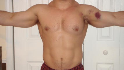
30yo male weight lifter who 10 days ago had a painful left shoulder injury after bench press. The next morning his left anterior chest wall and left upper arm were bruised and swollen. He went to see his PCP who diagnosed him with a muscle strain. 8 days later the bruising and swelling have resolved but he still cant move his shoulder and comes to the ED.
http://321gomd.com/wp-content/uploads/2015/01/pec-major-tears.jpg
The pec major attaches to the humerus and originates from the sternum and clavicle
Injury is usually due to tendon rupture off the humerus but can also occur at the muscle tendon junction or within the muscle belly itself.
Injury is becoming increasingly common due to the popularity in power lifting sports.
Mechanism: excessive tension on a maximally eccentrically contracted muscle.
Patients will complain of pain and weakness of the shoulder.
PE: Swelling and bruising to anterior medial arm. Palpable defect and deformity or anterior axially fold (may be hidden by swelling).
Weakness and pain with adduction and internal rotation and forward flexion
Chronic presentations can be challenging to diagnose. Consider ultrasound
Non operative treatment may be indicated for partial tears (sling, ice, NSAIDs)
Operative repair of tendon avulsions is very successful. Patients age, occupation/activity level and location of injury and condition of tear are considered.
Sesamoid Injuries
Unlike other bones in the human body that are connected to each other at joints, sesamoid bones are only connected via tendons (or are imbedded in muscle).
The largest sesamoid bone is the patella.
2 small sesamoid bones lie on the plantar foot near the great toe
Sesamoid bones can fracture and the surrounding tendons can become inflamed (sesamoiditis)
Traumatic injury is usually due to hyperextension and axial loading
Sx: Pain located under the great toe on the ball of the foot (Gradual with sesamoiditis and acutely with a fracture).
There may be associated swelling and bruising. Pain with palpation, flexion and extension.
The medial/tibial sesamoid is larger, has great weight bearing status and is more commonly injured that its lateral counterpart.
In many people (10 - 25%) the medial sesamoid of the foot has two parts (bipartite). This finding is bilateral in 25% of people.
This may confuse some providers as it may appear to be a fracture
Look for a smooth contour to the bones and clinically correlate (bruising, soft tissue swelling, etc.) if it is an incidental finding.
Other radiographic clues include
1) The fractured sesamoid is usually slightly larger than the lateral sesamoid while the bipartite sesamoid has a much larger medial sesamoid than lateral sesamoid
2) The fractured sesamoid shows a sharp, radiolucent, uncorticated line between the two fragments while the bipartite sesamoid has two corticated components
3) The fractured sesamoid fragments often fit together like pieces of a puzzle while the bipartite sesamoid has two components that do not fit together snugly
4) Other means to differentiate the two involve MRI and bone scanning
Treatment involves a stiff-soled shoe or applying a cushioning pad or J-shaped pad around the area to relieve pressure.
It may take months for the pain to subside.
http://www.apfmj-archive.com/afm5_3/afm50.htm#F1
Show References
Plain films are commonly used to screen children for pelvic fractures or dislocations following blunt torso trauma
The sensitivity of this common screening practice is unknown
A recent study looked at this question.
Of 451 patients with pelvic fractures or dislocations, 382 had AP radiographs. Injury was correctly identified in 297 patients (sensitivity 78%).
The sensitivity was greater in the sicker subgroups :92% for those requiring operative intervention and 82% for those with hypotension
Plain AP pelvic radiographs should have a limited role in the sole evaluation of children with blunt torso trauma.
They should be incorporated in the assessment of hemodynamically unstable children and those in whom the clinician is not planning on otherwise obtaining an abdominal/pelvis CT.
Show References
Medication-overuse headache (MOH) is one of the most common chronic headache disorders
Worldwide prevalence of 1 2%
Characterized by chronic headache and overuse of different headache medications
Withdrawal of the overused medication is the treatment of choice
A 2014 study looked at adolescent patients treated in a headache clinic with chronic post traumatic headaches (concussion headaches)
77 had chronic post-traumatic headache of 3-12 months' duration
54 of 77 (70.1%) met criteria for probable medication-overuse headache.
After the OTC medicine was stopped 68.5% had resolution or improvement !!
Excessive use of analgesics postconcussion may contribute to chronic post-traumatic headaches in some adolescents.
Sometimes the advise of "just keep taking the motrin and it'll get better" isnt the answer
Show References
Radiographs of the sacrum and coccyx in the emergency department (ED) have no quantifiable clinical impact, according to a study published in the American Journal of Roentgenology.
Researchers from Emory University Midtown Hospital and Morehouse School of Medicine in Atlanta, GA, sought to determine the yield and clinical impact of sacrum and coccyx radiographs performed in the ED.
Sacrum and coccyx X-rays performed on 687 consecutive patients over a six-year period in level-1 and level-2 trauma centers (4 total hospitals). The patients’ mean age was 48.1, 61.6% were women. The images were categorized as positive for acute fracture or dislocation, negative, or other.
The researchers then analyzed:
• Follow-up advanced imaging in the same ED visit
• Follow-up advanced imaging within 30 days
• New analgesic prescriptions
• Clinic follow-up
• Surgical intervention within 60 days
The researchers found positive results in 58 of the 687 patients, a positivity rate of 8.4%.
None of the 58 positive cases had surgical intervention.
There was no significant association between sacrum and coccyx radiograph positivity and analgesic prescription or clinical follow-up among the patients evaluated at the level-1 trauma centers.
However at the level-2 trauma centers, 34 (97.1%) of 35 patients with positive sacrum and coccyx radiographs received analgesic prescriptions or clinical referrals. Negative cases were at 82.9%.
Of all cases, 39 patients (5.7%) underwent advanced imaging in the same ED visit and 29 patients (4.3%) underwent imaging within 30 days.
“Sacrum and coccyx radiography results had no significant correlation with advanced imaging in the same ED visit,” the authors wrote. “There was no significant difference in 30-day advanced imaging at the level-1 trauma centers, but there was at the level-2 trauma centers.”
The researchers concluded that routine sacrum and coccyx radiography should not be part of ED practice and that patients should be treated conservatively based on clinical parameters.
Show References
https://www.youtube.com/watch?v=sCFOObsx_W4
What is their risk of MI???
Anger outbursts are bad for your heart. Out of 300 patients with an acute MI, just over 2% reported losing their temper within 2 hours of the event. A review of nine studies of rage and cardiovascular events all found an increase in cardiovascular events in the 2 hours preceding an anger outburst. Examples included arguments at home, at work or by road rage. Compared with their usual anger levels, the relative risk of heart attack from a fit of rage was 8.5.
What about those of us who are just fanatics, I mean fans....A recent study of World Cup soccer found that the intense strain and excitement of viewing a dramatic soccer match more than doubles the risk of acute heart attack, particularly in men with known coronary heart disease. This was regardless of the outcome of the match!
Show References
Exercise and the heart
Exercise increases the risk of sudden cardiac death (SCD) acutely.
Exercise decreases the risk of SCD in the long term.
Regular physical activity (even as little as 15 mins/day) reduces the risk of cardiovascular disease (CVD).
Up to 15% of MIs occur during or soon after vigorous physical exercise. This is typically in sedentary men with coronary risk factors.
In a 1993 study, in the first hour after heavy exertion, risk of heart attack rose more than 100-fold from baseline for habitually inactive persons. However, for frequent exercisers, this risk rose less than three-fold. Think of snow shoveling after a winter storm.
Both the Physicians’ Health Study and the Nurses’ Health Study show that the risk of SCD during exertion is reduced by habitual exercise.
If you are physically active, stay active. If you are not active, you should be because exercise has innumerable personal benefits. However, it is important to start gradually Some individuals at higher risk need to start under the guidance of a physician.
Show References
Orthopedic documentation
1) Document location with specificity and laterality.
2) Document the location with as much specificity as possible
-Name of specific bone and specific site on bone (Shaft, head, neck, distal, proximal, styloid)
3) Document fractures as open/closed, displaced vs. non-displaced, routine or delayed healing,
-Orientation of fractures, such as transverse, oblique, spiral
- Document intra-articular or extra-articular involvement
4) For a particular injury, a complete note will include mention of the following
The joint above (e.g. for shoulder injuries this would be the neck, for hip injuries - the back)
The joint below
Motor (e.g. for arm injuries document the distal median, radial and ulnar motor innervation)
Sensory
Vascular
Skin (for all fractures document intact overlying skin esp. when covering with a splint)
Compartments (a simple mention of compartments are grossly soft/not tense will suffice)
*Especially relevant for forearm and tib/fib injuries
Metacarpal Fractures
* Localize fracture to head, neck or shaft (neck most common)
5th metacarpal most commonly fractured
* Note amount of angulation, shortening and the presence of malrotation
*Treatment is based on which metacarpal is fractured and the location of the fracture
*The amount of acceptable angulation varies by the digit involved
For example for index and long finger - acceptable angulation of the shaft is 10-20 degrees and neck is 10 to 15 degrees
Whereas for the 5th digit - acceptable angulation for the shaft is 40 degrees and neck is 50 degrees
Pearls
No degree of malrotation is acceptable (document the absence of this!)
Strongly suspect fight bite injury with abrasions/lacerations overlying metacarpal heads
Highly prone to infection given the proximity to the joint capsule
Consider lacerations over metacarpal fractures as open fractures (do not close/discuss management with hand surgery re timing of washout. Many prefer delayed fixation for suspected infections )
Document integrity of the extensor tendon (can be lacerated and retracted)
A meta-analysis of 74 randomized trials with a total of 58,556 patients was recently published in the Lancet that looked at the effectiveness of NSAIDs in the treatment of osteoarthritis (OA) pain.
Briefly, their conclusion was that:
- Acetaminophen is ineffective as a single-agent in the treatment of OA.
- Diclofenac 150 mg/day had best evidence to support it as the most effective NSAID available presently with respective to its effectiveness in relieving pain and improving function.
- They found no evidence that treatment effects varied over the duration of treatment ( no tolerance)
You can find the article here http://www.thelancet.com/journals/lancet/article/PIIS0140-6736%2816%2930002-2/abstract
Show References
Femoral neck fracture
- The most commonly missed hip fracture
We typically think of the presentation of the displaced fracture severe pain, writhing in the bed, unable to ambulate, limited ROM
* However, patients with nondisplaced fractures (15 20%) may walk with a limp
* Occurs primarily in the elderly & osteoporotic population after a fall directly onto the hip
* Look for a cortical step-off in the femoral neck (w/ foreshortening)
* A patient with a minimally displaced fracture may only complain of hip, knee, or groin pain and may be able to walk (albeit with a limp)
* Almost 9% of hip fractures are radiographically normal (Nondisplaced or impacted fractures)
* Fractures which were initially nondisplaced, may become displaced upon re-presentation
* Remember the limitations of plain x-ray in the evaluation of femoral neck fractures!
* Because of the significant complication of overlooking a femoral neck fracture, MRI has become the recommended imaging modality of choice for a patient with a high suspicion for a femoral neck fracture, despite normal plain radiographs of the hip
Achilles tendon rupture
More common in
men, ages 30 - 40yo, s/p steroid injections, fluoroquinolone use, and episodic athletes "weekend warriors
Mechanism: usually during an athletic endeavor, sudden forced planar flexion or violent dorsiflexion of a plantar flexed foot
Location: Usually occurs 4 to 6 cm ABOVE the Achilles calcaneal insertion (hypovascular region)
Patient will report a sudden pop, gunshot like sound
History: Will report heel and calf pain and weakness/inability to walk
Physical examination: Palpable gap, weakness with plantar flexion, + Thompsons test
https://www.netterimages.com/images/vpv/000/000/007/7714-0550x0475.jpg
Consult orthopedics and splint in resting equinus
http://img.medscape.com/fullsize/migrated/408/535/mos0216.01.fig5b.jpg
Spondylolysis
Prevalence 3-6% in the general population (Higher in athletes)
Location: L4 (5-15% of cases) & L5 (85-95% of cases)
Population: More likely in the skeletally immature athlete due to the vulnerability of the immature pars interarticularis to repeated stress
Symptoms: Lumbar pain worse with extension
Higher risk sports: Gymnastics, diving, weightlifting, wrestling
Treatment: Bracing and activity modification, physical therapy
- Good results in 80% with conservative management allowing return to play.
- Those who fail benefit from iliac crest bone grafting and posterolateral fusion.
-Return to play is controversial in this group
Please review th images below for anaomy and imaging appearence
http://orthoinfo.aaos.org/figures/A00053F01.jpg
http://www.sonsa.org/images/spondylolysis.jpg
http://www.physio-pedia.com/images/2/22/Spondylolysis_x_ray_.docx.jpg
Show References
Recommended follow-up for common orthopedic injuries
The Salter Harris Classification System is used in pediatric epiphyseal fractures. The higher the type of fracture the greater the risk of complications and growth disturbance.
Some common exam facts about Salter Harris Fractures are:
- The type II fracture is the most common.
- The small metaphyseal fragment in Salter Harris type II and IV fractures is called the Thurston Holland Sign.
- Type III and IV fractures often require open reduction and internal fixation due to the fracture extending into the joint.
- Type V fractures may appear normal, but the epiphyseal plate is crushed and the blood supply is interrupted.
The Classification system as listed by Type:
- Type I: A fracture through the physeal growth plate. Typically can not be seen on x-ray unless they growth plate is widened or displaced..
- Type II: A fracture through the physeal growth plate and metaphysis.
- Type III: A fracture through the physeal growth plate and epiphysis.
- Type IV: A fracture through the epiphysis, physeal growth plate and metaphysis.
- Type V: A crush injury of the physeal growth plate.
For Maite, a helpful mnemonic is SALTR , Slipped (Type I), Above (Type II), Lower (Type III), Through (Type IV), and Ruined or Rammed (Type V)
A image of the fractures can be found on FP Notebook at http://www.fpnotebook.com/ortho/fracture/ephyslfrctr.htm
Sever's disease also known as calcaneal apophysitis, is the most common cause of heel pain in the young adolescent (ages 8 to 12).
It can be thought of as the Achilles tendon equivalent of Osgood-Schlatter's disease (patellar tendon insertion pain).
It is a non inflammatory chronic repetitive injury.
Commonly seen bilaterally in up to two -thirds of cases.
Patients will complain of activity related pain to the heel.
There may be tenderness and local swelling at the Achilles tendon insertion.
Radiographs are not necessary for acute cases.
Treat with activity modification, heel raise, physical therapy.
Protracted Recovery from Concussion
Age and sex may influence concussion recovery time frame
Methods: 266 adolescent athletes presenting to a sports medicine concussion clinic
Female athletes had a longer recovery course (P=0.002) and required more treatment interventions (p<0.001).
Female athletes were more likely to require academic accommodations (p<0.001), vestibular therapy (P<0.001) and medications (P<0.001).
Be aware that not all concussion patient subgroups with concussions recover in the same manner. Further study is needed to support whether female adolescent athletes require unique management and treatment guidelines.