University of Maryland School of Medicine logo
Emergency Medicine
All Departments
UMSOM Assistant

UMEM Educational Pearls

  • Home
  • Departments
  • Emergency Medicine
  • CME
  • UMEM Educational Pearls
  • CME
  • Courses and Conferences
  • Free Open Access Medical Education
  • UMEM Educational Pearls
Previous |  1 |  2 |  3 |  4 |  5 |  6 |  ... |  239 |  Next

Title: Should we start home beta-blocker for patients with sepsis

Category: Critical Care

Keywords: beta-blocker, sepsis, mortality, hospital length of stay. (PubMed Search)

Posted: 10/6/2026 by Quincy Tran, MD, PhD (Updated: 10/8/2026)

In this retrospective study of 4635 patients, continuing oral beta blocker within 48 hours of presentation for patients with sepsis was associated with a reduced risk of all-cause mortality within 90 days of hospital admission (hazard ratio 0.77; 95% CI, 0.61–0.98; p = 0.03), and a shorter hospital stay (incidence rate ratio 0.39; 95% CI, 0.38–0.41; p < 0.001).

Therefore, it is reasonable to consider Beta-blocker if patients are adequately resuscitated.

Show References

Christian-Miller N, Lee KT, Pashun R, Ballut K, Prescott H, Cascino T, Taylor SP, Ansari S, Sjoding MW, Nagle M, Admon AJ. Continuing vs. Withholding Home Beta-Blockers at Admission for Suspected Infection: A Target Trial Emulation. Crit Care Med. 2026 Sep 1;54(9):2350-2360. doi: 10.1097/CCM.0000000000007229. Epub 2026 Jun 9. PMID: 42262489

Attachments

  • 2026_10_05 Christian-Miller oral beta blocker for sepsis-6ac4834dec2d9.docx (16 Kb)


Title: Ultrasound Signs of Cholecystitis

Category: Ultrasound

Keywords: POCUS, abdominal pain, gallstones, cholecystitis (PubMed Search)

Posted: 10/5/2026 by Alexis Salerno Rubeling, MD (Updated: 10/8/2026)

Emergency Physicians have a high specificity and moderate sensitivity for detecting cholecystitis with point-of-care ultrasound. 

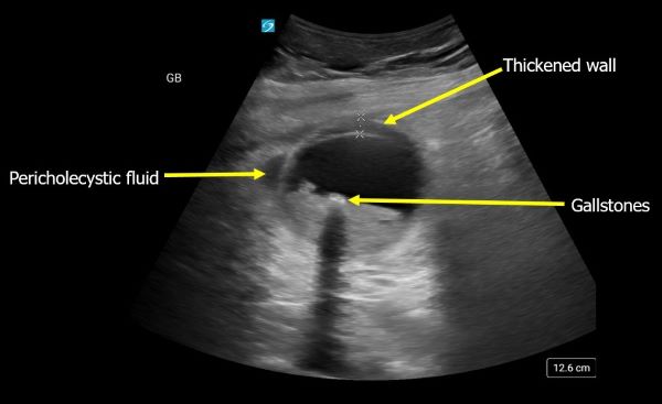
Signs of Cholecystitis on Ultrasound include: 

  • Sonographic Murphy's sign = The patient has pain when the ultrasound probe is directed over the gallbladder and compressed 
  • Pericholecystic Fluid = Fluid around the gallbladder 
  • Increased Gallbladder Wall Thickness = A wall thickness > 3 mm measured on the anterior gallbladder wall 
  • Most patients will have gallstones. Acalculus cholecystitis can occur with patients who are chronically ill, critically ill, on TPN or immunocompromised.  
Show References

Wilson SJ, Thavanathan R, Cheng W, Stuart J, Kim DJ, Glen P, Duigenan S, Shorr R, Woo MY, Perry JJ. Test Characteristics of Emergency Medicine-Performed Point-of-Care Ultrasound for the Diagnosis of Acute Cholecystitis: A Systematic Review and Meta-analysis. Ann Emerg Med. 2024 Mar;83(3):235-246. doi: 10.1016/j.annemergmed.2023.09.005.


Title: BCVI screening efficacy

Category: Trauma

Posted: 10/4/2026 by Robert Flint, MD (Updated: 10/8/2026)

How well do the Denver and Memphis screening criteria identify blunt cerebral vascular injury? 
From 2021:
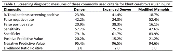
Not very well!  Blunt injury to the head and neck may be enough to prompt you to order a CTA of the neck.

Click to see Memphis criteria which overlap Denver

Show Additional Information

  • base of skull fracture with involvement of the carotid canal
  • base of skull fracture with involvement of petrous temporal bone
  • cervical spine fracture (including subluxation, transverse foramen involvement, and upper cervical spine fracture)
  • neurological exam findings not explained by neuroimaging
  • Horner syndrome
  • Le Fort II or III fracture pattern
  • neck soft tissue injury (e.g. seatbelt sign, hanging, hematoma)

Show References

https://thetraumapro.com/2021/09/01/screening-for-bcvi/


Title: PECARN Pediatric Cervical Spine Prediction Rule (2024)

Category: Pediatrics

Keywords: PECARN, cervical spine injury, blunt trauma, pediatric trauma, cervical imaging, CT reduction, decision rule (PubMed Search)

Posted: 10/2/2026 by Jeremy St. Thomas, MD (Updated: 10/8/2026)

One-liner 

A large prospective PECARN study developed and validated a highly sensitive clinical prediction rule to safely reduce unnecessary cervical spine imaging in children with blunt trauma.  

Background: Pediatric cervical spine injury (CSI) is rare (<1% of blunt trauma), but missing an injury has devastating consequences. Existing adult rules (NEXUS, Canadian C-Spine Rule) are not well validated in children. 

Methods: This multicenter prospective observational study enrolled over 22,000 children with blunt trauma across PECARN hospitals. Investigators identified clinical variables associated with CSI and derived a risk-stratification rule. 

Results: The rule demonstrated very high sensitivity (~94%) and an extremely high negative predictive value (>99.9%) for clinically significant CSI. High-risk factors (e.g., GCS 3–8, focal neurologic deficits) warrant CT, whereas intermediate-risk factors may be evaluated with plain radiographs. Children without identified risk factors generally do not require imaging. 

Limitations: Observational derivation/validation study rather than an interventional trial. Clinical judgment remains important in atypical presentations. 

Clinical Relevance: This is likely to become the pediatric equivalent of the PECARN head trauma rule and should reduce unnecessary CT imaging while maintaining patient safety.

Take-home Points 

  • First large, validated pediatric-specific CSI rule.  
  • Helps identify children who require CT versus radiographs versus no imaging.  
  • Expected to reduce radiation exposure.  
  • May become the new standard for pediatric cervical spine evaluation.

Show References
Leonard JC, et al. Lancet Child Adolesc Health. 2024.  

DOI: 10.1016/S2352-4642(24)00104-4.


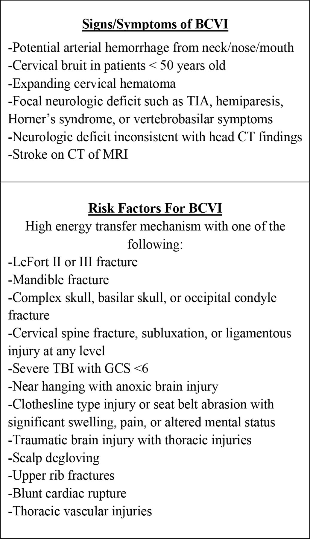
Title: Denver criteria for blunt cerebral vascular injury

Category: Trauma

Posted: 10/1/2026 by Robert Flint, MD (Updated: 10/8/2026)


Title: Metformin Associated Lactic Acidosis: MALA

Category: Toxicology

Keywords: Metformin, lactic acidosis, MALA (PubMed Search)

Posted: 9/30/2026 by Kathy Prybys, DO

Metformin, a biguanide, is the most widely used and recommended first line treatment for type 2 diabetes due to its efficacy and  low hypoglycemic risk.  It's mechanisms of action include inhibition of mitochondrial complex I electron transport  and oxidative phosphorylation (causing inhibition of hepatic gluconeogenesis and increases insulin sensitivity) and decreased intestinal absorption of glucose. It does not stimulate pancreatic insulin secretion so does not cause hypoglycemia even in overdose (except in massive dosages > 60 grams and rare cases). 

Metformin’s most serious toxicity is due to lactic acidosis. Metformin associated lactic acidosis (MALA) is a rare but life-threatening condition with incidence of 1 in 30,0000 and high mortality rate of 30-50%. Lactic acidosis can occur in both acute overdose and chronic therapeutic use in predisposed individuals. MALA is a serious condition with  that requires early recognition and aggressive treatment to increase survival and should be considered in all patients on metformin who present with lactic acidosis.

Show Additional Information

Metfromin associated lactic acidosis (MALA) occurs due to reduced lactate clearance caused by metformin’s inhibition of the mitochondrial electron transport chain causing a shift to anerobic metabolism (pyruvate -->lactate) and is at highest risk of occurrence when combined with other co-morbid states that cause lactate accumulation due to reduced lactate clearance (hepatic disease) or increased lactate production (hypotension, dehydration, sepsis, decompensated heart failure, hypoxia) and heavy alcohol use. The most significant risk factor for MALA is renal insufficiency, as metformin does not undergo hepatic metabolism but is almost entirely eliminated unchanged by renal excretion. Diminished renal excretion of metformin significantly prolongs its elimination half-life, leading to increased blood and tissue levels. 

Metformin carries a FDA black box warning for lactic acidosis in patients with liver and kidney impairment and should not be prescribed for patient’s with GFR < 30 ml/min.  Remember to discontinue metformin in patients at risk for lactic acidosis and AKI (sepsis, dehydration, administration of radiologic contrast).

In acute overdose, toxicity can be delayed for many hours. Observation for 12 hours is recommended to rule out lactic acidosis.

Diagnostic criteria for MALA include high AGMA with pH < 7.35 (in studies mean 7.02) and lactic acid level out of proportion > 5 (in studies mean >14) in patients using metformin. Lactic acid levels are usually higher than what would be considered survivable for other disease states.

Treatment of MALA:

  • STOP Metformin
  • Aggressive fluid hydration (consider D5W with sodium bicarb) 
  • Vasopressors
  • Ventilatory support (consider HFNC O2/ avoid intubation unless medically necessary)
  • Treat other causes of lactic acidosis 
  • Early nephrology consultation for intermittent hemodialysis (preferred) or continuous renal replacement therapy

Definitive Indications for hemodialysis include: Lactate >20,  pH < 7.0 , comorbid conditions (shock, renal insufficiency, liver failure), decreased level of consciousness, lack of response to supportive measures.

CNS depression is a marker of severe toxicity due to cerebral mitochondrial inhibition and depletion of ATP,  focal cerebral lactate accumulation,  systemic acidosis , and should prompt evaluation for the possibility of rare hypoglycemia.

Show References

Do Patients Die with or from Metformin-Associated Lactic Acidosis (MALA)? Systematic Review and Meta-analysis of pH and Lactate as Predictors of Mortality in MALA. Blumenberg A, Benabbas R, Sinert R, et al. J Med Toxicol. 2020 Apr;16(2):222-229. doi: 10.1007/s13181-019-00755-6. Epub 2020 Jan 6. PMID: 31907741; PMCID: PMC7099117.

See KC. Metformin-associated lactic acidosis: A mini review of pathophysiology, diagnosis and management in critically ill patients. World J Diabetes. 2024 Jun 15;15(6):1178-1186. doi: 10.4239/wjd.v15.i6.1178. PMID: 38983827; PMCID: PMC11229964.

Metformin-Associated Lactic Acidosis (MALA): When a Common Drug Turns Catastrophic. Kessler LE, Sachdeva D, Singhal R, et al. Cureus. 2025 Aug 3;17(8):e89292. doi: 10.7759/cureus.89292. PMID: 40904982; PMCID: PMC12404583.

Time to development of metformin-associated lactic acidosis. Theobald J, Schneider J, Cheema N, DesLauriers C. Clin Toxicol (Phila). 2020 Jul;58(7):758-762. doi: 10.1080/15563650.2019.1686514. Epub 2019 Nov 6. PMID: 31691608.

https://www.extrip-workgroup.org/metformin

Extracorporeal treatment of metformin associated lactic acidosis in clinical practice: a retrospective cohort study. van Berlo-van de Laar IRF, Vermeij CG, van den Elsen-Hutten M, et al.  Eur J Clin Pharmacol. 2020 Jun;76(6):815-820. doi: 10.1007/s00228-020-02857-5. Epub 2020 Mar 13. PMID: 32170333; PMCID: PMC7239820.


Title: Atropine in Unstable Bradycardia

Category: Critical Care

Keywords: unstable bradycardia, AHA, ACLS, atropine (PubMed Search)

Posted: 9/29/2026 by Kami Windsor, MD

Background: The 2025 AHA guidelines changed their recommended initial IV atropine dose for unstable bradycardia from 0.5 mg to 1.0 mg, while the 2025 ERC guidelines continue to recommend an initial dose of 0.5 mg. A 2025 review found limited comparative studies and noted that using 0.5 mg as an initial dose seems “clinically reasonable" [1].

Now: One tertiary care center performed a retrospective propensity-score matched study [2] comparing the 2 groups of patients with unstable bradycardia who had received an initial dose of 0.5 mg versus 1 mg in their ED between 2020 and 2025.

  • Primary outcome = First-Dose Success (reaching a heart rate >50 bpm within 3 minutes without additional therapy). 
  • Notably excluded were patients with noncardiac secondary bradycardia (hyperkalemia, drowning, trauma, etc.) and those with pacemaker failure, amongst others.

Results in the propensity score-matched cohort:

  • First-dose success was significantly higher in the 1.0 mg vs the 0.5 mg group (45 vs 25%; p=0.007)
    • NNT of 6 (95% CI, 3-18)
  • The 1.0 mg group had lower rates of:
    • Additional atropine requirement (26 vs 48%, p=0.003)
    • Other rescue therapy requirement (51% vs 74%, p=0.002)
    • ICU admission (53% vs 74%, p=0.005).
  • No difference between groups in 30-day mortality or subsequent permanent pacemaker placement

Bottom Line: Acknowledging the small size & retrospective nature of the study, it appears that an initial dose of 1mg IV atropine helps us work smarter--not harder--in the stabilization of patients with unstable bradycardia.

Show References

  1. Yokose M, Sangawa M, Shiomi H, et al. Initial Dose of Intravenous Atropine for Patients With Symptomatic Bradycardia?- A Scoping Review. Circ Rep. 2025;7(11):1044-1050. Published 2025 Sep 27. doi:10.1253/circrep.CR-25-0169
  2. Kokulu K, Sert ET, Akar EH, Gül M. Initial atropine dose of 0.5 mg versus 1 mg in unstable bradycardia: A propensity score-matched retrospective cohort study. Am J Emerg Med. Published online August 2, 2026. doi:10.1016/j.ajem.2026.07.065


Title: Beyond Withdrawal: Naltrexone for Alcohol Use Disorder

Category: Pharmacology & Therapeutics

Keywords: naltrexone, alcohol use disorder, substance use (PubMed Search)

Posted: 9/28/2026 by Lena Carleton, MD (Updated: 9/28/2026)

Naltrexone, an opioid antagonist, is an FDA-approved first-line medication for alcohol use disorder. It is available as a daily oral formulation or a monthly extended-release intramuscular injection. Naltrexone does not treat acute alcohol withdrawal; instead, it is used to reduce cravings, heavy drinking, and support abstinence. Patients can continue to drink while taking it without a disulfiram-like reaction. Common adverse effects include self-limited nausea, headache, and insomnia. It is contraindicated in patients with current opioid use, acute hepatitis, or liver failure.

Show References

Krist AH, Bradley KA. Addressing Alcohol Use. N Engl J Med. 2025;392(17):1721-1731. doi:10.1056/NEJMcp2402121.

Naltrexone hydrochloride tablet, film-coated [prescribing information]. Chartwell RX, LLC; updated June 19, 2026. DailyMed.


Title: Finding more than you are looking for

Category: Trauma

Posted: 9/27/2026 by Robert Flint, MD (Updated: 10/8/2026)

This institution  implemented universal CTA of the neck to evaluate cerebral vascular injury in blunt trauma patients. In the 531 patients, 6% had cerebral vascular injury and 23% had incidental findings of which 22% required follow up. Another example of a test ordered has potential unanticipated downstream effect.

Show References

Kummer KA, Ambroise G, Warfield J, Srivastava AK. Incidental findings on head and neck CTA obtained for evaluation of patients with traumatic injuries. Trauma. 2026;0(0). doi:10.1177/14604086261481678


Title: Analgesia for anterior shoulder dislocations

Category: Orthopedics

Posted: 9/26/2026 by Brian Corwell, MD

Anterior shoulder dislocations are commonly seen during an ED shift.

During a busy shift, the choice of analgesia may be a strong consideration.

Analgesia for reduction can be provided by intra-articular lidocaine injection or intravenous sedation among other options. 

How do these 2 analgesic options compare?

Show Additional Information

A 2022 systematic review of 12 RCTs (630 patients).

327 in Lidocaine group

303 in IV sedation group

Patients  aged 15 years or older 

Outcomes of interest included a successful reduction, adverse events, ED length of stay, pain scores, procedure time, ease of reduction, patient satisfaction, and cost.

Conclusions: 

There was no difference in reduction success between groups

As one would expect, significantly lower adverse events with lidocaine

As one would expect, shorter ED length of stay with lidocaine

No difference in pain scores post-analgesia during reduction

No difference in ease of reduction

Patient satisfaction was greater with sedation.

**This likely reflects being awake and processing the experience vs. being asleep and waking up to the finished product.

Conclusions

Intra-articular lidocaine may have similar effectiveness as IV sedation in the successful reduction of anterior shoulder dislocations in the ED with fewer adverse events, shorter ED length of stay, and no difference in pain scores or ease of reduction but may be associated with lower patient satisfaction.

Consider intra-articular lidocaine as an effective alternative to IV sedation for reducing anterior shoulder dislocations.

Show References

Sithamparapillai A, Grewal K, Thompson C, Walsh C, McLeod S. Intra-articular lidocaine versus intravenous sedation for closed reduction of acute anterior shoulder dislocation in the emergency department: a systematic review and meta-analysis. CJEM. 2022 Dec;24(8):809-819.


Title: Optimizing ED Care for Migration-Affected Patients

Category: Administration

Keywords: Migrant, Emergency Department (PubMed Search)

Posted: 9/23/2026 by Mercedes Torres, MD (Updated: 9/23/2026)

The emergency department may be the only point of access to the health care system for migrants.  As emergency physicians, we face the challenge of balancing our ethical obligations to our patients with our responsibilities to employers, the government, and/or law enforcement. In consideration of the mandate of EMTALA and this complex role within an evolving political landscape, this publication offers four principles to guide best practices when caring for migration-affected patients in the ED. 

Click the link to review these four principles which focus on ensuring equitable care for all migration-affected patients.

Show Additional Information

Four Principles to Support Equitable ED Care for Migration-Affected Patients:

1. Practice evidence-based clinical care to reduce bias.

2. Be aware of local and federal policies and migration factors that may influence patient care trends.

3. Target screening for population-specific risks and tailor patient care services.

4. Establish community partnerships and interdisciplinary care teams.

See article for more detail with examples of each principle in action.

Show References

Stanifer K, Smith-Bellille T, Bloem C, Willis J, Aurrecoechea A.  Beyond Borders: Advancing Equitable Care in the Emergency Department for Migration-Affected Patients. The Journal of Emergency Medicine. Vol 89, 2026, Pages 109-114, https://doi.org/10.1016/j.jemermed.2026.08.005.


Title: "Everything in Excess is Opposed to Nature"

Category: Critical Care

Posted: 9/22/2026 by Caleb Chan, MD (Updated: 10/8/2026)

Question

The following is an actual case. 

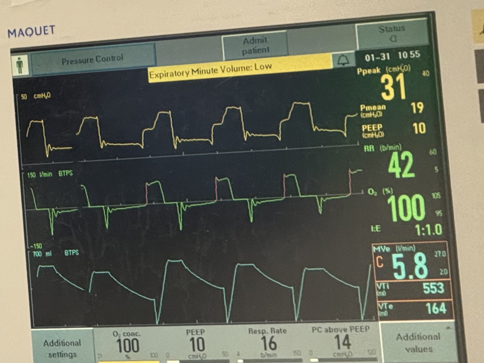
An ED patient without any history of asthma or COPD is intubated to facilitate an emergent MRI. After the MRI is completed (and without abnormalities). The patient is breathing  synchronously and comfortably on minimal PS settings. Further sedation is weaned to facilitate extubation in the ED as the patient has no need for continued mechanical ventilation and no ICU requirements. All of a sudden, you are emergently called overhead to the patient's room because the patient is “blue” and hypoxic. 

These are the ventilator waveforms. 

The patient did not receive any contrast or bronchospastic medications. The ETT is clear and without obstruction.

What is going on?
Show Answer
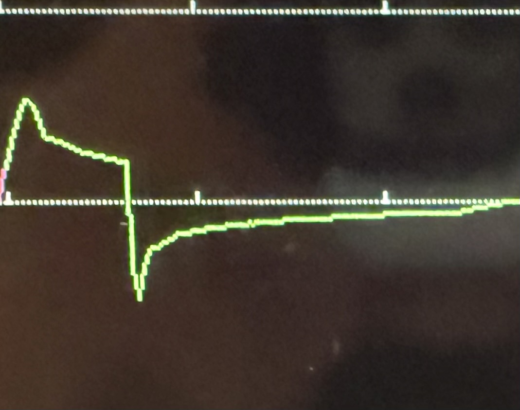
There are a lot of issues that are apparent on this ventilator waveform. However, the focus for this pearl is the expiratory waveforms which show significant expiratory obstruction (compare to normal below). The patient is getting inspiratory volumes (VTi 553 mL in light blue bottom right corner), but limited expiratory waveforms (VTe 164 mL) . Given that the patient has no history of asthma, COPD, or reason for acute obstruction, the concern was that patient effort (as patient became more awake with decreased sedation) was actually causing expiratory obstruction. Sedation was increased and the obstruction resolved. 

The patient was subsequently diagnosed with excessive dynamic airway collapse, a condition where the posterior membrane of the tracheobronchial tree weakens and bulges anteriorly into the airways (this can sometimes be seen on expiratory CT chests). This movement is exaggerated during forced exhalation due to increased intrathoracic pressure and causes a more severe expiratory obstruction pattern compared to passive exhalation. Consequently, this can lead to a cycle of agitation, increased work of breathing, and breath-stacking/accumulation of intrinsic PEEP. 

Of note, EDAC often gets misidentified as “COPD” or “asthma,” and given its mechanism, does not respond to bronchodilators or steroids. The weakening of the posterior membrane is a chronic process and hence EDAC does not develop acutely, it simply becomes clinically apparent in an agitated intubated patient. Hence, the treatment for this patient was a sedative to calm the patient while preserving respiratory effort (i.e. dexmedetomidine) and extubating the patient to remove the exacerbating stimulus (being intubated). 

Extubating to NIPPV could be considered as a bridge granted it does not cause more agitation and increased work of breathing.

In summary, EDAC is a cause of expiratory obstruction that should  be considered in patients with evidence of obstruction and increased of work of breathing, particularly if they don’t have a history of COPD/asthma or reason for bronchospasm.


Title: A missed opportunity

Category: Cardiology

Posted: 9/20/2026 by Robert Flint, MD (Updated: 10/8/2026)

This commentary points out the missed opportunity to improve chronic hypertension care in the ED. A substantial number of patients seeking care for asymptomatic hypertension and an even larger cohort seen for other complaints who are also chronically untreated  hypertensives leave the ED without a prescription for antihypertensives. ACEP and AHA say we should be treating these people and arranging follow up.

Show References

N. E. Harrison and P. Levy, “ Chronic Uncontrolled Hypertension in the ED—We Hold This Truth to Be Self Evident,” Academic Emergency Medicine 33, no. 9 (2026): e70409, https://doi.org/10.1111/acem.70409.


Title: Perspectives and Preferences of Underrepresented in Emergency Medicine Resident Applicants: A Qualitative Study

Category: Administration

Posted: 9/19/2026 by Kevin Semelrath, MD (Updated: 10/8/2026)

This small qualitative study demonstrated that representation and hearing from other URiM residents is an important factor in recruitment during EM residency application.

Show Additional Information

M. Suh, B. Torres, A. Bhananker, et al., “ Perspectives and Preferences of Underrepresented in Emergency Medicine Resident Applicants: A Qualitative Study,” AEM Education and Training 10, no. 3 (2026): e70176, https://doi.org/10.1002/aet2.70176.


Title: Predictors of clinically significant underlying etiologies in children presenting with acquired torticollis

Category: Pediatrics

Keywords: torticollis, neck pain, fever (PubMed Search)

Posted: 9/18/2026 by Jenny Guyther, MD (Updated: 9/18/2026)

This was a retrospective review of children < 18 years who presented to the pediatric emergency department with torticollis in a single center over a 6 year period.

Clinically significant underlying etiologies included infection, tumor, or atlantoaxial rotary fixation requiring surgery (AARF).

352 children with a median age of 4.8 years presented with torticollis and 59 were found to have a significant underlying etiology.

Children with infectious causes were younger and more likely to be febrile and have an elevated CRP.  Infections included retropharyngeal abscess, lymphadenitis and cervical spine osteomyelitis.  AARF was associated with prolonged symptoms, older age, midline tenderness and restricted motion.  Tumor related cases often featured additional red flag symptoms such as headache and vomiting and symptom duration ranging from 5 days to 3 months.

Show References

Shemer M, Noyman S, Rimon A, Cohen N. Predictors of clinically significant underlying etiologies in children presenting with torticollis. Paediatr Child Health. 2026 Jan 12;31(2):111-119. doi: 10.1093/pch/pxaf113. PMID: 41821840; PMCID: PMC12976673.


Title: Arterial complication after hip fracture

Category: Trauma

Posted: 9/17/2026 by Robert Flint, MD (Updated: 10/8/2026)

This case report describes a 2 week delayed hemorrhage from a lacerated profunda femoris artery after an intertrochanteric fracture that requires ORIF. The patient represented with anemia, pain and swelling in the surgically repaired hip. Pulses were still present. The authors conclude: 

“The profunda femoris artery is the vessel most commonly involved in vascular complications associated with proximal femoral fractures because of its close anatomical relationship with the lesser trochanter. Delayed presentations may occur weeks after injury as repeated mechanical irritation from a displaced bone fragment leads to pseudoaneurysm formation and subsequent rupture.”

Show References

https://doi.org/10.1177/14604086261481188


Title: Is EMS documentation of bystander CPR accurate?

Category: EMS

Keywords: 911, dispatch, CPR, bystander (PubMed Search)

Posted: 9/16/2026 by Jenny Guyther, MD (Updated: 10/8/2026)

The national cardiac arrest registry includes information on bystander CPR.  An EMS clinician currently enters bystander CPR (B-CPR) information into the prehospital care report.  The accuracy of this documentation has not been validated.  This study compared EMS documented B-CPR rates to the bystander CPR rates on the 911 audio recordings of the calls in Birmingham Alabama.

Show Additional Information

EMS documentation in the Birmingham CARES registry recorded a B-CPR rate of only 12.3%, while review of corresponding 9-1-1 audio recordings identified a substantially higher rate of 27.5% — more than double the EMS-documented rate.

Of 236 adult non-traumatic out-of-hospital cardiac arrest (OHCA) cases analyzed, 56 cases (23.7%) had disagreements between EMS documentation and 9-1-1 audio review, while 180 cases (76.3%) were concordant. 

In 46 of the 56 discordant cases, 9-1-1 audio confirmed that B-CPR was performed but EMS did not document it. The majority of these involved callers who initiated CPR but discontinued it before EMS arrival, making it invisible to arriving crews. Only 7 of these 46 appeared to be outright EMS misclassifications.

In 10 cases, EMS documented B-CPR but the 9-1-1 audio did not support it — all involved calls that ended before EMS arrival without cardiac arrest recognition or CPR instruction by the dispatcher

Bottom line: Sustained telecommunicator CPR instruction matters through EMS arrival,  as early caller discontinuation of CPR was the primary driver of undercounting, and better dispatcher engagement could both improve CPR continuity and documentation accuracy.

Show References

Coute RA, Smith T, Nathanson BH, Richardson JD, Ferguson WC, Strickland JD, von Schweinitz B, Jackson EA. Discrepancies in Bystander CPR Documentation: Comparing the Birmingham CARES Data with 9-1-1 Audio Review. Prehosp Emerg Care. 2026;30(3):409-413. doi: 10.1080/10903127.2025.2584506. Epub 2025 Dec 9. PMID: 41247107; PMCID: PMC12969134.


Title: SUSPECT Cardiogenic Shock: A Mnemonic to Help Make the Diagnosis

Category: Critical Care

Keywords: cardiogenic shock; cardiology; shock (PubMed Search)

Posted: 9/15/2026 by Shoshana Rudin, MD (Updated: 10/8/2026)

Cardiogenic Shock = a cardiac disorder that results in both clinical and biochemical evidence of sustained tissue hypoperfusion (1).

Cardiogenic shock has high mortality (approx 35-40% in many studies). Mortality worsens at higher stages of cardiogenic shock severity (2,3). Early recognition of cardiogenic shock is critical in order to initiate treatment and transfer patients to an appropriate level of care in a timely manner. 

As an ED physician, you need to SUSPECT cardiogenic shock so that you can ensure appropriate definitive management of your patient (4).

SUSPECT = Symptoms/Signs; Urine Output; Sustained Hypotension; Perfusion; EKG/Echo; Congestion; Triage

Take a look at the Additional Materials to break down this mnemonic in more detail and learn what might make you SUSPECT CS!

Show Additional Information

So what do you need to think about to SUSPECT CS?

Let's break down that mnemonic in a little bit more detail.

Symptoms/Signs:

  • Signs of Shock (Hypoperfusion): AMS, confusion, rapid pulse, cold and clammy extremities, delayed capillary refill
  • Signs of Congestion: Elevated JVP, crackles, rales, lower extremity edema, narrow pulse pressure (<25% of SBP)
  • Symptoms: Chest pain or pressure, chest pain equivalents, orthopnea, paroxysmal nocturnal dyspnea

Urine Output:

  • Oliguria or anuria
    • <0.5 mL/kg/h (roughly <30 mL/h in a 60 kg adult)
  • Place a foley (most accurate hourly UOP), or measure UOP into a urinal, bedpan, Purewick, or condom cath

Sustained Hypotension:

  • SBP <90, MAP <65 for >30 min
  • Or… relative hypotension! >30 mmHg decrease from baseline 
  • Or… anyone who needs pressors or mechanical support to maintain SBP >90
  • Not every patient with cardiogenic shock will have hypotension (yet)!

Perfusion:

  • Lactate: >2 
  • LFTs: ALT >200 U/L or >3x upper limit of normal 
  • Renal function: AKI, AKI on CKD, or Cr >2x upper limit of normal (very concerning)
  • Acidosis: pH <7.2 (very concerning), or metabolic acidosis without another clear etiology
  • You don't need all of the above to be concerned for cardiogenic shock

EKG/Echocardiogram:

  • EKG: STEMI? Arrhythmias? New conduction abnormalities (AV block, LBBB or RBBB)?
  • POCUS: Visually estimate EF, evaluate for LV and/or RV dilation and systolic dysfunction, look for regional wall motion abnormalities
  • More detailed echo: assess for valvular pathology, more accurate quantification of EF (LVOT-VTI, Simpson’s Biplane, etc)

Congestion:

  • Assess for presence or absence of congestion based on physical exam and hemodynamics
    • LV congestion: think about pulmonary edema
    • RV congestion: think about extremity edema, cardiac ascites, elevated JVP, hepatic dysfunction
    • Biventricular congestion: all of the above!

Triage:

  • ICU admission!
    • AMI-CS (Acute MI causing cardiogenic shock): activate cath lab
    • HF-CS (decompensated heart failure, either acute or chronic, causing cardiogenic shock): call heart failure team (if available at your hospital) to discuss advanced therapeutics
    • Choose cardiac ICU if available
  • Activate shock team (if available) to discuss advanced therapeutic options
    • No shock team? Call your ICU, call your on-call cardiologist, and talk through options!
  • Consider transfer to a higher level of care if: 
    • You don’t have access to a cath lab or advanced level of cardiology services
    • You don’t have a cardiac ICU or a MICU with capacity to do advanced hemodynamic monitoring 
    • Your patient might need mechanical circulatory support
    • Your patient might need evaluation for surgical treatment options (CABG, valve repair/replacement, etc)

Proposed Classification of Cardiogenic Shock Levels of Care (originally figure 3, Sinha et al)

Consider the resources that your hospital has available when deciding if/where to transfer a cardiogenic shock patient to a higher level of care.

Show References

1. Waksman R, Pahuja M, van Diepen S, et al. Standardized Definitions for Cardiogenic Shock Research and Mechanical Circulatory Support Devices: Scientific Expert Panel From the Shock Academic Research Consortium (SHARC). Circulation. 2023;148(14):1113-1126. doi:10.1161/CIRCULATIONAHA.123.064527

2. Baran DA, Grines CL, Bailey S, et al. SCAI clinical expert consensus statement on the classification of cardiogenic shock: This document was endorsed by the American College of Cardiology (ACC), the American Heart Association (AHA), the Society of Critical Care Medicine (SCCM), and the Society of Thoracic Surgeons (STS) in April 2019. Catheter Cardiovasc Interv Off J Soc Card Angiogr Interv. 2019;94(1):29-37. doi:10.1002/ccd.28329

3. Naidu SS, Baran DA, Jentzer JC, et al. SCAI SHOCK Stage Classification Expert Consensus Update: A Review and Incorporation of Validation Studies. JACC. 2022;79(9):933-946. doi:10.1016/j.jacc.2022.01.018

4. Sinha SS, Morrow DA, Kapur NK, Kataria R, Roswell RO. 2025 Concise Clinical Guidance: An ACC Expert Consensus Statement on the Evaluation and Management of Cardiogenic Shock. JACC. 2025;85(16):1618-1641. doi:10.1016/j.jacc.2025.02.018


Title: Number One Cause of Death in Pregnancy in the US?

Category: Obstetrics & Gynecology

Posted: 9/14/2026 by Jennifer Wang, MD

Bottom Line: Move over, post-partum hemorrhage, because cardiovascular causes reign supreme! In a new study based on CDC data from 2005-2024, cardiovascular causes were the number one cause of death in pregnancy overall (well, except for COVID…).

Show Additional Information

This study looked at 20 years of data, based on 14000 deaths recorded in CDC's Pregnancy Mortality Surveillance System and found that cardiovascular causes (which include cardiomyopathy, coronary artery disease, valvular disease, and arrhythmias among others) were the number one over cause of death in pregnancy, causing about a quarter of deaths.

Cardiovascular causes led every five year period from 2005 to 2024 except 2020-2024, when infection/sepsis was number one, primarily because of COVID-19, which personally caused almost 60% of pregnancy deaths secondary to infection/sepsis.

Immediately following cardiovascular causes comes infection/sepsis, which causes around 17.8% overall, then other non-cardiovascular medical causes (12%), and then hemorrhage, at only around 11%.

That's not to say that hemorrhage isn't still a big killer - it is, but it's important for emergency providers to keep in mind that pregnancy doesn't do away with all other diseases - a pregnant patient can have as many non-pregnancy problems as they dang well please, whether that's CAD, kidney disease, PEs, or your good, old-fashioned COVID, so always keep looking!

Show References

Njie F, Hollier LM, Gibson C, et al. Trends in Pregnancy-Related Mortality in the United States: Pregnancy Mortality Surveillance System, 2005-2024. Obstet Gynecol. Published online August 7, 2026. doi:10.1097/AOG.0000000000006396


Title: Hydrofluoric acid exposure from Li battery fires

Category: Toxicology

Keywords: hydrofluoric acid exposure (PubMed Search)

Posted: 9/13/2026 by Robert Flint, MD

There is increasing evidence that burning lithium-ion batteries produce hydrofluoric acid (HF) and other fluoridated compounds.  HF is a weak acid and thus it can penetrate deeper into tissue before causing complications. HF exposure causes irritation to the mucosa/skin and leads to pain out of proportion to the appearance of the burn/exposure.  These exposures present a unique risk to first responders and care should be taken to wear appropriate PPE during these events. Regular medical gloves do not protect from exposure. Respiratory protection must be worn to prevent inhalation. Click for treatment options.

Show Additional Information

Treatment is:

-decontamination- remove contaminated clothing and copious irrigation. To prevent secondary exposures, chemical resistant gloves and masks should be worn. Regular medical gloves do not prevent exposure! 

-For skin exposure, calcium gluconate gel applied and massaged into the skin deactivates the HF and corrects the local electrolyte imbalances.

-For pulmonary exposure, nebulized 2.5% calcium gluconate 

-For high level exposures, be aware that there are significant electrolyte shits that can lead to arrythmias and cardiac arrest. Again, calcium is the antidote.

Show References

  1. Cherry Song, Michael Marano, Robin Lee, Christina Lee, Mukosolu Ndubisi, Mostafa Elbahrawy, Ayodeji Folarin, 954 Hydrofluoric Acid Fumes Associated with Electric Vehicle Lithium Ion Battery Fires, Journal of Burn Care & Research, Volume 46, Issue Supplement_1, March/April 2025, Page S365, https://doi.org/10.1093/jbcr/iraf019.485
  2.  Larsson, F., Andersson, P., Blomqvist, P. et al. Toxic fluoride gas emissions from lithium-ion battery fires. Sci Rep 7, 10018 (2017). https://doi.org/10.1038/s41598-017-09784-z
  3.  Stephen Keelan, Maria Murphy, Mark Abrahams, Odhran Shelley, Sharon Kennedy,
    Hydrofluoric acid inhalation injury after electric bike battery fire,
    Journal of Plastic, Reconstructive & Aesthetic Surgery,Volume 118, 2026, Pages 424-427, ISSN 1748-6815, https://doi.org/10.1016/j.bjps.2026.04.035


Previous |  1 |  2 |  3 |  4 |  5 |  6 |  ... |  239 |  Next
University of Maryland School of Medicine logo
  • Departments
  • Programs
  • Research Centers
  • Institutes
  • Graduate Education
  • CIBR Cores
  • Offices of the Dean
    • University of Maryland, Baltimore
    • University of Maryland Medical Center
    • Medical Alumni Association of the University of Maryland
    • Health Sciences and Human Services Library
University of Maryland School of Medicine
655 W. Baltimore Street
Baltimore, MD 21201
Contact Us

© University of Maryland School of Medicine

    • Webmaster
    • Web Accessibility
    • Site Index
    • UMB Hotline
    • Employment
    • Map